




![Physiologie animal en Arabe, Transport membranaire [Actif et Passif].](https://i.ytimg.com/vi_webp/Rd5WCnrSbgs/maxresdefault.webp)


















Voltage-gated sodium channels have two gates (activation and inactivation) that control sodium influx during action potentials, transitioning through three configurations: closed but capable of opening at rest (-70 mV), open during depolarization (-55 to +30 mV), and closed and inactivated during repolarization; voltage-gated potassium channels have a single slower gate that opens later to repolarize the membrane, with potassium efflux occurring when the membrane potential returns from +30 mV to -80 mV.
Voltage-Gated Na and K Channels | Human Physiology Explained
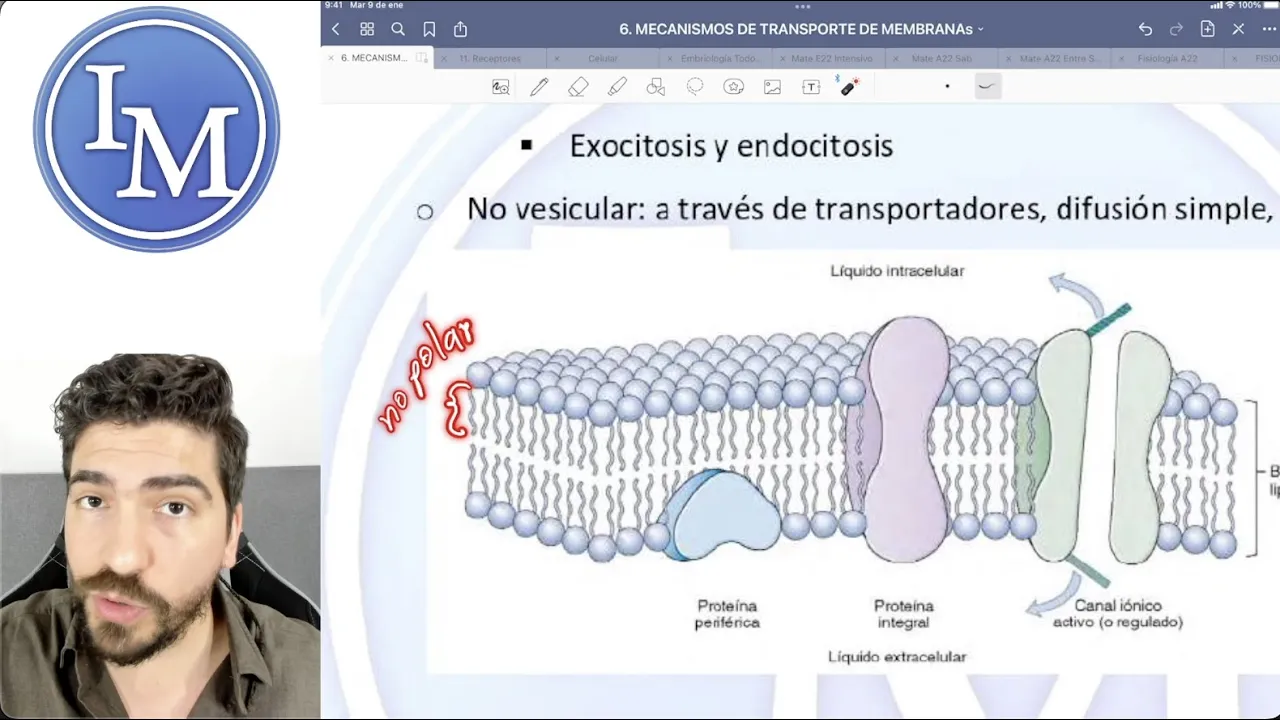
Added:Understanding of the structure and selectively permeable nature of the neuronal cell membrane (lipid bilayer and transport proteins).
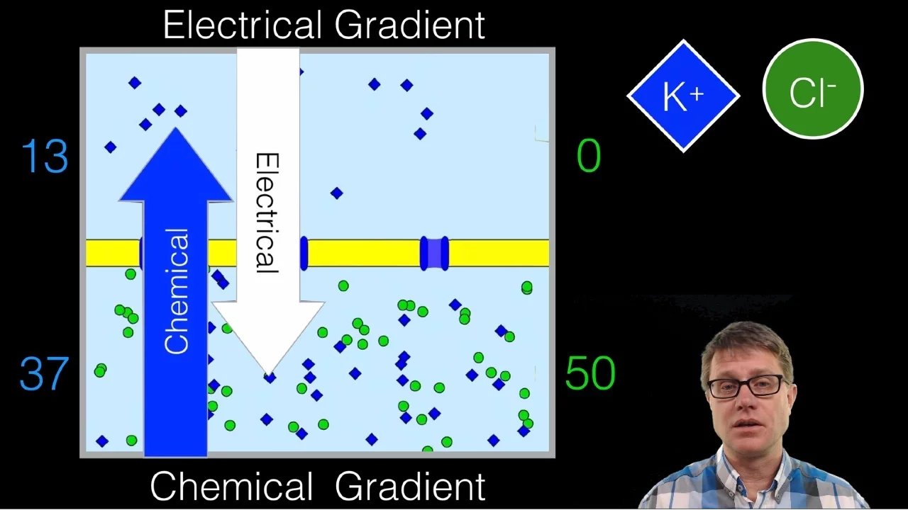
Concept of electrochemical gradients, including the differences between concentration (chemical) gradients and electrical gradients.
The resting membrane potential, specifically how it is established and maintained by the Sodium-Potassium Pump (Na+/K+ ATPase).
Basic anatomy of a neuron, including dendrites, soma, axon, and the axon hillock where action potentials are initiated.
Mechanisms of action potential propagation along the axon, comparing continuous conduction in unmyelinated axons to saltatory conduction in myelinated axons.
The physiological significance of absolute and relative refractory periods in ensuring one-way propagation of nerve impulses.
Synaptic transmission, including how the action potential triggers voltage-gated calcium channels to release neurotransmitters into the synaptic cleft.
Clinical applications and pharmacology, such as how local anesthetics (e.g., lidocaine) or neurotoxins (e.g., tetrodotoxin) block voltage-gated sodium channels.
39.4K views0likes10:58Original Release: 2015-01-06
>>Dr. Ketchum: This is the third video lecture for neural integration, and in this video lecture we’re going to be focusing on the voltage-gated channels. So in order to understand how a neuron responds to its environment, you have to understand how voltage-gated channels can change their configuration, and then ion permeabilities (through those open channels) can change the membrane potential. But you need to understand the background behind or the fundamentals of how these voltage-gated channels work. So in this outline, our focus is to look at the types of channels in which we’ve already talked about leak channels, but we’re going to be focusing on gated channels. And so then once we focus on our gated channels we'll spoke— we will focus on specific types of gated channels as well.
So if you recall, I mentioned earlier that this particular slide you’d be seeing a lot of this semester. And so we’ve already talked about our leak channels and the fact that leak channels are always open. Now we’ve already talked about—in terms of gated channels—chemically ligand-gated channels. So what you should do after we discuss voltage-gated channels is you should compare and contrast what you learned about chemically gated or ligand-gated channels to what you’re going to learn about voltage-gated channels. So the figure on the right here illustrates voltage-gated channels for you. And so these voltage-gated channels are going to open in response to a change in the membrane potential. So when the membrane potential changes, a voltage-gated channel can open. Voltage-gated channels do undergo conformational changes and voltage-gated channels do illustrate the concept of specificity, which we’ve talked about on several occasions now. Please compare and contrast chemically or ligand-gated channels to voltage-gated channels.
So we want to look at where these channels are located on the neuron or in the membrane of the neuron, and various structures that make up the neuron. And so first off for leak channels, leak channels once again are always open, and they’re located along the plasma membrane of the entire neuron. So you would find leak channels in the plasma membrane of the dendrites, the cell body, the axon, the axon terminal. The ligand-gated channels are going to be found primarily on the cell body (we’ll use “cb” for cell body) and the dendrites.
And so you’ve already seen when you studied excitatory postsynaptic potentials and inhibitory postsynaptic potentials, you’ve already studied these ligand-gated channels that are located on the cell body and the dendrites. And so we also have sodium voltage-gated channels and we have potassium voltage-gated channels. These are located at the axon hillock and along the axon. There’s a higher concentration of these sodium voltage-gated channels and potassium voltage-gated channels at the axon hillock. And the function for these channels is to initiate and propagate the action potential. We have a third type of voltage-gated channels and those are called calcium, calcium voltage-gated channels. These are located primarily at the axon terminal. So when you study the synapse and events that occur at the synapse, you will learn about voltage-gated channels and their activity at the synapse.
So first let’s focus on the voltage-gated sodium channels, and then we will return our focus to the voltage-gated potassium channels. The voltage-gated sodium channels have two gates. And that’s very important to remember. There are the inactivation gate and there is the activation gate. It’s very important that you understand how each of these channels works, or each of these gates rather, works. Both gates make up the voltage-gated sodium channel. The inactivation gate on your figure looks like the ball and chain. So when it’s closed, it’s bound to a receptor. And the inactivation gate responds to what the activation gate does. So the voltage-gated sodium channel activation gate is voltage sensitive. Notice we did not say the inactivation gate is voltage sensitive; it is not, it responds to what the activation gate does. So if the activation gate is voltage sensitive, that means that it will open in response in a change in a membrane potential.
So first off, let’s look at the configuration of the voltage-gated sodium channel when your neuron is at rest. So remember the resting membrane potential for a neuron is negative 70 millivolts. In this particular situation, the activation gate is closed and the inactivation gate is open. Remember we called the inactivation gate “the ball and chain,” so you can see how it looks in this diagram. Remember that the inactivation gate has a receptor that when it’s closed, it’s bound to that receptor. If the inactivation gate is going to close, you have to activate the receptor in order for the inactivation gate to close.
So in this configuration, the activation gate is closed, the inactivation gate is open—we call that configuration closed but capable of opening. What that means, and this is a very important note, this means that your neuron is capable of responding to a stimulus.
So when your gates are in this configuration and you place you hand on the hot stove, your neurons can sense the hot stove and you will remove your hand from the hot stove. So this is the closed but capable of opening configuration. So the neuron will respond to a stimulus.
That’s the first configuration for the voltage-gated sodium channels.
The next configuration is the open or activated configuration. So here this is when the membrane potential is from negative 50 or negative 55 millivolts to positive 30 millivolts. So when the membrane potential is between negative 55 and positive 30 millivolts, we have an open or activated voltage-gated sodium channels. So notice the configuration of the gates.
The activation gate is open, the inactivation gate is open, but the inactivation gate is triggered. And what we mean by triggered is that it’s triggered to close. It is not closed, but it is triggered to close. When the activation gate opens, that triggers the inactivation gate to close. But it’s not closed, it takes a couple of milliseconds for the inactivation gate to actually close. So in this case when both the acti—both the activation and the inactivation gate is open, sodium can move into the cell. And when it moves into the cell, it’s moving based on it’s electrochemical gradient. The last configuration for the voltage-gated sodium channels is the closed and not capable of opening. In other words, the channel is inactivated. When you think about this, think about the neuron and the fact that the neuron cannot respond to a stimulus when the channels are in this configuration. So if you put your hand on the hot stove and your voltage-gated sodium channels are in this configuration, your neuron will not respond to that situation. Luckily for us it doesn’t happen, [laughs] and we will explain that later.
Okay, so let’s look at what the gates are doing in this closed and not capable of opening configuration. So here the activation gate is open and the inactivation is closed. Remember that it was triggered to close when the activation gate opened. And now when it’s physically closed, it’s bound to a receptor. So in this particular case, you have the inactivation gate closed and any time you have any gate closed, your ion—such as sodium in this case—cannot move through the channel. So you cannot have sodium influx when your chann—when your gates are in this configuration. Now this configuration occurs when your membrane potential goes from a positive 30 millivolts to a negative 70 millivolts. So we will be using this information when we talk about the ionic basis for the action potential, which will be the next video.
So we also have the voltage-gated potassium channels that are very important in an action potential as well. And when you look at the voltage-gated potassium channels, you will note that there is only a single gate. So we don’t have a particular name for the gate, but there is only one gate. Voltage-gated potassium channels are slow. This inactivation gate is also a tad bit slow. So it gets triggered, but even though it’s triggered, it doesn’t close for a few milliseconds. So back to the voltage-gated potassium channel.
So we said that the voltage-gated potassium channel has one gate, therefore, it has no name and it is slow. So when the voltage-gated potassium channel is closed, this is when your membrane potential goes from negative 70 to positive 30. This is the point in time where these voltage-gated potassium channels are closed. When the channel is closed, potassium ions cannot move through the channel. So potassium ions cannot ent—cannot exit the cell.
When the voltage-gated potassium channel is open, which occurs when your membrane potential is—goes from positive 30 to a negative 80 millivolts. When this channel is open, potassium can leave the cell, or in other words we have potassium efflux, and what’s driving that efflux is the electrochemical gradient. So for both potassium and for sodium, their movement is based on electrochemical gradients. In this case potassium is leaving the cell, taking its positive charge with it. Which is a very important concept that we will return to when we discuss the ionic basis for the action potential, which will be the next video for you to watch.
Up Next
7:50
Resting Membrane Potential: Definition and Nernst Equation
@osmosis
851.9K views•2016-12-13

50:12
Bacterial Communication: Quorum Sensing and Biofilm Formation Explained
@JHUAAP
12.6K views•2011-06-28

5:35
Enteric Nervous System Explained: The Gut's Brain | Neurobiology Lecture
@alumniu6029
438 views•2018-09-12

7:08
Bacteriophages: Earth's Deadliest Killers and Future Antibiotics
@kurzgesagt
34.6M views•2018-05-13
Related Study Plans & Knowledge Roadmaps
Structured learning paths in Biology
biology
RNAi: Biogenesis, RISC & Gene Silencing
01Molecular Biology Foundations: DNA to Protein
02Introduction to RNA Interference (RNAi)
03Molecular Biogenesis of miRNA and siRNA
+ 2 more learning modules
10h5 modules
Explore Roadmap
biology
Mycorrhizae: Signaling, Biophysics & Ecology
01Foundations of Mycorrhizal Symbiosis
02Molecular Signaling and Root Colonization
03Biophysics and Bioenergetics of Nutrient Exchange
+ 1 more learning modules
11h4 modules
Explore Roadmap
biology
Circadian Clocks: TTFL, SCN & Periphery
01Foundations of Molecular Biology & Gene Expression
02Introduction to Chronobiology and Actograms
03The Core TTFL: Molecular Architecture of the Clock
+ 3 more learning modules
14h6 modules
Explore Roadmap
biology
Population Genetics: Math, Drift & Selection
01Genetic & Probability Foundations
02The Hardy-Weinberg Principle and Baseline Models
03Mathematical Modeling of Natural Selection
+ 3 more learning modules
18h6 modules
Explore Roadmap