





![[#2] INTRODUÇÃO À FISIOLOGIA DO SISTEMA NERVOSO: CÉLULAS DA GLIA E NEURÔNIOS | MK Fisiologia](https://i.ytimg.com/vi_webp/AlWtsInlvMY/maxresdefault.webp)





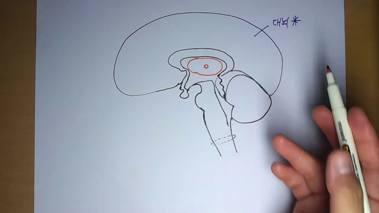


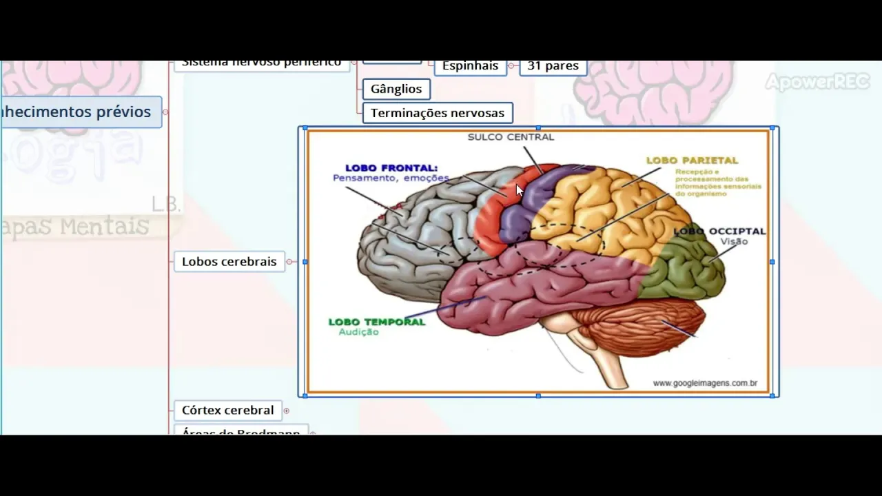






This video provides an introduction to brain anatomy by labeling key structures in a coronal brain section, including the temporal and parietal lobes, corpus callosum, fornix (part of the Papez Circuit), thalamus, substantia nigra, hippocampus, insula, caudate nucleus, red nucleus, internal capsule, cerebral crus, and putamen, demonstrating how different brain regions are spatially organized and interconnected.
Brain Anatomy Introduction: Structure & Function | Soton Brain Hub
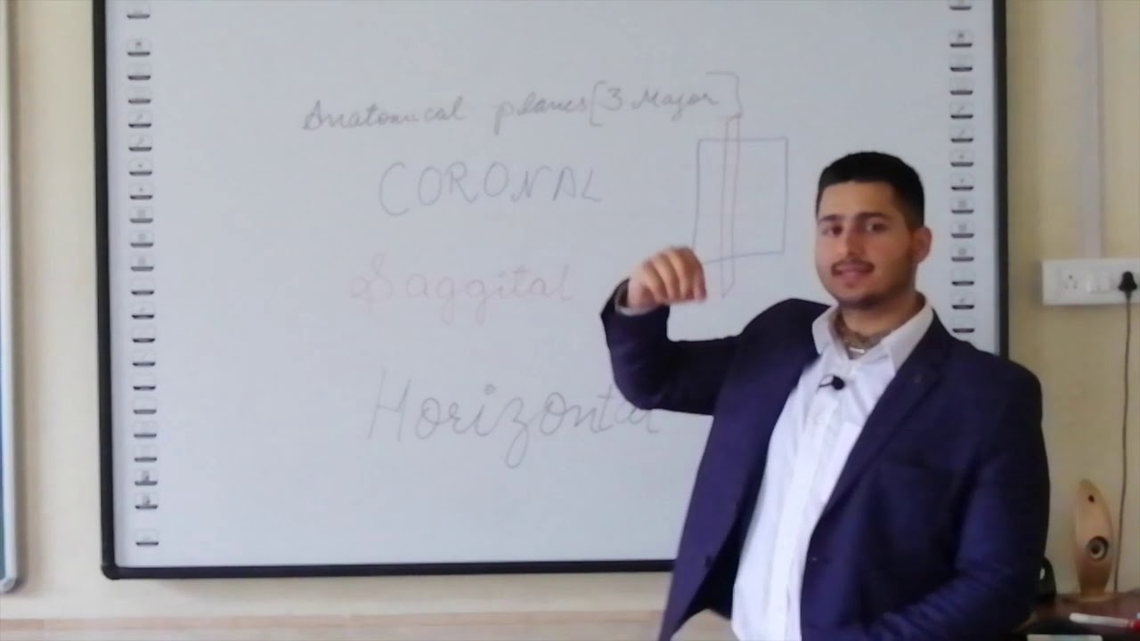
Added:Basic anatomical terminology, including directional terms such as anterior/posterior and dorsal/ventral, to understand spatial relationships in the body.
The fundamental division of the human nervous system into the Central Nervous System (CNS) and the Peripheral Nervous System (PNS).
An understanding of cellular-level nervous system components, specifically the basic structures and functions of neurons and glial cells.
The core concept of neural communication, including how electrical impulses and chemical neurotransmitters transmit signals.
Advanced neuroanatomy, focusing on deep-brain structures such as the limbic system, basal ganglia, and brainstem nuclei.
Cortical localization and functional mapping, including Brodmann areas and the sensory and motor homunculi.
Clinical neuropathology, exploring how structural damage (e.g., stroke, traumatic brain injury) correlates with specific cognitive or physical deficits.
An introduction to neuroimaging modalities (such as fMRI, MRI, and CT scans) used to visualize brain structure and function in clinical and research settings.
9.5K views66likes5:11Original Release: 2015-06-28
[Scott] OK, what we've got here is a coronal section of the brain, and we’re going to do our best to label it so we’ll start of by just labelling some of the lobes. We can see here that the view is quite posterior, so we have to bear in mind parts of the cortex that we’re going to see at this level here. So around this section here, this is going to be the Temporal Lobe. And this section round here is going to be the Parietal Lobe. OK, so we have a Temporal Lobe this side and this side, a Parietal Lobe here and a Parietal Lobe there. We have the Longitudinal Fissure in the mid-line just here. We have the body of the Lateral Ventricle in here. This structure in green is the Corpus Callosum. And the structure in red on each side here is part of the body of the Fornix, this is the major outflow from the hippocampus and part of the Papez Circuit: so body of Fornix there. We’re going to clear these diagrams now so we can start again and label some other structures. So we've also got the Thalamus in here. We have these stained areas in here of course: the Substantia Nigra. In here in each Temporal Lobe the small section of coiled brain there is the Hippocampus.
And in here we have part of the middle Cerebellar Peduncle which is of course in the Pons. Here we have the Medulla and it’s worth pointing out we’ve got an in-folding region of cortex on each side and that’s known as the Insula, like that. Let’s then create some more room for ourselves and have a look at some of the other structures. We've got a couple of structures in here: this is the head of the Caudate Nucleus- part of the Basal Ganglia. We also have the Red Nuclei in here like this-this is the Red Nucleus which is part of the Midbrain. We have the posterior limb of the Internal Capsule which is running down here on each side. So that is the posterior limb of the Internal Capsule (I.C. for Internal Capsule) and in continuation with that we have part of the Midbrain called the Cerebral Crus or Crus.
So that’s part of the Midbrain as well, Corticospinal Tract fibres will run anteriorly through there. It’s probably also worth pointing out that this space just in here is the inferior horn of the Lateral Ventricle. And…and on top of that we have one last structure to name, I'm going to use blue for this: we have part of the Basal Ganglia- I'm not going to use blue for this that’s a silly idea, because that’s too close to the Cerebral Crus. So I'm going to use this yellow colour just to highlight another part of the Basal Ganglia in here called the Putamen. OK, that’s it!
[Andrew] Thank you very much for watching, and remember to subscribe to Soton Brain Hub for more videos to help explain the mysteries of the brain!
Up Next
10:40
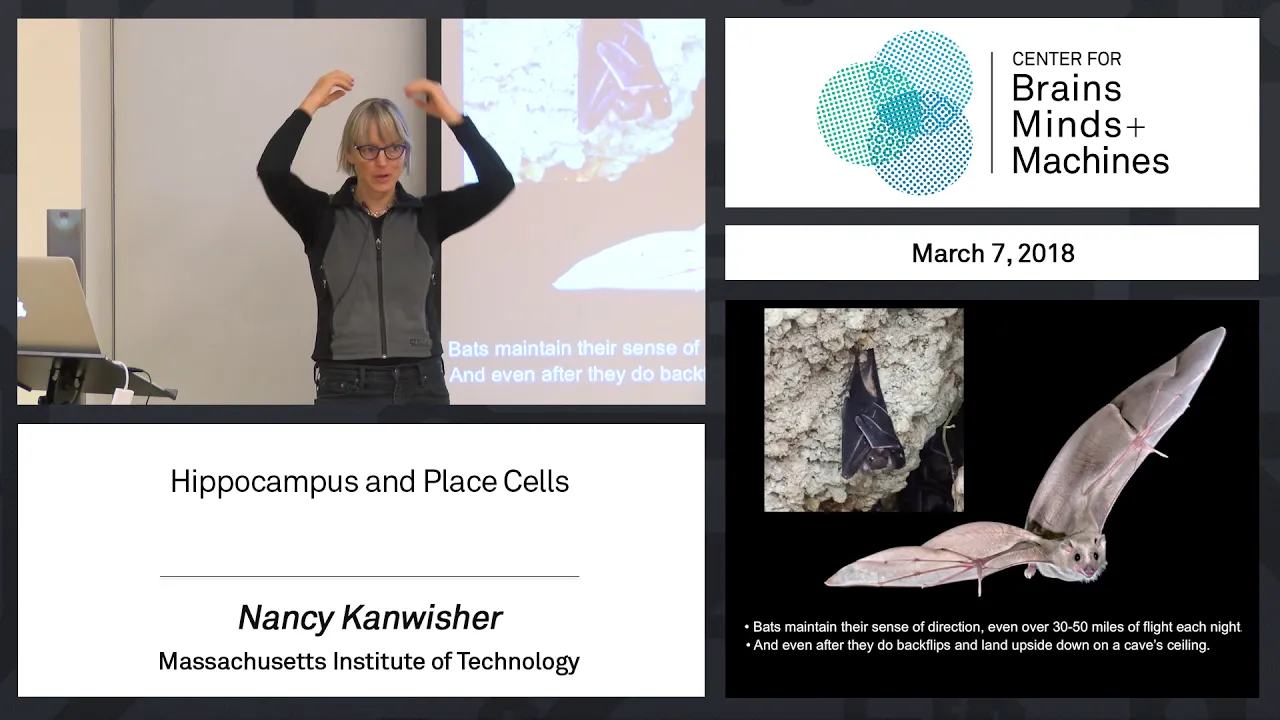
The Hippocampus and Place Cells: Neural Maps of Spatial Navigation
@MITCBMM
19.3K views•2018-05-22

21:55
Deciphering Neural Circuits Controlling Anorexia in Mice | Richard Palmiter
@AllenInstitute
2.3K views•2011-10-10

5:52
Hippocampus Anatomy, Function & Pathology | Neuroscience Tutorial
@SotonbrainhubUk1
3.7K views•2021-08-01

13:03
How Exercise Benefits Your Brain: Science Explained
@TED
11.4M views•2018-03-21
Related Study Plans & Knowledge Roadmaps
Structured learning paths in Neuroscience
neuroscience
Appetite Regulation: Brain Circuits & Hormones
01Foundations of Endocrinology and Brain Anatomy
02Introduction to Energy Homeostasis
03Peripheral Signals: Leptin, Ghrelin, and Insulin
+ 2 more learning modules
14h5 modules
Explore Roadmap
neuroscience
Neuroeconomics: Value, Risk & Brain Mechanisms
01Foundations of Neuroscience and Brain Anatomy
02Introduction to Neuroeconomics and Subjective Value
03Dopaminergic Prediction Errors & Reinforcement Learning
+ 3 more learning modules
18h6 modules
Explore Roadmap
neuroscience
Spatial Navigation: Cells, Grids & Brain Maps
01Foundations of Brain Anatomy and Memory
02Place Cells and Head-Direction Cells
03Grid Cells: The Brain's Coordinate System
+ 2 more learning modules
12h5 modules
Explore Roadmap
neuroscience
BCIs: Neural Decoding & Prosthetic Control
01Neuroscience Foundations: How the Brain Signals Intent
02Signal Acquisition: Recording the Brain's Activity
03Neural Decoding: Translating Brain Waves into Data
+ 2 more learning modules
25h5 modules
Explore Roadmap