BCIs: Neural Decoding & Prosthetic Control
Learning Goal: Understand the neuroscientific and engineering principles of brain-computer interfaces (BCIs), focusing on how motor intent is decoded from cortical signals, processed via decoding algorithms, and translated into prosthetic control and somatosensory feedback.
Prerequisites
- Basic algebra and introductory probability/statistics (matrix multiplication, Bayes' theorem).
- Introductory biology or neuroscience concepts (neuron anatomy, basic electrical properties of cells).
- Familiarity with signal representation (time vs. frequency domain).
Estimated Total Study Time
25 hours (includes video lectures, core readings, algorithmic deep-dives, and implementation exercises).
Module 1: Neuroscience Foundations: How the Brain Signals Intent
This module builds the biological foundation of the curriculum. You will explore the structure of the motor cortex, how individual pyramidal neurons generate action potentials, and how populations of these cells plan and execute voluntary movements. You will also learn about the differences between non-invasive cortical signal manifestations (scalp electroencephalography - EEG) and deep local field potentials.
Recommended Videos
Video 1: Motor Cortex

- Why this video: This video offers a clear anatomical breakdown of the primary motor cortex (M1, Brodmann’s area 4), premotor cortex, and supplementary motor area (SMA). It details the descending corticospinal tract and motor homunculus, which are crucial for understanding how localized neural populations control specific muscle groups.
- Knowledge Checkpoint:
- Identify the location of the primary motor cortex relative to the central sulcus.
- Differentiate between the functions of the primary motor cortex (M1), premotor cortex, and supplementary motor area (SMA).
- Describe how the motor homunculus maps different muscle groups across the precentral gyrus.
Video 2: EEG and Action Potentials

- Why this video: This video bridges micro-level and macro-level neural activity. It explains the biophysics of action potentials (depolarization and repolarization phases) and details how scalp EEG electrodes detect the spatial summation of post-synaptic potentials from thousands of aligned pyramidal cells, rather than individual action potentials.
- Knowledge Checkpoint:
- Explain why single action potentials are invisible to non-invasive scalp EEG sensors.
- Describe the role of apical pyramidal cells in generating the dipoles detected by EEG.
- Define the difference between excitatory post-synaptic potentials (EPSPs) and action potentials.
Video 3: The Motor Cortex and You

- Why this video: Utilizing clean visual animations, this video details how motor intent travels down the central nervous system. It provides an intuitive look at how five core motor regions cooperate to plan, coordinate, and fine-tune movement before sending descending signals to the peripheral nervous system.
- Knowledge Checkpoint:
- Trace the path of a motor signal from cortical planning to muscular execution.
- Explain how contralateral control dictates how motor intent in the left hemisphere influences the right side of the body.
Module 2: Signal Acquisition: Recording the Brain's Activity
In this module, you will transition from biology to neural engineering. You will study how various recording modalities capture brain activity. This includes non-invasive electroencephalography (EEG) and highly invasive penetrating microelectrode arrays (specifically the Utah Array). You will also learn about signal conditioning, hardware filters, impedance matching, and biological tissue reactions like glial scarring.
              [Brain Activity]
                     │
     ┌───────────────┴───────────────┐
     ▼                               ▼
[Invasive: Utah Array] [Non-Invasive: EEG]
- Action Potentials - Synaptic Dipoles
- Spatial homing: ~100μm - Scalp attenuation
- Risks: Glial scarring - Filters: High/Low/Notch
Recommended Videos
Video 1: Behind the Brain Chip: An Inside Look at Blackrock Neurotech

- Why this video: This video provides an inside look at Blackrock Neurotech (historically intertwined with the Utah Array's manufacturing). It demonstrates how penetrating microelectrode arrays capture high-fidelity single-unit and multi-unit action potentials directly from the cerebral cortex.
- Knowledge Checkpoint:
- Describe the physical dimensions and structural composition of the standard Utah Array.
- Explain why invasive arrays are required to record high-frequency individual action potentials (spikes) versus local field potentials (LFPs).
- Contrast the signal-to-noise ratio (SNR) of invasive microelectrode arrays with scalp EEG.
Video 2: Melanie Segado -- Brain Implants & Mind Reading
- Why this video: This video focuses on the chronic tissue-electrode interface. It covers the immune response initiated by rigid silicon arrays, which leads to encapsulation by glial cells (astrocytes and microglia) and can degrade recording performance over time.
- Knowledge Checkpoint:
- Define "glial scarring" and explain how it affects the electrical impedance of recording electrodes.
- Explain why rigid silicon probes cause mechanical mismatch within soft brain tissue.
- Identify the historical lifespan limitations of typical Utah Array implants in clinical research patients.
Video 3: Basics of EEG-based Brain-Computer Interfaces

- Why this video: This video outlines the standard engineering pipeline of a non-invasive EEG-based BCI. It covers the essential stages of signal acquisition, including differential amplification, bandpass filtering, feature extraction, and machine learning classification.
- Knowledge Checkpoint:
- Diagram the four stages of the classical EEG processing pipeline.
- Explain the purpose of a differential amplifier in scalp EEG recording.
- Differentiate between time-domain and frequency-domain feature extraction.
Video 4: Understanding Frequency Filters in 10 Minutes

- Why this video: EEG signals are highly susceptible to environmental noise. This video explains how high-pass, low-pass, and band-stop (notch) filters are applied to isolate physiological signals (2–120 Hz) from slow DC drift and high-frequency noise, such as 50/60 Hz powerline interference.
- Knowledge Checkpoint:
- Explain how a high-pass filter eliminates low-frequency drift caused by sweat or respiration.
- State the typical cutoff frequency used to eliminate powerline hum in both North America (60 Hz) and Europe (50 Hz).
- Describe how a low-pass filter prevents high-frequency muscle artifacts (electromyography - EMG) from corrupting the EEG signal.
Module 3: Neural Decoding: Translating Brain Waves into Data
This module explores the mathematics of neural decoding. You will study how populations of neurons code movement directions, how to compute population vectors, and how to apply state-space models like the Kalman Filter to reconstruct continuous trajectories (e.g., hand or cursor coordinates).
Deep-Dive: Mathematical Formulations of the Decoders
To address the limitations of high-level pop-science overviews, you must master the fundamental mathematical models of neural decoding:
1. Population Vector Algorithm (PVA)
The movement direction vector is estimated as a weighted sum of the preferred directions of individual neurons:
where is the baseline-subtracted firing rate of neuron at time , and represents the unit vector pointing in the neuron's preferred direction of movement (determined during calibration).
2. Kalman Filter (Linear State-Space Model)
The Kalman Filter estimates the latent kinematic state (e.g., position, velocity) from observed neural firing rates :
- State Equation (Motion Model):
- Measurement Equation (Observation Model):
Where:
- represents the state transition matrix (physical system kinematics).
- represents the measurement matrix mapping kinematic states to expected firing rates.
- and are zero-mean Gaussian noises representing model uncertainty and sensor/firing rate variance.
During decoding, the filter alternates between a Predict step (estimating state using kinematics) and a Correct step (refining the estimate using incoming neural spikes scaled by the Kalman Gain ).
Recommended Videos
Video 1: Brain and Behavior - Motor System

- Why this video: This video introduces directional tuning curves in individual cortical neurons. It explains how population vector analysis combines the noisy preferred directions of multiple neurons to reliably decode a single intended movement direction.
- Knowledge Checkpoint:
- Define "directional tuning" and describe the shape of a typical neuronal tuning curve (usually approximated via cosine tuning).
- Perform a simple population vector calculation given three neurons with known preferred directions and firing rates.
- Explain why recording from more neurons improves the directional accuracy of a decoded vector.
Video 2: RI Seminar: Steven Chase: Cognitive factors in neural prosthetic control
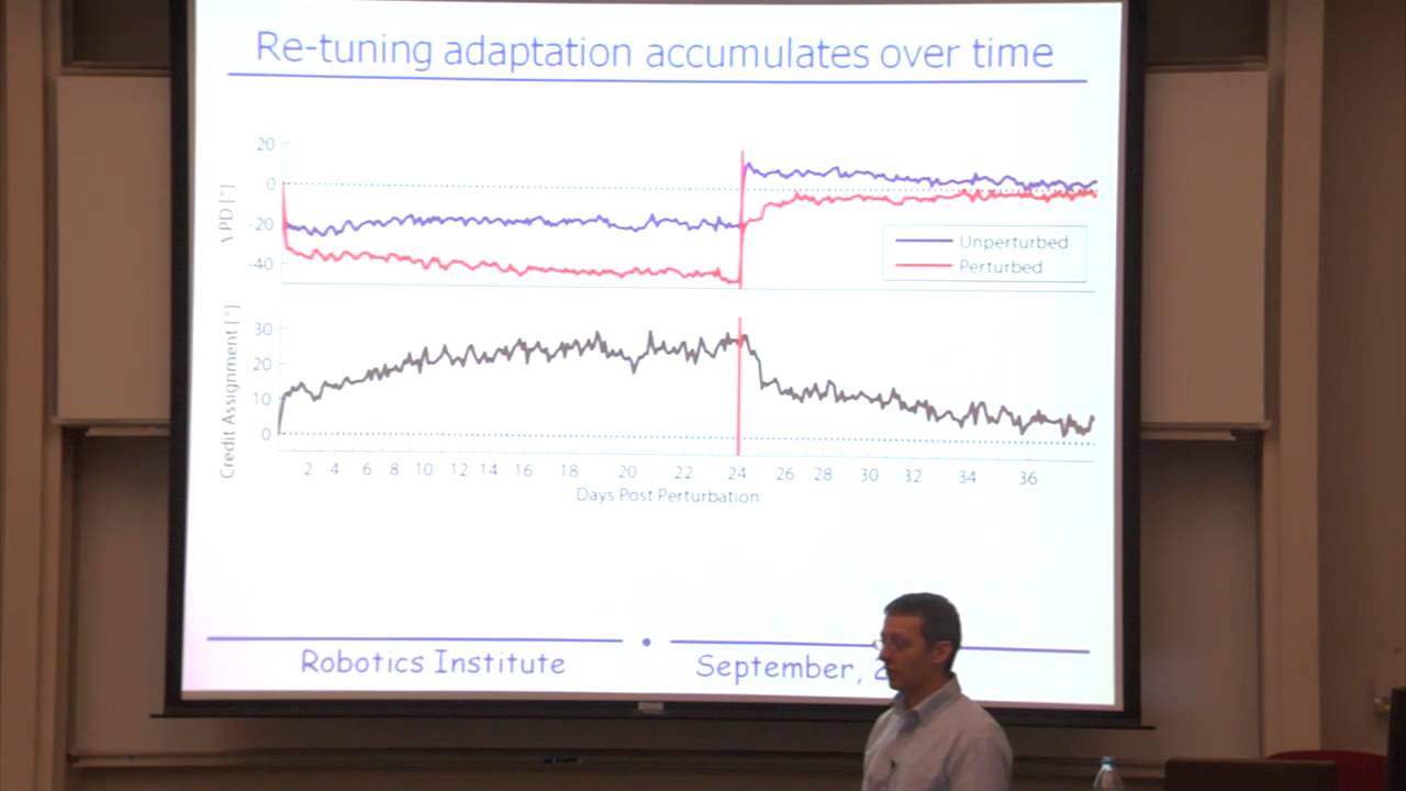
- Why this video: This lecture excerpt focuses on the state-of-the-art decoder in neuroprosthetics: the Kalman Filter. It explains the relationship between the state transition equation (kinematic tracking) and the measurement equation (mapping neural firing rates).
- Knowledge Checkpoint:
- Write down the state transition and observation equations of a Kalman Filter.
- Identify what the variables , , , and represent in a BCI trajectory reconstruction task.
- Explain why a Kalman Filter generally outperforms static linear decoders (like the Wiener Filter) during continuous cursor control.
Video 3: Intuitive Intro to Kalman Filter (Part 1)

- Why this video: This video explains how the Kalman Filter balances mathematical predictions with noisy real-world sensor measurements. It covers how the algorithm weights model certainty against sensor variance to update its state estimate.
- Knowledge Checkpoint:
- Explain how the Kalman Gain () acts as a weighting factor between model predictions and new observations.
- Describe the two alternating phases of the Kalman Filter: the Prediction step and the Correction step.
- Explain how a covariance matrix represents system uncertainty in multivariate states.
Video 4: Next Generations: Machine Learning for BCI
- Why this video: This lecture explores modern machine learning applications in BCIs. It distinguishes between discrete classification (e.g., motor imagery classification for communication) and continuous regression (e.g., decoding joint angles for robotic reach-and-grasp systems).
- Knowledge Checkpoint:
- Contrast classification with regression within the context of BCI decoding.
- Describe how motor imagery (such as imagining left/right hand movement) alters sensorimotor rhythms (mu and beta bands) over the motor cortex.
- Identify one challenge of applying deep neural networks (e.g., CNNs or LSTMs) to raw neural data, such as non-stationarity.
Module 4: Prosthetic Control: Actuating Devices and Robotic Limbs
Once neural intent is decoded, it must be mapped to control external hardware or software. This module explores cursor trajectory reconstruction, multi-degree-of-freedom robotic control, closed-loop visual feedback systems, and functional electrical stimulation (FES) to restore movement to paralyzed limbs.
┌──────────────────┐ Neural Spikes ┌──────────────────┐ │ Motor Cortex ├──────────────────────>│ Kalman Filter │ └────────▲─────────┘ └────────┬─────────┘ │ │ Decoded Kinematics │ Visual/Haptic Feedback ▼ │ ┌──────────────────┐ └─────────────────────────────────┤ Robotic Arm │ └──────────────────┘
Recommended Videos
Video 1: Windows Cursor Control Using a Brain Computer Interface
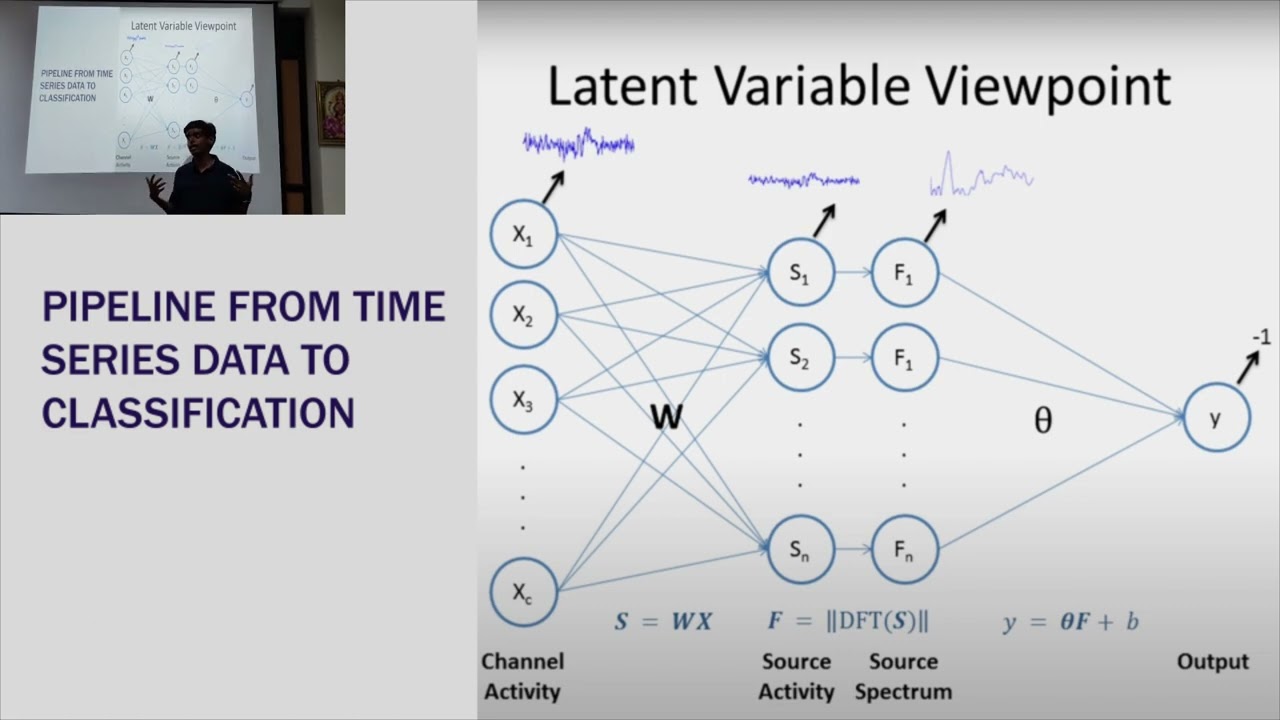
- Why this video: This video demonstrates how non-invasive EEG signals are mapped to a computer screen to control a cursor. It covers the extraction of oscillatory band power features from the sensorimotor cortex and details the calibration routines required for users to master 2D control.
- Knowledge Checkpoint:
- Explain how spatial filters (such as Common Spatial Patterns, or CSP) are used to isolate motor imagery features in multi-channel EEG.
- Describe how real-time visual feedback allows the user's brain to adapt to decoder errors.
- Distinguish between asynchronous (self-paced) and synchronous (cue-based) cursor control designs.
Video 2: Thought control of robotic arms using the BrainGate system

- Why this video: This video details the clinical BrainGate trials. It demonstrates how individuals with tetraplegia can control multi-joint robotic arms to perform daily tasks like reaching, grasping, and drinking from a cup, highlighting the transition from 2D cursor control to 3D physical workspace control.
- Knowledge Checkpoint:
- Identify the degrees of freedom (DoF) required to perform basic reach-and-grasp maneuvers with a robotic arm.
- Explain how clinical researchers map decoded velocity signals to physical robotic actuators.
- Identify the safety parameters required in physical human-robot interaction systems controlled by BCIs.
Video 3: Re-engineering Brain-Machine Interfaces to Optimize Control and Learning

- Why this video: Decoders often experience performance degradation over time due to shifts in neural tuning or electrode movement. This video demonstrates Closed-Loop Decoder Adaptation (CLDA), which continuously updates the decoder's parameters in real-time as the user practices, helping maintain high performance.
- Knowledge Checkpoint:
- Define "decoder drift" and explain why a fixed calibration mapping eventually fails.
- Explain how closed-loop decoder adaptation (CLDA) uses target intent assumptions to update model parameters on-the-fly.
- Describe how co-adaptation occurs when both the user's brain and the machine's decoding algorithm learn simultaneously.
Video 4: Karen Moxon, Ph.D. — The Relationship Between A.I., Machine Learning and Brain Health
- Why this video: This lecture explains how neural signals can bypass injured spinal cords to control Functional Electrical Stimulation (FES) systems. By stimulating nerves in paralyzed limbs, these systems help restore voluntary control to the user's own body instead of an external robot.
- Knowledge Checkpoint:
- Define Functional Electrical Stimulation (FES) and describe its clinical applications in spinal cord injuries.
- Contrast the physical control of a robotic prosthetic limb with the biological control of an FES-reanimated native limb.
- Explain how closed-loop feedback operates when stimulating muscle groups directly.
Module 5: Somatosensory Feedback: Feeling Through the Machine
A bidirectional BCI records motor commands and writes sensory information back to the brain. This module covers the neurobiology of tactile sensations and details how Intracortical Microstimulation (ICMS) stimulates target neural populations in the primary somatosensory cortex (S1) to restore touch and proprioception.
Deep-Dive: Intracortical Microstimulation (ICMS) & Spatial Somatotopy
To address the lack of deep technical video resources in the pool, you must master the fundamental principles of writing sensations back to the brain:
1. Spatial Somatotopy of the Somatosensory Cortex (S1)
The primary somatosensory cortex (Brodmann's areas 3, 1, and 2), located in the postcentral gyrus, is organized somatotopically. Different body areas are mapped to specific locations along the cortical surface. By placing microelectrodes into the hand and digit representations (lateral aspect of S1), researchers can target specific areas to produce localized sensations (referred to as projected receptive fields or phosphenes/sensory units).
2. ICMS Pulse Parameters
Microstimulation involves injecting tiny currents into the cortical extracellular space to depolarize nearby afferent fibers. The stimulus profile consists of charge-balanced, biphasic pulse trains:
- Cathodic Phase: Depolarizes the local neuronal membrane to trigger action potentials.
- Anodic Phase: Balances the electrical charge to prevent tissue damage and electrode corrosion.
Current (μA) ▲ │ ┌──────┐ Biphasic Pulse Train │ │Anodic│ ├─────┼──────┼───────► Time (μs) │ │ │ │ ┌───┘ └───┐ │ │ Cathodic │ ▼ └──────────────┘
To encode different tactile sensations, researchers modulate several parameters:
- Pulse Frequency (Hz): Typically ranges from 10 to 300 Hz. Increasing the frequency generally increases the perceived intensity of the sensation (frequency coding).
- Pulse Amplitude (μA): Typically limited to to prevent excitotoxicity. Increasing the amplitude recruits a larger volume of neurons, which can alter both the perceived intensity and spatial area of the touch (spatial recruitment).
- Pulse Width (Duration, ): Typically 200–400 per phase. This controls the total charge injected per phase:
where is amplitude and is duration.
Recommended Videos
Video 1: Neuralink Launch Event

- Why this video: This event clip demonstrates somatosensory cortex mapping. It explains how stimulating specific locations within S1 corresponds to localized touch sensations in different body parts, such as the digits, illustrating the primary spatial map used to write sensory feedback back to the brain.
- Knowledge Checkpoint:
- Describe the spatial organization of the somatosensory cortex relative to the motor cortex.
- Explain how microstimulation can generate artificial sensations in a phantom limb or paralyzed hand.
- Explain why biphasic, charge-balanced currents are necessary to prevent tissue damage.
Video 2: 7 CRAZY Robot Avatars Controlled By VR
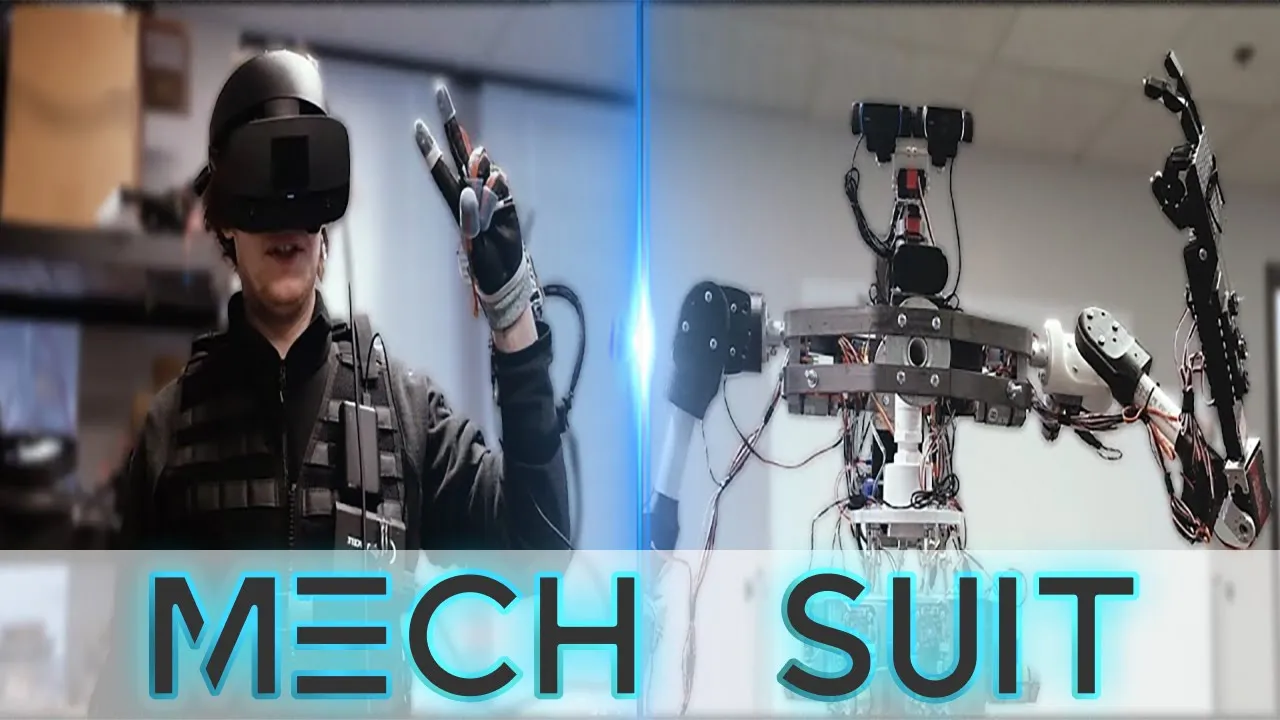
- Why this video: This segment details Nathan Copeland’s clinical trial (conducted by UPMC and the University of Pittsburgh). Nathan became the first person to experience microstimulation-induced tactile sensations through a BCI, showing how sensors on a robotic hand can trigger electrical pulses in S1 to simulate touch.
- Knowledge Checkpoint:
- Explain how pressure sensors on a robotic finger are mapped to S1 microstimulation parameters.
- Identify the sensory qualities (e.g., warmth, pressure, vibration) reported by patients undergoing microstimulation.
- Describe how real-time somatosensory feedback improves a user's performance during grasp tasks.
Video 3: Interfacing the Brain with a Computer for Spinal Cord Injury (and everything else)by Dr. Adam Sachs

- Why this video: This clinical lecture highlights the importance of bidirectional interfaces. It explains why open-loop, motor-only BCIs often feel clumsy, and demonstrates how integrating both recording arrays in M1 and stimulating arrays in S1 creates a high-performance closed-loop system.
- Knowledge Checkpoint:
- Define a "bidirectional brain-computer interface" (bi-BCI).
- Contrast how motor planning in the frontal lobe interacts with sensory integration in the parietal lobe under healthy physiological conditions.
- Explain how sensory feedback helps prevent object slippage during robotic manipulation.
Video 4: Richard Dawkins and Richard Dawkins AI answer questions about the future!

- Why this video: This discussion segment introduces the conceptual future of writing information directly to the brain. It provides an intuitive look at how artificial stimulation can replicate natural somatic inputs, laying the foundation for advanced, bi-directional neural interfaces.
- Knowledge Checkpoint:
- Contrast the sensory experiences of artificial cortical microstimulation with biological tactile transduction.
- Explain the safety and ethical considerations of writing electrical signals directly to human sensory networks.
Course Map
This flowchart maps the logical structure and dependencies of the five modules, tracing the path from raw brain signaling to machine output and sensory return.
Key People Index
The following researchers and clinical pioneers have significantly shaped the hardware, algorithms, and applications covered in this curriculum:
- Dr. Richard Normann (University of Utah)
- Context: Inventor of the silicon-based microelectrode array known as the Utah Array. His work established the foundation for modern invasive multi-channel recording in primates and humans.
- Dr. Miguel Nicolelis (Duke University)
- Context: Pioneer in multi-electrode recordings, population vector analysis, and non-human primate research. His work demonstrated that monkeys could use motor cortex signals to control robotic arms, leading the way for modern clinical brain-machine interfaces.
- Dr. Steven Chase (Carnegie Mellon University / Robotics Institute)
- Context: Expert in the cognitive mechanisms of neural prosthetic control, focus on how the brain adapts to decoder modifications, and developer of advanced state-space Kalman Filter applications.
- Dr. Karen Moxon (UC Davis)
- Context: Pioneer in using machine learning and neural interfaces to restore voluntary control of paralyzed limbs via functional electrical stimulation (FES) systems.
- Nathan Copeland
- Context: The first human clinical trial participant to receive microelectrode implants in both the motor cortex (for robotic arm control) and the somatosensory cortex (for ICMS tactile feedback). His participation demonstrated the viability of chronic, bidirectional clinical BCIs.
- Ian Burkhart
- Context: A prominent clinical trial participant who worked with the Battelle Memorial Institute and the Ohio State University. He utilized an invasive Utah Array to control a custom functional electrical stimulation (FES) sleeve, successfully reanimating his own paralyzed arm and fingers.
Final Self-Assessment
To evaluate your mastery of the material, complete this final self-assessment.
- Action Potential Biophysics: Can you describe the flow of sodium () and potassium () ions that occurs during an action potential, and explain how the resulting extracellular currents summate to produce a signal detectable by EEG?
- Non-invasive Filtering: Can you explain why a notch filter centered at 60 Hz (or 50 Hz) is commonly applied to raw EEG recordings, and describe the potential impact of high-pass filtering on slow-wave DC cortical potentials?
- Invasive Hardware Challenges: Can you explain how the immune response to a rigid silicon microelectrode array leads to glial encapsulation, and identify how this change in tissue impedance affects the signal-to-noise ratio (SNR) over time?
- Preferred Direction Decoding: Can you calculate a 2D movement vector using the Population Vector Algorithm, given the preferred directions and baseline-subtracted firing rates of four motor cortex neurons?
- Kalman Filter Mechanics: Can you write down the state transition and measurement equations of a Kalman Filter and explain how the Kalman Gain updates state estimates during continuous cursor trajectory reconstruction?
- Classification vs. Regression: Can you explain when to use a classification algorithm (e.g., Support Vector Machines) versus a regression model (e.g., Kalman Filter) in a BCI system?
- Closed-Loop Adaptation (CLDA): Can you describe how a CLDA system updates a decoder's parameters in real-time, and explain why this co-adaptation helps prevent performance degradation?
- FES vs. Robotics: Can you contrast the control dynamics and sensory feedback loops of a multi-joint robotic arm with those of a native limb controlled via a Functional Electrical Stimulation (FES) sleeve?
- S1 Somatotopy: Can you identify the cortical region targeted to produce tactile sensations in the index finger, and describe how the postcentral gyrus is organized relative to this region?
- ICMS Safety & Coding: Can you explain why microstimulation requires biphasic, charge-balanced waveforms, and describe how modulating pulse amplitude versus pulse frequency alters the user's perception of touch?