









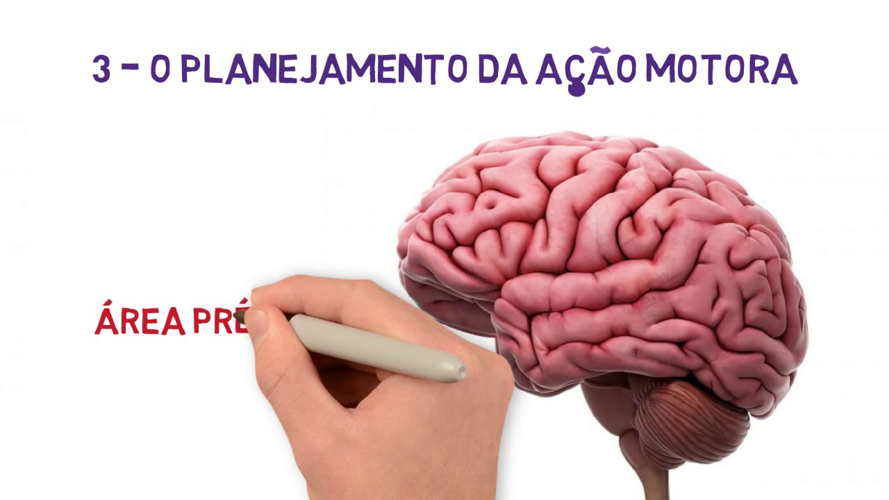





The motor cortex, located in the frontal lobe just anterior to the central sulcus, consists of three main subareas: the primary motor cortex (Brodmann area 4) which executes motor actions through the motor homunculus with plasticity allowing skill improvement; the premotor area which forms motor images, sets posture, and contains mirror neurons for learning through imitation; and the supplementary motor cortex which plans and organizes motor sequences and bilateral muscle contractions. Additional specialized areas include Broca's area for speech production, the eye field for voluntary eye movements, the head rotation area, and the hand skills area which, when damaged, causes motor apraxia.
Motor Cortex: Primary, Premotor, & Supplementary Areas Explained | CNS Physiology
Added:Basic neuroanatomy of the cerebral cortex, particularly the anatomical location and significance of the frontal lobe and the central sulcus.
Fundamentals of neural signaling, including action potentials, synaptic transmission, and motor unit recruitment.
The functional distinction between afferent (sensory) and efferent (motor) pathways within the nervous system.
The concept of somatotopy, specifically how different parts of the body map to specific regions of the cerebral cortex (the cortical homunculus).
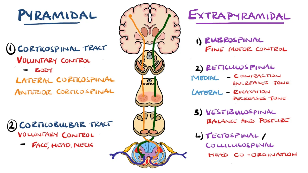
The structure and function of descending motor pathways, specifically the corticospinal (pyramidal) tract and extrapyramidal tracts.
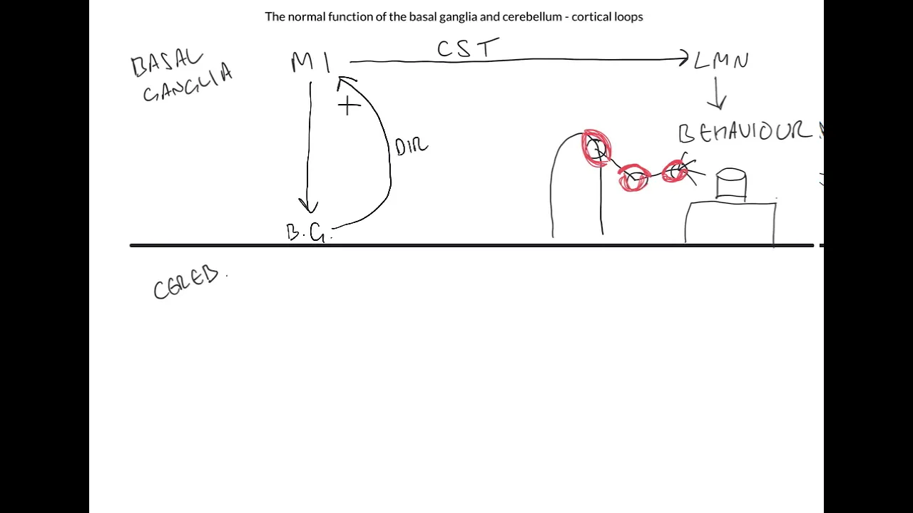
The regulatory roles of the basal ganglia and cerebellum in planning, initiating, and refining motor movements.
Clinical pathologies associated with motor cortex damage, such as Upper Motor Neuron (UMN) syndrome, apraxia, and Broca's (expressive) aphasia.
Modern biomedical applications, including neuroplasticity rehabilitation techniques and Brain-Computer Interfaces (BCIs) that decode motor cortex signals.
7.6K views212likes12:08Original Release: 2022-05-27
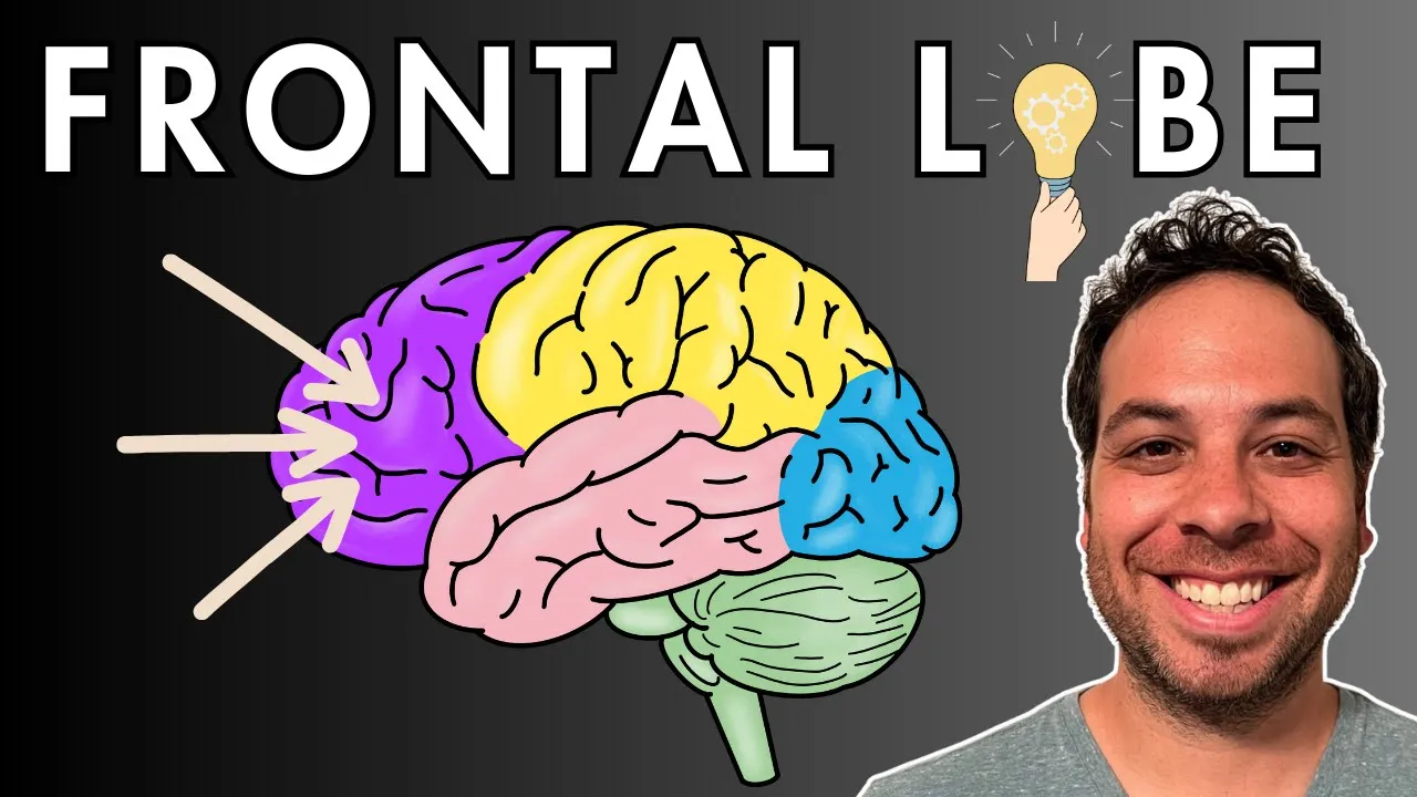
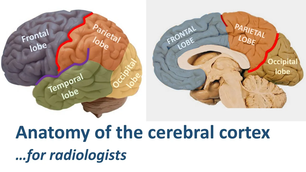
[Music] hello everyone welcome to my youtube channel physiology learning in today's video we are going to discuss about motor cortex so we are discussing our cns lectures wherein we started with the sensory system and we have completed a sensory system in the previous video in the sensory system we saw that the receptors are here and they are going to the brain and reaching the brain and the impulse is perceived so the sensory system we took it from the receptors to the central nervous system whereas in case of motor system the brain has to decide first what action it has to do and then it will send its reasoning tracks and finally the action can be performed so we'll be starting with our motor cortex where the processing is happening for any action to be done so let's dive into our discussion of the motor cortex so coming to today's topic we will be discussing under these sub headings that is the primary motor cortex pre-motor cortex supplementary motor area then finally the specialized areas in the motor cortex so first coming to the location of sub-areas in the motor cortex motor cortex as a whole it is represented by all the divisions that we see are in the learning objectives so where is it located the majority of motor cortex is primarily located in the frontal lobe so it is located in the frontal lobe in our discussion of sensory system itself what we saw where is the sensory cortex located we saw that in the brain there is a major sulcus what is this major sulcus that is nothing but a central circus yes very good this is our central sulcus just behind the central sulcus what was the cortex we saw just behind the central surface we saw that it was the somatosensory cortex whereas just in front of the sulcus what do we have just in front of the sulcus we have our motor cortex what is sulcus and gyri it is pretty simple sulcus is depressions so whenever any depressions are seen in the brain they are called as sulci whereas this elevated portions are called as gyrus so we will be representing that each specific loop on their locations so first coming to the primary motor area as you can see here this is the central sulcus and primary motor area is just in front of the central sulcus and this elevation is called as pre central gyrus since it is an elevation we call it as a gyrus so this area represents our pre central gyrus and this is where the primary motor cortex is located now coming to the next area which is the pre-motor area the pre-motor area is just located in front of the primary motor cortex it is located just in front of the primary cortex then what where is the supplementary area located the supplementary area is located more on the medial side like here we are seeing the outer surface of the brain but supplementary area is primarily located on the medial side and there is one more specific sulcus called as cingulate sulcus in the medial surface the medial surface of the brain if you draw there is a sulcus going like this which is called as cingulate sulcus just above the cingulate sulcus we have our supplementary area so these three areas that is a primary motor cortex the supplementary area and the pre-motor area are the major components of the motor cortex whereas there are some additional areas these are called specialized areas because they have noticed that these areas specifically perform some kind of actions what are those areas first coming to here that is the word formation it is written it is a brocas area this broca's area will study in detail in the language and speech also so this area is broca's area and all these specialized areas are located just in front of the primary motor cortex and second coming to the eye field area here you can see that eye movements is written it is the eye field area then coming to third region which is head rotation as the name indicates it is going to helpful for the head rotation then finally the fourth area that is hand skills so these are four specialized areas which is specifically performing those functions we will discuss them in detail also so the primary quartrix is here the supplementary motor area is here and the specialized areas are just in front of the primary motor cortex so let's discuss our individual topics that is what is the role of primary motor cortex as the name suggests it is the one which is going to perform the action by sending the descending tracts so this area is also referred as em1 area m1 area the primary motor cortex and it comes under our broadband's area 4 in the previous session in summation system also we saw so many broadbands area and the motor cortex that is the primary motor cortex is broadman's area 4.
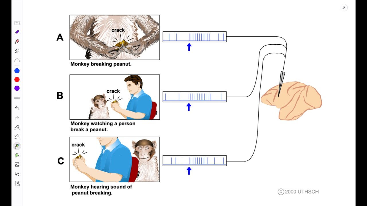
what is the location it is located just in the pre-central gyrus extending up to the central sulcus like we saw it in the sensory system there is something called a sensory homunculus where there is representation of the entire body entire body's representation is also present for the motor cortex also this is called as motor homunculus and here also the size is just proportional to the skill that is performed for example what is the maximum utility of the human organs it is the speech area that is the mouth and the other organs associated as well as the hand movements because majority of fine moments on all the object holding everything of a developed human being is done with the help of hands so the maximum representation is for the motor areas which are involved in speech as well as in hand movements so as we can see in this diagram this person is having the maximum representation of the vocal cords on those areas and as well as the hand because majority of the fine movements and grasping actions and most of the actions are done with the help of hand like we saw it in sensory system here also there is a property called as plasticity what is this plasticity plasticity means as and when the person is developing a skill that region of the motor homunculus can be improvised for example if somebody is a football player then this area's representation will start becoming larger and larger in the motor home and and if some area is cut for example a finger is cut what will happen to that area that area will be merged by the other finger so this kind of plasticity happens in the motor homunculus also now coming to the pre-motor area as the name indicates it is pre-motor area so there is something that is happening even before the motor area is getting the information what is it happening this area what it does is it receives the information from the sensory system and first it will form a motor image and what is this motor image for example if you want to pick a cup of coffee from a table first we have to think like how is the action performed like first we are doing it subconsciously but still there are these things are happening in the brain at a rapid rate what happens is first the distance between you and the coffee cup will be calculated and accordingly your actions will be done this primary motor image is formed in the pre-motor area and where is the location of free motor areas it is located just in front of the pre-central gyrus or the primary motor area and this pre-motor area also also got somatotopic representation of the map that is the representation of human body in the entire pre-motor cortex and what is the function they receive the information from the sensory system and conveys it to the m1 what is m1 it conveys it to the primary motor cortex it conveys it to the spinal cord and it always converts to the brainstem reticular formation it conveys the information to all these regions and it forms the motor image of the action that needs to be performed not only that it also sets up the posture for a planned auction for example if i want to extend my arm but suppose the object is too far what i have to do i have to set the posture like i have to bring my body first and set the portion for it then i will start extending my hand this setting up of the background posture for the action is done by the pre-motor area and they are involved in controlling the proximal limb the logic is pretty simple because the proximal side is the one which is involving in maintaining the posture they give the background setting of portion and the fine moments will be done with the help of distal limbs and fingers and it also has something called as mirror neurons what is this mirror neurons this mirror neurons are very very uh helpful for humans and lower animals also they these neurons what they do is suppose if somebody is performing an action we tend to see it and sometimes repeat and do the actions most of our learning skills are done like this only we observe somebody and repeat it and learn the skill and improvise it on a later stage this mirror neurons are specifically present in the premotor area and this helps to learn new things by imitation like you imitate somebody and learn the new skill so these are all the functions of pre-motor area now coming to the third important area that is supplementary motor area this supplementary motor area is just located to the superior to the cingulate sulcus and it also has a somatotopic map but it is less precise there is no exact representation but it also has something of a somatotopic map that like the primary as well as the pre-motor areas then what is its function it is helpful in organizing and planning the motor sequences it is helpful in planning the motor sequences and not only that it helps in bilateral contractions of the muscle most of our grasping actions for example if you want to hold it it is done bilaterally and it is very helpful for primitive animals which were climbing trees also for example it has to grasp and then it has to climb and this grasping action happens with bilateral contractions of both sides muscle so these are all the functions of the supplementary motor cortex now coming to the specialized areas in motor cortex coming to the most important area that is broca's area this area is also called as area of motor speech motor speech area because whenever we understand something that is done with the help of understanding is done with the help of wernicke's area whereas the word production is done with the help of broca's area we will discuss broca's area extensively in language and speech but here you just know that it is a motor area for speech and it is also located anterior to the primary motor cortex and just above it we have something called as eye field area this eye feel area helps to control the voluntary eye movements and they also help in blinking of the eyes if this area is damaged what will happen is voluntary eye movement will be lost in the person and blinking also will be lost in the person and coming and over another area which is head rotation this head rotation and the eye field area both of them they are in coordination and they help in movement of the eye as well as the movement of the head to see the moving object simultaneously and finding finally coming to the hand skills area this hand skill area is also just located in front of the primary motor cortex whenever any specific lesion happens here the person will not be able to perform any coordinated motor tasks that is called as motor apraxia so all these areas in the motor cortex are generating an image and a motor action which needs to be performed and what they do they send their impulses through the descending tracks and finally they will cause the motor action but the primary planning and everything as well as the motor image formation everything is done in the motor cortex itself i hope it's clear thank you for watching if you like the content give a thumbs up and share it to your friends thank you so much [Music]
Up Next

3:25
Motor Cortex Explained: Brain Anatomy & Movement Control
@davedoodles
11.7K views•2014-04-08

21:55
Deciphering Neural Circuits Controlling Anorexia in Mice | Richard Palmiter
@AllenInstitute
2.3K views•2011-10-10

4:10
Vagus Nerve (CN X): Anatomy, Nuclei & Functions Explained
@Alilamedicalmedia
305.2K views•2022-10-31

13:03
How Exercise Benefits Your Brain: Science Explained
@TED
11.4M views•2018-03-21
Related Study Plans & Knowledge Roadmaps
Structured learning paths in Neuroscience
neuroscience
Appetite Regulation: Brain Circuits & Hormones
01Foundations of Endocrinology and Brain Anatomy
02Introduction to Energy Homeostasis
03Peripheral Signals: Leptin, Ghrelin, and Insulin
+ 2 more learning modules
14h5 modules
Explore Roadmap
neuroscience
Neuroeconomics: Value, Risk & Brain Mechanisms
01Foundations of Neuroscience and Brain Anatomy
02Introduction to Neuroeconomics and Subjective Value
03Dopaminergic Prediction Errors & Reinforcement Learning
+ 3 more learning modules
18h6 modules
Explore Roadmap
neuroscience
Spatial Navigation: Cells, Grids & Brain Maps
01Foundations of Brain Anatomy and Memory
02Place Cells and Head-Direction Cells
03Grid Cells: The Brain's Coordinate System
+ 2 more learning modules
12h5 modules
Explore Roadmap
neuroscience
BCIs: Neural Decoding & Prosthetic Control
01Neuroscience Foundations: How the Brain Signals Intent
02Signal Acquisition: Recording the Brain's Activity
03Neural Decoding: Translating Brain Waves into Data
+ 2 more learning modules
25h5 modules
Explore Roadmap