Fear Circuits: Amygdala, Context & Extinction
Learning Goal: Analyze the neural circuits of threat processing and fear conditioning, detailing how the basolateral and central amygdala coordinate defense responses, how the hippocampus gates fear expression via contextual cues, and how prefrontal-amygdala pathways mediate extinction learning.
- Prerequisites: Basic introductory biology or physiology (recommended but not strictly required).
- Estimated Total Study Time: 12 Hours
Module 1: Introduction to Brain Anatomy & Neurobiology
This module builds a foundational understanding of the brain's structural anatomy and cellular communication. You will explore the limbic system, learn the physical components of a neuron, and analyze the electrochemical propagation of action potentials that allow brain regions to communicate.
Recommended Videos
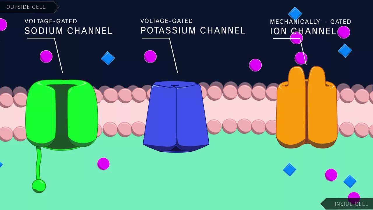
Why this video: This lecture from Harvard Extension School offers an exceptionally rigorous explanation of the ionic mechanics behind neural signaling. It visualizes the shift in membrane potential from resting state (-70mV) to threshold (-55mV), illustrating how voltage-gated sodium and potassium channels operate to propagate electrical signals. Understanding this process is crucial for mapping the synaptic plasticity that occurs during fear conditioning.

Why this video: Delivered as part of Stanford's human biology series, this video provides a concise anatomical overview of the limbic system. It establishes the high-level relationship between the amygdala (emotion and fear), the hippocampus (learning and memory), and adjacent structures like the septum. It acts as an excellent macroscopic map before diving into microcircuits.
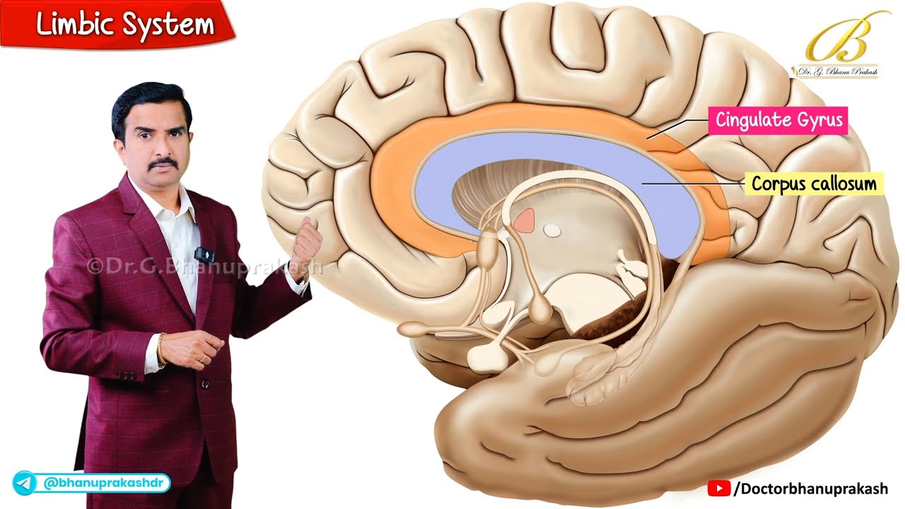
Why this video: This clinically focused breakdown reinforces limbic structures with highly organized, board-style anatomy drawings. It reviews how the hippocampus processes memory consolidation and how the amygdala integrates sensory inputs to govern emotional states, providing a bridge between structural biology and clinical relevance.
Why this video: This animated resource provides a clear visual demonstration of synaptic neurotransmission and the spatial summation of Excitatory Postsynaptic Potentials (EPSPs) at the axon hillock. It clarifies the role of the sodium-potassium ATPase pump in maintaining resting potential, helping you conceptualize how neurons fire in response to sensory stimuli.
Module 1 Knowledge Checkpoint
- Explain how a neuron's membrane potential shifts from -70mV to during depolarization, identifying the specific ion channels responsible.
- Diagram the physical relationships between the hippocampus, amygdala, and thalamus within the medial temporal lobe.
- Differentiate between graded postsynaptic potentials (EPSPs/IPSPs) and the "all-or-nothing" propagation of an action potential down the axon.
Module 2: Fundamentals of Fear Conditioning & the Amygdala
This module introduces the classical Pavlovian fear conditioning paradigm, where an emotionally neutral conditioned stimulus (CS) is paired with an aversive unconditioned stimulus (US). You will study the gross anatomy of the amygdala, focusing on the structural differences between its major subregions.
Recommended Videos
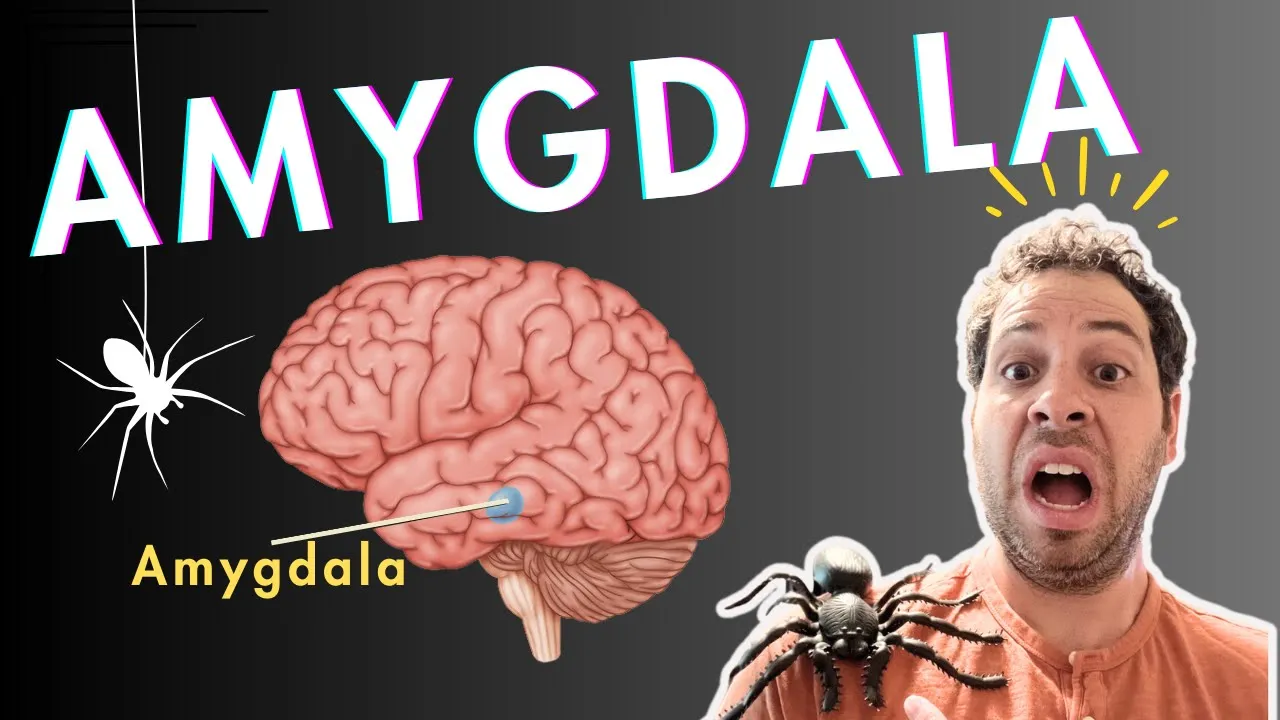
Why this video: This video effectively maps out the dual pathways of fear sensory input: the rapid, subcortical "low road" (direct from thalamus to amygdala) and the slower, highly processed "high road" (passing through the sensory cortex first). This visual model explains how the brain initiates survival reflexes before conscious awareness occurs.

Why this video: Taken from an MIT introductory lecture, this video highlights behavioral studies of fear processing. It explains the outcomes of bilateral amygdala lesions in animal models, demonstrating how damage to these regions abolishes the capacity to evaluate and respond to learned environmental threats.
Why this video: This technical workshop clip maps out the signal flow during fear conditioning. It tracks information arriving at the Lateral Amygdala (LA), transferring to the Basolateral Amygdala (BLA) where synaptic convergence drives associative learning, and finally outputting through the Central Nucleus (CeA) to launch downstream autonomic responses.

Why this video: Though very brief, this video features a molecular visualization of amygdala subregions. It emphasizes that the BLA is dominated by glutamatergic projection neurons, while the CeA is highly compartmentalized and contains GABAergic inhibitory circuits.
Video Pool Gaps & Independent Study Advice
The video pool contains limited standalone content covering the cytoarchitectural and functional divergence of the Basolateral Amygdala (BLA) versus the Central Amygdala (CeA). To supplement this, you are strongly encouraged to independently search academic databases (such as Google Scholar or PubMed) using these target queries:
"Basolateral vs Central Amygdala structural divergence neuroscience""Adolphs threat processing amygdala review"
Focus your self-study on how the BLA is structured like a cortex (layered, primarily glutamatergic projection neurons), whereas the CeA is structurally related to the striatum (primarily GABAergic medium spiny neurons).
Module 2 Knowledge Checkpoint
- Describe the differences in latency, accuracy, and evolutionary utility between the sensory "low road" and "high road" to the lateral amygdala.
- Identify the primary input and output nuclei of the amygdala during a standard Pavlovian tone-shock conditioning trial.
- Contrast the primary neurochemical profile of the BLA (glutamatergic) with that of the CeA (GABAergic).
Module 3: Microcircuits of Threat Processing and Defense Responses
This module explores the inner synaptic microcircuits of the amygdala. You will learn how sensory signals converged in the BLA project to the CeA, and how the CeA's subregions (lateral, ; and medial, ) orchestrate defensive states—such as freezing—via projections to the Periaqueductal Gray (PAG).
Recommended Videos

Why this video: This clip illustrates the application of optogenetics to dissect fear pathways. It explains how researchers use light to selectively activate or inhibit specific pathways, and introduces the concept of distinct "fear" and "extinction" neuron populations within the basolateral amygdala.
Why this video: While this cell paper abstract touches on appetitive behaviors, it provides excellent functional schematics of the BLA-to-CeA projection pathways. It demonstrates how distinct genetic populations of projection neurons in the BLA synapse onto localized target microcircuits in the CeA.

Why this video: Dr. Shansky introduces her research into prefrontal cortex projections to the periaqueductal gray (PAG). She discusses how these circuits coordinate freezing versus active "darting" defense responses, emphasizing the diversity of behavioral coping strategies during threat processing.

Why this video: This clip explains the bidirectional nature of central amygdala microcircuits. It describes how separate populations of interneurons act as "on" and "off" switches for fear expression, illustrating how mutual inhibition inside the CeA regulates defense behaviors.
Video Pool Gaps & Independent Study Advice
The video pool lacks granular animations of the synaptic connections between BLA pyramidal neurons, the Intercalated Cell Masses (ITCs), and the lateral () and medial () central amygdala divisions. To master these microcircuits, search independently for:
"Basolateral and central amygdala microcircuits fear conditioning lecture""Neural pathways of freezing response amygdala periaqueductal gray"
Focus your notes on how the serves as the primary output of the amygdala, projecting directly to the ventrolateral PAG (vlPAG) to trigger freezing, and how intercalated GABAergic cells (ITCs) act as gatekeepers that keep this pathway under tonic inhibition.
Module 3 Knowledge Checkpoint
- Explain how optogenetic tools permit causal dissection of BLA-to-CeA circuits, and how this differs from traditional lesion studies.
- Map the synaptic pathway from a BLA projection neuron to the ventrolateral periaqueductal gray (vlPAG), including intermediate steps within the CeA.
- Contrast the physical, behavioral, and cardiac features of the freezing response with active avoidance behaviors like "darting."
Module 4: Hippocampal Gating and Contextual Fear Conditioning
In this module, you will analyze how contextual fear conditioning differs from cued (sensory-specific) conditioning. You will study how the hippocampus encodes environmental and spatial representations and projects to the amygdala to gate fear expression according to context.
Recommended Videos
Why this video: This scientific panel discussion features neuroscientists debating memory retrieval pathways. It provides deep insight into how both the dorsal and ventral hippocampus cooperate during contextual fear conditioning, moving beyond simple spatial memory models to highlight emotional state gating.

Why this video: This clip provides a clear explanation of how high stress and elevated cortisol impair hippocampal function. When the hippocampus is compromised, the brain struggles to contextualize threats, leading the amygdala to overgeneralize fear responses to neutral environments.
Why this video: This short video shows a real-world scientific paradigm of contextual fear conditioning in action. It illustrates how mouse freezing behavior is quantified 24 hours post-conditioning to measure functional memory and context processing.
Video Pool Gaps & Independent Study Advice
The video pool does not contain a step-by-step diagram showing the precise synaptic projections of the Ventral Hippocampus (vHPC) to the BLA, nor does it detail how vHPC projection neurons modulate fear. For a complete understanding, search independently for:
"Hippocampus to amygdala pathway contextual fear conditioning""Maren contextual fear extinction gating vHPC"
Be sure to research how the vHPC provides direct excitatory input to BLA neurons, allowing a specific physical space (Context A) to activate or suppress the conditioned fear response.
Module 4 Knowledge Checkpoint
- Explain why a mouse with a complete bilateral lesion of the hippocampus will still freeze to a conditioned tone, but will fail to freeze when returned to the training context.
- Contrast the functional roles of the dorsal hippocampus (spatial and cognitive mapping) and the ventral hippocampus (emotional processing and anxiety gating).
- Explain how structural atrophy of the hippocampus in PTSD leads to the "overgeneralization" of fear responses across safe contexts.
Module 5: Prefrontal Regulation and Extinction Learning
This final module focuses on the prefrontal mechanisms that regulate learned fear. You will analyze the contrasting roles of the Prelimbic (PL) and Infralimbic (IL) cortices in regulating fear expression, and trace the prefrontal-amygdala pathway that drives extinction learning.
Recommended Videos

Why this video: This comprehensive, hour-long seminar by Dr. Gregory Quirk—a leading pioneer in fear extinction—is the cornerstone of this module. It provides a detailed look at prefrontal regulation of fear. Dr. Quirk breaks down how the prelimbic (PL) cortex drives fear expression while the infralimbic (IL) cortex drives extinction and active avoidance, highlighting how these structures regulate the amygdala.
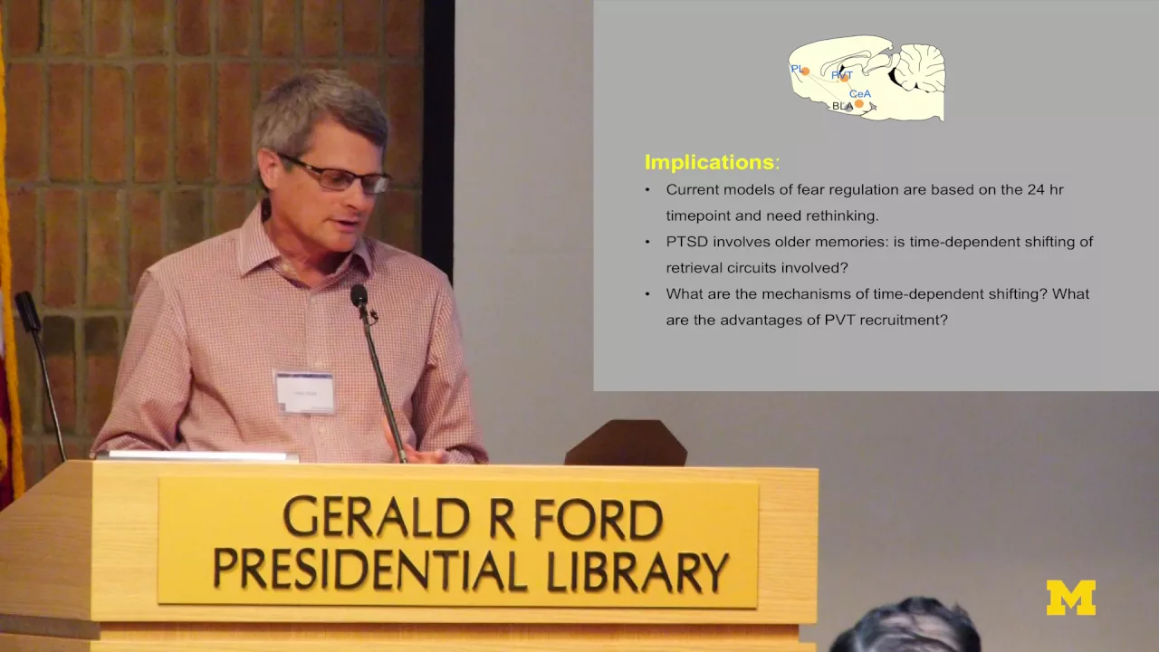
Why this video: In this shorter overview, Dr. Quirk discusses how fear circuits are dynamic. He explains how the structures mediating fear expression transition over time, illustrating that the brain uses different pathways to process immediate learned fear versus remote fear memories.

Why this video: Dr. Kerry Ressler provides an excellent translational talk explaining that fear extinction is not the passive erasure of old memories. Instead, it is an active, new inhibitory learning process that builds competition between prefrontal pathways and the original fear traces.
Video Pool Gaps & Independent Study Advice
While Dr. Quirk's lecture is highly detailed, you may want to seek out visual animations of the synaptic connections between the IL cortex and the intercalated cell masses (ITCs) of the amygdala. For further study, search independently for:
"Infralimbic and prelimbic cortex fear extinction neural circuits""ITC cells amygdala fear extinction"
Focus on how IL projections excite GABAergic ITCs, which in turn inhibit the CeM output to suppress the conditioned response during extinction retrieval.
Module 5 Knowledge Checkpoint
- Explain why fear extinction is classified as an active, new learning process rather than the passive unlearning of the original fear memory.
- Differentiate between the functions of the Prelimbic (PL) and Infralimbic (IL) cortices in fear processing.
- Diagram the pathway through which Infralimbic (IL) projection neurons excite intercalated cells (ITCs) to suppress output from the Medial Central Amygdala (CeM).
Course Map
This flowchart maps out the modules in this curriculum, demonstrating how basic neurobiology builds toward complex prefrontal regulation of fear.
Key People Index
- Dr. Gregory Quirk (University of Puerto Rico School of Medicine)
- Context: Features prominently in Module 5. A leading pioneer in fear extinction research, his lab was among the first to prove the functional split between the prelimbic (fear-promoting) and infralimbic (extinction-promoting) prefrontal cortices.
- Dr. Rebecca Shansky (Northeastern University)
- Context: Features in Module 3. A notable neuroscientist exploring sex differences in fear and stress responses. Her work highlighted alternate threat-response behaviors like "darting" alongside traditional freezing behaviors.
- Dr. Kerry Ressler (Harvard Medical School / McLean Hospital)
- Context: Features in Module 5. A prominent translation researcher focused on PTSD and fear extinction pathways. He translates basic amygdala microcircuit discoveries into clinical treatments for trauma survivors.
Final Self-Assessment
This comprehensive self-assessment evaluates your understanding of the entire curriculum. Complete these items to test your mastery of fear conditioning and regulatory circuits.
- Draw a diagram showing the changes in membrane potential during an action potential, labeling the threshold potential, depolarization phase, repolarization phase, hyperpolarization phase, and the active ion channels for each phase.
- Explain how sensory information (auditory tone) and nociceptive information (foot shock) converge on single cells in the lateral amygdala to trigger long-term potentiation (LTP).
- Map the path of visual threat signals through both the "low road" (thalamus-amygdala) and "high road" (thalamus-V1-amygdala).
- Differentiate between the anatomical structures of the Basolateral Amygdala (BLA) and the Central Amygdala (CeA).
- Map the flow of signals within the amygdala from the Lateral Amygdala (LA), to the Basolateral Amygdala (BLA), to the Intercalated Cells (ITCs), and finally to the Medial Central Amygdala (CeM).
- Explain how the Medial Central Amygdala (CeM) controls autonomic and behavioral threat responses via projections to the periaqueductal gray (PAG) and lateral hypothalamus (LH).
- Define contextual fear conditioning, and explain why hippocampal lesions disrupt contextual conditioning but do not impair tone-conditioned (cued) fear.
- Detail the physical pathway through which the Ventral Hippocampus (vHPC) projects to and gates the activity of BLA projection neurons.
- Contrast the behavioral and neurological roles of the Prelimbic (PL) and Infralimbic (IL) medial prefrontal cortices.
- Explain the neural basis of fear extinction retrieval, highlighting the role of the IL-to-ITC-to-CeM pathway in suppressing fear responses during exposure to a safe environment.