












![[WEBPALESTRA] OS DIFERENTES TIPOS DE TRANSTORNOS DE ANSIEDADE](https://i.ytimg.com/vi_webp/8SM9JRN5mfo/maxresdefault.webp)





The prefrontal cortex regulates fear expression through two distinct inhibitory circuits: parvalbumin-expressing (PV) interneurons in the infralimbic region promote fear extinction by disinhibiting principal neurons, while those in the prelimbic region promote fear expression; optogenetic manipulation demonstrates that PV interneuron inhibition is necessary and sufficient to drive fear expression, and contextual fear discrimination requires intact DMPFC-VLp pathways.
Neuronal Circuits Controlling Fear Behavior: Optogenetics Research
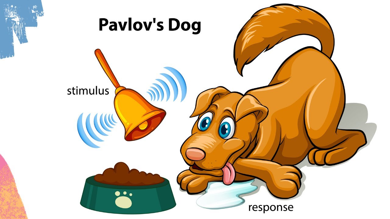
Added:Fundamentals of Classical Conditioning: Understanding Pavlovian fear conditioning, unconditioned/conditioned stimuli (US/CS), and the concepts of fear extinction and generalization.
Neuroanatomy of Fear Circuits: Familiarity with the key brain regions involved in emotional processing, specifically the amygdala, hippocampus, and prefrontal cortex.
Basic Principles of Optogenetics: How light-sensitive opsins (like channelrhodopsin and halorhodopsin) are genetically targeted to control specific neuronal populations with light.
Introduction to Electrophysiology: Understanding single-unit extracellular recordings and how they are used to measure the action potentials of individual neurons in awake, behaving subjects.
Clinical Translations to Anxiety and PTSD: Exploring how dysregulation in fear extinction circuits relates to post-traumatic stress disorder and how these circuits can be targeted for therapeutic intervention.
Synaptic Plasticity and Memory Consolidation: Investigating the cellular and molecular mechanisms (such as Long-Term Potentiation and Long-Term Depression) that stabilize fear memories or facilitate extinction learning.
Advanced Closed-Loop Optogenetics: Studying how real-time neural recording can be paired with automated optical stimulation to manipulate circuits dynamically based on ongoing brain activity.
Microcircuitry and Interneuron Diversity: Analyzing how specific inhibitory interneuron subtypes (e.g., parvalbumin or somatostatin-expressing cells) gate and modulate signal flow within fear networks.
854 views11likes1:41:28Original Release: 2014-08-13
So I will present a bit exotic uh presentation because I'm will talk mainly about auditory fear conditioning and the um the way we have approached the probing of the neonal circuits. So just to start I um uh I will turn the main question we are addressing in my lab. Uh so in this scheme you have a neonal circuit with different neonal elements excited training between neurons. Uh those secrets receive some sensory inputs. Uh these inputs are processed within the network and somehow uh a var output is produced. So the main question we are addressing in my lab is how to relate the plasticity that develops in touch surface uh with virtual learning and as I said just before uh we are using a really simple behavioral model that is the auditory conditioning uh for those who are not familiar with this uh it's a simple model in which animal is presented with conditioned stimulus like a tone that is repeatedly associated with unconditioned stimulus like a food shock. Um and after uh this occurrence the animal learn that the uh sound predict the food shock and on a test day the next day for instance when you present back the tone the animal will display a broad range of condition fear responses including a new mobilization reaction that is a good indicator of the the fear of the animal that is called a freezing response and we quantify that to evaluate the condition here.
So this is just the the conditioning the pairing between the CS atomic QS you get development of your reaction with time.
And interestingly if you present repeatedly the same tone but this time without any aggressive consequences uh in a different context than the conditioning context. you will induce a progressive decrease in condition fear responses and this phenomenon is has been labeled fear extinction. Next, if you um if you introduce a delay between this extinction phase and a retrieval phase where you present back the the sound, uh you will observe that not all the animal but alpha of the animal will show a recovery of condition responses. And this phenomenon is quite important because it tell us that extinction is not an eraser of the original memory trace but extinction is a new learning.
And during a tree goal there is a competition between uh the fear memory trace and the extinction memory trace.
And depending on the winner then show high fear recovery or low fear recovery. Um so which are the noral substrate of uh fear conditioning and extinction.
So this is a coronal slice from a mouse brain where you see uh here the amygdala and in fact has been shown that this region of the medial temporal lobe is really important for uh associative memories and in the context of auditory conditioning. uh the information related to the sound and the food shock reach the amygdala via two main roots directly from the sensor for the sensory or indirectly via the sensory cortex. What information are processed within the lateral and basil amigdala relay to the central amigdula that is the output station that project to brain stem and structure that have been involved in the genesis of uh freezing responses.
So if you make lesion or uh inactivation of this uh region uh you completely abolish condition fear in in memos and it has been shown also that the inactivation of one particular nucleus the basil amydala here also block the extinction of condition responses. So it's know from this data that uh the basolateral amigdala is involved in both fear conditioning and extinction.
uh and uh yeah is involved for the acquisition the storage and the extinction of condition memor so um this is just a clinical point I wanted to make about the prevalence of anxiety disorders among pathological conditions and you see that sorry uh that's anxiety disorder are highly prevalent uh pathological conditions and they uh they are like the um post-traumatic stress disorder order general generalized anxiety, panic disorder or OCD and all these pathologies uh are associated with the hyper activation of the amid. So the synopsis of my presentation today I will try to cover three topics. The first one is related to the encoding of your extinction behavior in basamala circuits.
The second is is related to more prefal secrets and the role of this prefontal secrets in the regulation of fear expression after commissioning. And the last one something that is not published uh related to the role of profal region in contextual fear generalization or discrimination. Uh so the I will focus mainly on the second part of the I mean this would be the longest part of my presentation and this part would involve a lot of optogenetics. I discuss a lot about optogenetic here. Uh for the first part, this work was done in uh was published in 2008 and at this time optogenetics were not really developed.
So I will uh discuss some uh different tools we have been using to address the uh which circuits are involved in fequisition and expression. Okay. And I start just with the encoding of your extinction behavior in basomic data sets. of course just interrupt me when whenever you want.
Um so as I said earlier the amygdala is involved in uh um extinction learning and this has been shown by pharmacological blockade. If for instance you inject an MDA receptor antagonist or mkinas inhibitors directly into the basol amigdala you block extinction learning. During my my PhD I was uh doing some C force experiment. CE force was used as a marker of internal activity in this case and we performed se force staining in the basil amida after extinction learning and we observed that upon extinction we there was a strong increase in the expression of the seos in a particular region the basil amida.
So this prompt us to uh uh work in much more detail about the basil amigdala and which were the neon secrets in extinction learning and to address this question we uh use single units recordings in uh behaving animals. So in principle the principle is is quite simple. Uh you put an electrode in the region of interest in this case it was the basil amigdala.
you posit the electrode around uh some neurons and you pick up the extracellular activity of those neuron that that are action potentials. So in this uh uh representation here you have uh there is a color code for the cells and you have superimposed several action potential that correspond to a single cell. So is extracellular recordings and you can see that um based on the on the cell the waveform shape is quite constant and based on this constancy you can establish cluster of action potential that correspond to single neurons and you can follow the changes in the activity of these neurons during your burial experiment. So we have performed uh single units recording in the basic.
These are the recording site in in this label and we have submitted the animals to auditory condition. So in this paradigm we use two different tones uh the blue one the CS minus that is the control sound that is not associated with the foot and the condition tone in red the CS that is associated with the foot. So uh before conditioning that is during the application shituation session if you measure the freezing responses you see that the presentation of the tones does not induce any particular freezing behavior but after conditioning the animal discriminate between the CS and the CS minus and CS plus uh and after extinction the fear is gone. So you can acquire the fear and reduce the fear. So we have recorded uh two main kind of neurons in the basil amigdala during this paradigm. The first kind of neurons we call them the fear neurons.
Uh what I'm describing here is the changes in neal activity in uh average population for presentation of the CS minus in blue or the CS plus in red and we are looking at the changes in activity before the tone presentation or after the tone presentation. So what you can see for these sphere neurons is that you get an increased activity of these neurons when you play the CS+ after conditioning. But these neuron are silent after extinction learning. They are either even inhibited. So this is the first category of neurons we recorded in the basil amydala. They can I have a question?
Yeah. Do you think these neurons always respond to um to the potential queue? Like if you would record from the same neuron but for so 24 hours later would they respond? So for this particular class of neuron I can tell you that uh if you uh test animal well the first thing is that you need to record the same neuron for several days. This is a this is an issue. But if you are if you manage to do that uh for this particular class of neuron we know that each time the animal display fees those neurons were activated.
So it's like a constant characteristic. Yeah that's exactly. And but when the fear is extinguished they do not respond anymore.
I have a related question. Yeah. What about allocation of new memories? So if you train the animals in a distinct task, diffuse your conditioning task with a different tone. Yeah. Do these neurons get reactivated in a new? So we haven't we haven't performed this experiment. So this is just a guess. I would say that uh uh probably the neurons are activated also to different conditioning session but we haven't tested it in particular. Um so this is they represent 15% of the neurons recorded in the And the second category are extinction label extinction neurons. In fact, they behave the opposite manner than the fear neurons. Those guy are activated only when the fear is low after extinction but not when the fear is high. And they also represent 15% of the neurons. So this is just 30%. We uh uh 50% of neurons recorded were not were silent. And uh yeah, sorry. uh would they respond to the context conditional context? So um to in this particular experiment we extinguish the animal in a different context than the condition context but this is not shown in this figure but we have after the end of extinction we have tested the animal in the original conditioning context and of course when you replace the animal in the conditioning context the fear recovers and of course you observe an activation of the fear neurons.
Okay, so this is just a summary of what I was describing. In gray, you have the uh freezing behavior during the two session of extinction. And in top of it, you have the changes in neonal activity for fear and extinction neurons. So when the fear is high, fear neurons are activated when the fear is low. And when the fear is low, extinction neurons are activated neurons.
So they are inversely correlated during free during high and low state. And the second thing you can see is that when their activity crossover, you have to a change in a significant change in in behavior. Okay. So to address the specificity of your extinction neurons, we design the following experiment. We perform a discriminative extinction. The question here was uh to know if uh the extinction and fear neurons belong to separate class of neuron or if they are the continent of the same class of neuron. So what we have done we have conditioned animals to two different tone the CS1 and the CS2 both were associated with the food shock but we only extinguish one.
uh so we extinguished the first one and what we did we present just simply presented the second town at the end of the extinction. So this is what is depicted here. You have the arbituation you have the fear response to the CS1 that we extinguish and the fear response to the CS2 that was not extinguished and of course we recorded fear and extinction neuron through this paradigm. So what about extinction neurons? As expected uh they first we concentrate on the CS1. This is the early extinction and the late extinction. The extinction neurons are activated when the fear is low during late extinction. In contrast, fear neurons are activated when the fear is high on the on the early extinction but not when the fear is low. So what about when what what is going on when we presented the CS2 at the end of the extinction? This is what we observed is that each as soon as you switch from CS1 to CS2, you switch back the activity of the extinction neurons, they become silent.
And in contrast, the uh fear neurons that were silent start to be activated. Okay.
So the next question we asked at this time uh was whether or not uh fear neuron extinction neurons were inhibitary neuron or excitator reproduction neurons and how they were integrated into the neon secretary of fear conditioning and extinction. And to address this question we use the following strategy. Again as I told you before at this time in 2008 there was no uh the optogenetics were not really developed. So how can you address how can you evaluate whether a neuron project to a structure that is it is a excitatory neuron or uh uh and and where exactly? So we designed the following experiment. Let's say that in the red you have the f neurons we have recorded and those neurons they can uh project to remote structure like the prefal cortex of the of the hypocmpus or they can receive inputs from these structures. So if you uh activate with electrical stimulation the fibers in the remote structure you can drive two kind of neon responses. The first one is called an antidromic response where you induce a spike in the fibers that travels back to the soma. This is an antidromic spike. Or if you stimulate the fibers that project to these sphere neurons for instance, you will generate a orthodromic spike. And the difference between the two is that there is a signapse in between for the autoromic spike. And these signups uh induce a delay uh after a delay in uh introduce a delay and then the action potential that you generate will never occur arrive at the same time. So there is a jitter due to this signups and this is clearly not an example of an automic response. So if you got this kind of response you know that uh the neurons you are recording receive projection from the rem structure and depending on the latency you can evaluate more or less if it is a monosaptic or a poly synaptic connection with antidromic stimulation and antidromically generated spike is much more interesting because there is no signups. So it means that each time you will stimulate the antidromically generated spike will always arrive at the same time. And another charact characteristic of antidromic spikes is that they follow high frequency stimulation. So this is an example of at 200 hertz. And you see that you get the first spike related to the first stimulation, the second one to the second stimulation and the to the third one with always the same latency in between.
So when you get this uh particular responses, you are sure that the neurons you are recording from project to the structure you are stimulating. So this is a way to evaluate how a neuron can uh whether or not a neuron project to different structure. So because we were in the basil amydala if you identify neurons that project to remote structure, they are likely to be excitatory neurons. Based on using this strategy, this is the result we got. We stimulated the the ventral campus and the medial prefal cortex because those two structure are heavily connected with the basil amigdala and for f neurons we observe that uh they receive strong input from the hippocmpus and they project to the prefrontal cortex. In contrast um the extinction neurons are not connected with the inocmpus but they um project and receive input from the profontal cortex.
So something I didn't mention of course um to realize this experiment what you need to do is to first record the neurons during the behavioral experiment and then uh we slightly anesthetize the animal and we stimulated the uh the prefal cortex or the hypocmpus while recording from the neurons identify as fear and extinction. Okay. So using this strategy we were able to show that both here and extinction neurons are excited three neurons and that they are differentially connected with the prefal cortex and the hypocmpus. Okay. So the next u thing we uh did in this experiment was to um try to bring some causality. Again there was no genetic available at this time. So it was not easy to target the f extinction neurons. So what we have decided is to use a pharmacological approach. Uh we use uh we inactivated uh in the basil amigdala by yontroic injection of mucimol that is a gaba a receptor agonist and um and to do so we use the following techniques. So uh we perform uh the ytoic injection with this device that we built. In fact, you have a glass pipet that was filled with mucimol and uh around the tip of the uh the pip pipet, we posit some electroility of the neurons. And the first experiment we did was to evaluate whether the mucal injection [Music] was inhibiting neonal activity and that's exactly what you see. This is multi-unit activity before the injection. And as soon as you do the mucual injection, you see that you reduce completely the uh you don't see any any more spikes and this for two hours. So we have a temporal window of two hours uh to inhibit uh the neon activity within the basil amidala. So this is the kind of injection we were able to do a really targeted injection and we uh we use for controlling uh the diffusion we use the fur on so you can precisely control injection sites uh um and this is the burial result we got at this time. So this is a simple conditioning experiment with two extinction session. We have the control group in blue that is just the fur injected animals. So if we concentrate on the control group first.
After conditioning, they display IP responses. You can get extinction with time. And in comparison, if you inject the mucimol before the second extinction in the group in red, the mucimal injected animals, you see that you completely block extinction. And this was uh not an effect due to a lesion because the next day in a drug-free situation if you reexpose the animal to the sound they they they got ext they extinguish the field. Okay. So this demonstrate that the basil amigdaline activation completely block extinction neurons. So from those data we have shown that there is sub population of neurons in the basila that specifically encode fear and extinction behavior.
Uh these data are interesting but they uh also raised uh other question and the the two question we were interested in are the following one uh what is the anatomical connectivity of human extinction neurons the pre size one not only the MPFC but which region of the prefrontal cortex for instance and uh is there any mean by which we could specific specifically manipulate those fear and extinction neurons doing fear behavior.
So to address this question what you would like to do is to label the neurons to inject the D in the neuron and and and here we have a problem because we use extracellular recording so we cannot label the neurons. So how to turn around this question we develop the following strategy. So what do we know about fear and extinction neurons? The first thing first thing we know is that fear neurons project to the prefrontal cortex and extinction neurons project and get input from the prefrontal cortex.
So what we have done we have used a retrograde tracer injected in the prefrontal cortex to back label uh the cell bodies of the neurons projecting to the MPFC and uh because um different region of the prefrontal cortex are known to differentially modulate fear behavior.
Uh in particular there is the dorsal region or the prenyic area that is known to when you stimulate this region you induce fear behavior. In contrast, the infraramic region is known to be involved in extinction. If you activate those guy was this structure here, you induce low fear responses. So obviously we injected the retrograde in the PL and BIL. And the second thing we know is that uh fear neurons are activated when the fear is high after conditioning and extinction neurons are activated when the fear is low after extinction after extinction or during extinction. So in complement to the retrograde tracer injection we perform C4 imostaining in injected animals. And here you have an example of a retro labling of amydala neuron in red here coupled with C force detection here. So these neurons um has been activated during a particular behavioral task and is project to the NPFC. So this is the strategy we have used to identify fur and extinction neurons. So these are the result for the seforce experiment. Um so we of course we quantify the collabing of the retroid and the se force and we did that after conditioning and after extinction. So first when we uh evaluate the total population of prefontal neurons including PL and IL productive neurons uh we observe that we get a strong C force expression after conditioning and after extinction. But now if we if we separate into PL and IL, we observe that uh the neurons from the amygdala that project to the PL were strongly activated after conditioning but not after extinction. Whereas the extinction the sorry the neurons that project to the intralic area were strongly activated after extinction and to a lesser extent after f conditioning. So these two uh uh population the PL projecting or the IL projecting uh could correspond to fear and extinction. That was the the idea of this experiment. So um we at this and this paper was just published uh this year uh beginning of the year uh it has been a long story. This was a followup of the the Fon extinction paper we published in 2008. But this time there was autogenetic available. So uh to perform those autotogenetic manipulation of uh the PL and IL protecting neurons we use the following strategy. So we injected a virus we basically we injected two virus that uh uh bring create the cre re combination to the sum of neurons in the BNA. So they are retrograde viruses. If you inject it in a targeted region, they will travel back to the soma and label the neurons. So we have used a combination of two. I will discuss that a bit later. We have used her simplex virus expressing re combin or the calf 2 virus that is a canine adino associated virus. Both are transported retrogradely. And here you can see uh you know if you see that you see the labing of the neurons in the braic data that project to the pl to the So this is an example of uh neurons we were able to drive uh using this strategy. So here you see each line correspond to uh sorry each dot correspond to action potential and each line correspond to one one trial for um of course we have complemented the the injection here in the basil amigdala with a credependent virus that was expressing both the channelopsin and the aloropsin.
So we could manipulate the neurons in two different ways uh in the same preparation. So of course in blue you will activate the channelopsin in yellow the aloropsin and we identify neurons that were we were able to drive neurons or to inhibit neuronal activity and u we have done also we have coupled this injection with recordings of the neurons. So we have identified extinction neurons and fear neurons and uh of not of course but uh this was um an output of the of the story is that all the neurons that project to the IL or I will put it in a different way. All the identify extinction neurons were neurons projecting to the all fear neurons were neurons projecting to the prenic area.
So uh and then we of course we manipulated the IL and PL protein neurons with autotogenetics uh and these are the behavioral result.
It's just a summary but in essence uh following extin if we manipulated the IL and PL proteing neurons during extinction learning we got an effect the next day. Uh and I will concentrate here on the inhibition of the neonal activity. If you inhibit the neonal activity of extinction neuron or IL protein neurons, you induce an increase in fuel responses the day after extinction. In contrast, uh if you inhibit the activity of fear neurons, you induce a decrease of condition responses after extinction.
So these demonstrate that uh the optogenetic manipulation of plyic and infrolic protecting um basil amigdala neurons oppositely affect long-term extinction memories. Okay. So uh this was the first part about the fear and extinction neurons and I will switch now to the prefal cortex and I first before doing that would be scheme. So to summarize what we have have shown so far uh so you have here an extinction neurons in the basilic data we know they project differentially to the uh pyic or the the dorsal profile cortex or the ventral um infrolic area. So extinct neurons project to the preamic extinction neurons do the uh infraic and we know that the stimulation of these two areas uh prod prod use different output. If you activate the PL, you induce high fear. If you activate the IL, you induce low fear. So we know that the prefer cortex is involved in the regulation of fear behavior. But what is not known is which are the neonal elements the particular neonal elements in particular for the pomicaria that are involved in the regulation of your expression. So this is just what I was saying. So if you perform inactivation of the MPFC uh you block fear expression. Um if you perform electrical stimulation of the plyic area you induce fear behavior and uh from the group of great work in in Puerto Rico they have shown that uh you have neurons which neonal activity correlate with fear expression in the picaria.
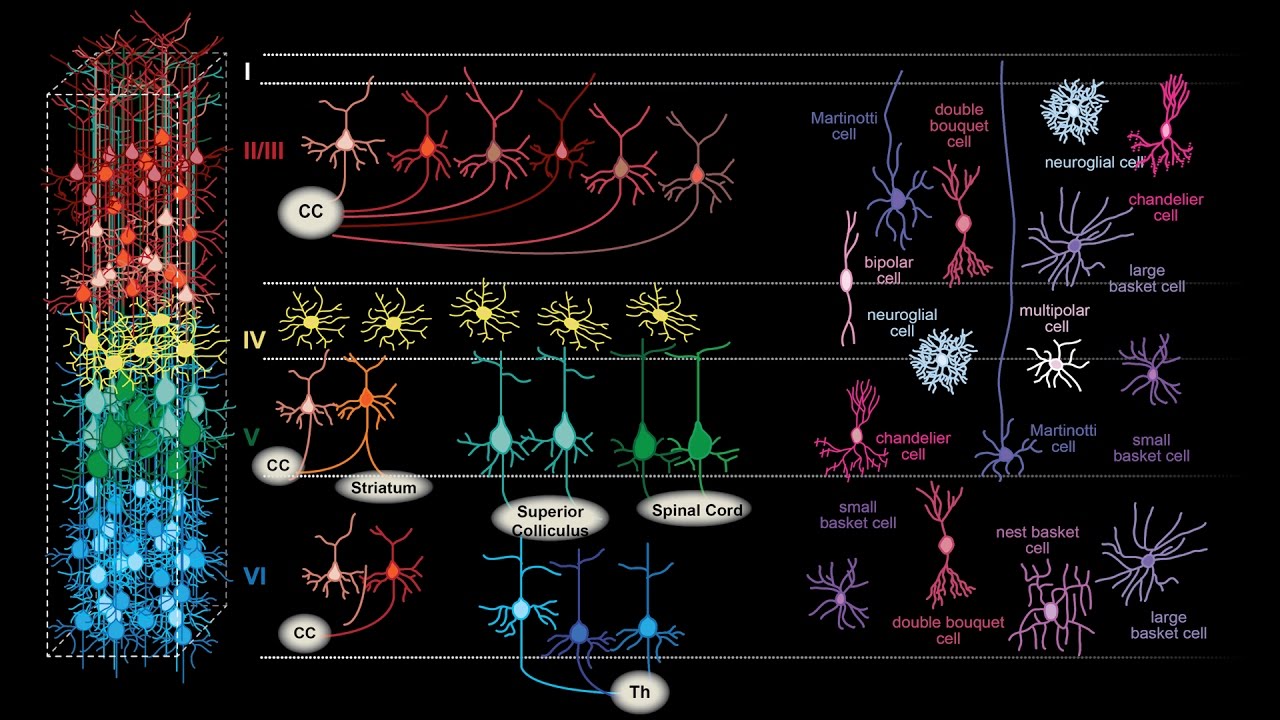
So this is the background uh and as I said the particular neonal elements involved in the pimic area are completely unknown. So that was the question we were addressing in the in the study right now. So like in any other cortices the profontal cortex contains both excitatory neurons in red here and a myriad of inviting neurons that represent 20% of the cell population. And I will focus particularly on uh the inhibitory neuron in blue that are called the basket cell and the chandelier cell because they target the soma of the excitatory neuron and the axon lock and uh with these properties they can precisely control the act the output activity of the principal neurons. So they are uh really interesting candidate because they can block or uh allows the the neurons to fire.
So we know from uh we know from anatomical studies that the pyramidal excitatory neuron in the prefrontal cortex they project to both the basolateral amigdala and the periodical gray and to many other structure but I'm just mentioning the one that are involved in fia behavior the basil amigdala and the periodical gray uh as I said there is different population of interurons that regulate the activity of principal at different level at the s at the level as well and uh the basket and the shand cell that control the cell body are known to express parallel they are p expressing inter neurons.
So we uh in this study we asked the question uh of what was the role of prefontal inry in neurons during condition behavior and in particular of the role of the uh PV expressing in the neurons. So again we use single unit recording at the start. This time it was not implanted in the amydala but the dorsal profile cortex. And again we were able to follow the activity of single neurons during during the session. The behavior was the same. We used the classical fear conditioning with an habituation session where we presented the control sound the CS minus or the CS plus. We conditioned the animal to the CS plus only and we have two day of extinction extinction one extinction two. After extinction two you get a reduction a significant reduction of condition few responses. And if you test the animal in the extinction context seven days later you get a recovery of condition theory species. So this is the classical behavior and we recorded the activity of uh inter neurons and pure inter neurons during each of the session. So this is where we have done the recordings in the dorsal prefal cortex the singulate area and the pyic area. We were able to record around 700 neurons in these structures and among those neurons around 67% were tone reactive neurons. The other one were just not reactive. So the first thing we did we tried to separate the neuron into putative excitatory neurons or putative inhibitary interurons using electrofphysiological criterion. The first crit was the firing frequency of the neuron. Uh the second one was the spike al width al width and the third one was the curve the area under the curve that is supposed to correspond to the after hyper polarization from spikes recorded recorded intraceller. So using this strategy this is a completely unbiased uh methodology.
We are not we are not selecting the neurons that belong to each class is an automatic automated process and you can clearly see two cluster of neurons in blue the one that correspond to the putative excitatory neuron and in red the one that correspond to the putative inhibitary inter neurons and characteristic of the putative inhibitary inter neuron is that uh the al spike width is shorter than for um putative excitatory neuron and we observe also So really fast spiking neurons in the putative tree cluster the putativon cluster here. So using this strategy um among the um the 493 tone reactive neuron 29% were classified as 10 neurons and the remaining were classified by excitatory neuron and this roughly correspond to what has been described in the literature about the proportion of excitatory and in between neurons. So for the the for this particular study as I said we focus on the inhibitory inter neurons here and uh we wanted to know what was the firing pattern of the neurons during behavior. So for this we use unbiased meth methodology called the principal component analysis to identify the firing pattern of the neuron in response to the tone presentation.
This is for inter neurons. You have the tone onset here. And this is the two main patterns we observe for the initial inter neurons uh patterns we call the type one and type two. Type one the neurons are excited following the tonal set and type two they are strongly inhibited. This is an example of such neuron the excited one type one or the strongly inhibited one the type two. And this is the population of neurons we recorded. So we got 68 type one inter neurons putative inter neurons and uh 15 putative type two inter neurons. So as you can see on this uh graph they uh those two type of interons display antagonistic fing patterns that's something I will come I will come back to that in in a moment. Okay so what are the characteristic of these uh type one and type two inter neurons. First we wanted to know whether they were segregated within the simulate or the premaria but we found that they were uh present everywhere in the simulate and the p. So there was no particular layers where those neurons were present or not but they were everywhere in the singulate and picaria.
We look at the firing rate to discriminate between type one and type two inter neuron. And we observe that type two inter neurons display fast firing patterns in comparison to type one inter neurons. So this distinguish what we call the regular versus the fast fighting inter neurons. And I remind you that I mentioned that in the past that we observed within the um the two cluster of excitatory and in between neurons we observe some neurons displaying a high firing rate and basically this is those guys here. So again we have the type one and the type two in neurons and uh we went a bit further into the analysis. we look at whether or not they were phase locked to uh uh to some oscillation uh because this phase locking is characteristic of some inter neurons. So for type one this is a theta osillation and on top of it you have the firing activity of the neuron. What we observe for type one inter neuron is that there were not phase lock to the oscillation to theta oscillation. In contrast, we observed that the type two interurons were phase lock to the peak of the osillation. So this is a difference between type one and type two. Type two inter are phase lock to t oscillation.
And this is more visible on this spectrogram where you clearly see uh the locking of the neuron to a particular band that is in the 8 herz range. Uh that is the pa oation and from the literature this has been described in the cortex as being uh as corresponding to parvaline parvaline expressing interance and this to nonparaline expressing interance. So using just those uh criteria we have a a clue that type two might be a PV expressing interance but not type one.
And of course to identify uh this in a more precise manner we switch to adoptogenetic strategy this time. So we use um mice that express the cre combinese under the control of a paralumin promoter the creep mice in which we uh we perform a conditional expression of arurodopsin. So we injected the credependent virus in the prefrontal cortex uh to label with arurodopsin only the PV interons. So I won't go into the detail of optogenetics because uh lex uh provided a clear description this morning about how it works. I will just uh um discuss what what we observe here.
So this is an example of a type two interuron that was identified based on those criterion and this is the control situation. We don't have light. So each line again is a a trial where we have the neonal activity and you see that under the control condition there is nothing of course but as soon as we switch on the light that is uh um we um inhibited PV interance we were able to identify this guy as a PV inter so the uh sorry so this is one example but we did that for a certain amount of neurons we have n of five here where we were able to uh identify the type two interurons as being uh PV expressing inter neurons and none of the type one inter neurons uh was showing any inhibition indicating that type one are not uh PV expressing interurons. So from this optogenic experiment we were able to say that the type one and type two inter neurons we were we recorded from the MPFC where uh the type two were PV expressing inter neurons. Uh so when I'm saying that uh type two are PV expressing in neurons I'm not saying that all PV are type two internals. I will explain this in a detail. So we know that the type one are type one inter neurons are not are not PV expressing inter neurons and um among the PV interurons we were able to identify what some were type two interons but other were not identified. So what I'm saying here is that um oh here sorry what I'm saying here is that the type two inter neurons are a particular class of PV interurons. Uh you have several uh class of PV interurons only the type two class of inter neurons were inhibited during tone presentation. Those guy that were also PV interurons were not inhibited during tone presentation. So type two are a particular class of um a functional class of PD expressing interments. Okay, this is just some um control we provide for uh the the some anatomical controls. What we have done here we have injected the viruses expressing the HT in PV3 mice and we wanted to control that all the sale labeled were PV neon. So we call label PV and you have the overlap here and in fact we observe that 99.9% of um the uh GFP positive neuron that is the neuron expressing RT were also PV expressing in neurons and we have done some slice physiology experiment to evaluate um whether the expression of RPOPS in those PV neurons will change their firing pattern.
So maybe we can just we were able to uh record currents in activated or inhibited neurons using channelopsin or archeropsin. Uh but this is maybe the more critical experiment we have done.
We have recorded from GFP neurons.
Uh so PV expressing just GFP, PV expressing channelopsin or PV expressing HTT receptor and we have performed in we have performed current injection and measure the firing frequency of those neurons and there was no difference between um those three category of PV neurons in um in following the current injection.
So they behave the same way than the neurons that were not infected with archeropsy. That was an important control for us. Okay, I mentioned earlier that type one and type two display antagonistic firing patterns. So this suggests that maybe type one could inhibit type two inter neurons. And how did we address this question? We uh use the following strategy. uh the first thing we have done we uh compare the um the um the activation and the inhibition latency of those neurons. So in type one you have the uh the excitation in type two the inhibition and you see that there is a kind of perfect match between the latency of those two neurons indicating that type one could inhibit type two and 10 neurons but we were not uh this was not enough for us. What we wanted to do is to record in the same animal from type one and type two inter neurons. And on both guys what we on both those animals we have evaluated the firing latency of type one and type two inter neurons. And we observe that type one inter neurons fires always before type two inter neurons suggesting that they can inhibit type two inter neurons. Okay. So this was just a characteristic. We know now that type one are nonp expressing inter neurons, type two are PB expressing inter neurons and the question was uh what was their behavior during presentation of the CS plus or the CS minus. So this is again the two examples type one and type two uh and this is the response to the tone presentation in blue the CS minus the control tone or in red the CS plus. So you see that for the type one those guys were strongly activated to the CS plus in comparison to the CS minus and for the type they were strongly inhibited to the CS plus in comparison to the CS minus. This is the quantification of it.
We have on the same graph in gray the freezing behavior during the CS minus and CS plus. So low fear high fear and in top of it you have the changes in yal activity during the first 200 millisecond for type one and type two inter. So for the type one you see there is a almost perfect match between uh the behavior and the change in neural activity. When the fear was low the neurons were not activated. When the fear was high they were activated and the type two were invertly correlated with freezing behavior. That is when the fear was low they were not inhibited but when the fear was high the type two was strongly inhibited.
So this is just the conclusion. Yeah.
How would you comment that you have quite a robust reaction to So as you have seen maybe in the uh if I go back just to that um okay this is the behavior uh in fact you see that you have some residual freezing to the CS minus they are not at zero they're at around 20%.
So some animals may display higher physic responses to the CS minus. So it's why it's why you see this uh uh this response to the CS minus uh in this example here. So I'm not saying that the animal do not freeze at all during CS minus.
They could freeze a bit. Right? So it's it's why you get this uh this uh slight increase. But the the point is that we know we have a significant difference in freezing behavior between CS minus and CS plus and this is evident also in terms of the activity.
Alternatively proposion so we we thought about this question that's a really good point.
Um something you can see here uh especially for the type two in um can we see it here? Okay. So um you see that in the first is it in fact in the first 10 millisecond the blue here is really there is a strong inhibition to the CS minus right and in fact if you consider the 10 millisecond after the tonal set there is no significant difference between the uh inhibition induced by the CS minus and the CS plus and we think that this is reflecting attention processes what is occurring within the 10 millisecond but what is occurring later is not related to attention because you have a differential effect depending if you present the CS minus or the CS plus but definitive definitely we think there is some attentional processes going on here especially in the in the few 15 millcond after the tonet.
So the question here is uh okay we have the firing pattern of the neurons and the question we wanted to address is whether uh this was a reflection of the freezing behavior or if it's or if this was causing the freezing behavior but that was the question does Tony evoke neural activity in type two interference mediate behavior or the rel and the approach we uh used to address this question was the following one we are not using pure tones In our protocol we are using a series of 27 pips. So the the tone goes like pep during 30 seconds. Of course during this tone presentation we can uh extract the freezing period when the animal is mobile and of course we can extract the pips that are inside the freezing period outside the freezing period or just before the freezing period. The idea here is being that if the changes in yal activity predict the changes in fear behavior, this should be obvious when selecting the first beep before the freezing episode. So using this strategy, if you consider the pips inside the freezing period, you can find back what we observe for the type two that is an inhibition. If you take the pips outside of the freezing period, there is no inhibition. But interestingly, if you take the first peak before the freezing period, you can already find the inhibition suggesting that uh the changes in neuronal activity in type two intanguron preede and predict condition behavior. So this is just the quantification and this is the conclusion from that. So this is the first approach we use to this question.
The second approach was a noctogenetic approach.
So now what I will discuss is the optogenetic manipulation of PV interurons. And again when I'm when I'm saying u optogenetic manipulation I want to say that we manipulated the activity of the PV interance within a 200 millisecond to match perfectly their firing pattern. We are not inhibiting or activating the PV continuously. It's really precisely to match their firing pattern.
So uh what we have done first is the manipulation of PV the inhibition of PV interance using this is the behavioral protocol we have used uh we have a preconditioning session a fe conditioning session 24 hours later a post conditioning session and again 24 hours later an extinction session the first manipulation we have done is during the preconditioning session without any conditioning. If you inhibit the PV in 10 neurons, this is the the effect we observe. We induce fear behavior without any conditioning. The second manipulation was we conditioned animals and we uh performed an extinction. So the fear was gone and at the end of the extinction when the fear was gone we inhibited the PV interurons and the result of that was an increase in freezing behavior in comparison to control animals that was infect were infected only with GFP. We did the opposite manipulation. Uh we activated the PV interurons. So this time we conditioned animal. We had a postphere conditioning test during which we manipulated the neuron and the 24-hour test later on. So this is the result following conditioning the animal freeze to 80%.
And if you activate the type the PV the one we think are type two interance you reduce condition responses significantly. And the next day when we test animal there was no more difference between uh the presentation of the CS indicating that this effect was really transient. So those two experiment demonstrate that the inhibition of PV expressing in neurons is both necessary and sufficient uh to drive expression.
So this is the interpretation we have.
Uh there could be a second interpretation that could be more problematic. In fact um when we inhibit the PV interance we induce this freezing behavior and we think this is freezing behavior. But someone could say okay this is not freezing behavior. You're just altering the mobility of the animal. So this is just a motor reaction. It has nothing to do with their conditioning. So that was important to control. And to control for that we uh used the uh different behavioral task that is a place avoidant task uh where we manipulated the PV inter. So we inhibited the PV interance. So the animal were pre-exposed on day one to a twochamber task maze and they could freely explore the compartment one or two and following the first day we selected their pre the compartment where they spend most of their time and this was labeled as the preferred compartment. Uh so the next day what we have done each time the mass was entering the preferred compartment we inhibited the PV interurons and we observed the barrier effect and what we observed is that from day one to day from day one to day two when you uh inhibit the PV interurons during day two you then avoid the compartment in which the PV was stimulated indicating that the PV inhibition per se does not induce motor alteration because in this case was avoiding the compartment.
So this rule out the uh the possibility I was mentioning before that this effect could be due just to uh motor impairment. So what is the functional relevance of this prefontal inhibitory circuit? So this is just the model we we had in in mind this time. um you have two condition the CS minus condition and the CS plus condition. When you play the CS minus this is associated with a low fear state. When you play the CS plus this is associated with a high fear state. And this is the three types of neurons we have recorded in the MFC type one and type two and also the putitive excitatory neurons. So during the CS minus condition when the fear is low uh the putative the type one are not activated. So the type two are not inhibited and there is a tonic inhibition from type two to the production neurons. When the fear is high, when it is induced by the CS plus, we observe that we have an increased activation of type one that inhibit type two in 10 neurons. And this uh inhibition induce you sorry uh because those guy are inhibited, they cannot inhibit anymore the projection neurons and this phenomenon is called a disinhibition process. You remove an inhibition onto some population to make them activated.
So this is the model and this is the actual result when we look at the production neurons recorded. We observe that during the CS presentation the um production neurons the excitatory neuron were uh strongly activated in response to the CS plus and much less to the CS minus. So consistent with the this inhibitary process we have in mind.
So we use also optogenetic to demonstrate that the PV interurons directly contact the principal neurons. So uh this is um an example of a neuron here during CS presentation.
This is an excitatory neuron. You see that when you present the CS you get an activation in this neuron. If you associate the tone presentation with the activation of PV inter neurons you block this activation. And this is the total population. So this clearly demonstrate that PV interurons can directly control the activity of uh principal neurons in the prefontal context. We did the opposite strategy to evaluate a bit more the disinhibitory process. Uh we use archopin stimulation.
Again this is an example. This is a pyramidal neuron during no light condition. Nothing and a pyramidal neuron during light inhibition of PV inter neurons. If you inhibit the PV inter neurons, you disinhibit the principal neuron. And this is the entire population and this is with a really sharp latency less less than 2 millisecond indicating that this effect is. So the conclusion from this is that inition of PV expressing inter neurons disinhibits prefer principal neurons during fear expression. Okay. So uh maybe you have observed uh I was describing this uh principal neuron pattern. So you see that the neuron is activated at the tonal set but there is like a rebound of activity just after here and here. It's maybe more evident on this example where we compare the activation of the putitative excitatory neuron to the CS minus and the CS plus again NCS minus is associated with low fear. CS+ is associated with. So what you see you see clearly the disinhibition here on the CS plus onset but also that the neuron is oscillating uh in the in this portion of the graph. So this is more uh visible on this example where we recorded nine neurons in a single animal during CS minus and CS plus presentation. During CS minus when the fear was low the neurons are not doing a lot of things but when you present the CS plus you see that those neurons are activated and they start to oscillate. So because the oscillation of uh principal neurons has been linked somehow to the local field potential or the os certain at has been linked sorry to certain some oscillation uh in certain frequencies and because here you can see that the oscillation occurs every 10 millisecond every 100 millisecond consistent with theta modulation. So we evaluated the chance in the local field potential using the same recordings in the animal and we compare first the CS minus and the CS plus. So this is the local field potential filter in filter in the A to 12 volts range in the T theta range I TA during CS minus presentation you don't see anything anything occurring in the LFP but during CS plus presentation you see that you have a transient increase in the amplitude of the oscillation just after the tone on set this is for a single pip here but if you look at the 27 pips we have been using for our presentation This is just the super imposition of the LFP for the 27 bits. You can see that there is nothing occurring in the CS minus condition when the fear was low. But when the fear is high, we observe that there was a resetting of the oscillation. That is that you have a transient increase in the amplitude that always occur at the same time. And of course this uh was linked to the firing of the neuron just in the peak of the oscillation.
This is the methodology we have used to uh to quantify the resetting of the oscillation. We have calculated the variance of appearance of the first peak or the second pick. In case of low variance, this indicate that the peak always occur at the same time. So we have a low variance for the CS plus condition and a high variance for the CS minus. And this is the quantification of the the resetting to the CS plus. you have a low variance for the CS. So this is just to uh just indicate that the tone plantation during state induce a phase resetting of ta oillation. So you probably know that uh ta osulation have been strongly linked to the hypocmpus. So the obvious question we had here whether or not uh this detaillation recorded in the prefrontal cortex was coming from the hippocmpus. So in other words is it uh locally generated or is it generated in the hippoc campus and to address this question we uh use again a targeted inactivation of uh the region involved in the genesis of ttoillation that is the medial septum.
We perform uh this is the canula tract and we injected the muros mucimon mucimo in the medial septtop and we performed the following experiment with fear conditioned animals. We tested the animal before mucimal injection during mucimal injection or after mucimal injection and these are the behavioral results. Following conditioning all the animals were conditioned independently of the group. So the mucimal had no effect on the behavior.
However, when you look at the uh local field potential in the hypocmpus, we observe a strong reduction of the amplitude of the oscillation in the hypocmpus. So basically what I'm saying is that when we inactivated the medial septum, we reduce the theta oscillation. Okay, so this was the effect and now we evaluated the resetting of the oscillation in the prefrontal cortex to see if this manipulation will alter the resetting of the oscillation. and we didn't observe any effect of the manipulation of the medial septum. So the um resetting of the oscillation was still there uh while the doing the the reduction of theta osillation in the hypocmpus suggesting that this t theta resetting that the ta observed in the prefal cortex does not come from the uh hippocmpus but could be rather uh locally generated and we uh we we made the following observation. we uh manipulated the PV interons and we look at the effect of this manipulation to the local field potential and this was really an interesting observation. This is the 27 local field potential after uh in the control condition there is no light and when we inhibited the PV interons we observe that we got the resetting of the oscillation. So here we are manipulating the PV in the prefrontal cortex. So we induce the resetting of the oscillation and really interestingly if you increase the length of uh the light pulse you increase the number of oscillation. So this clearly indicate and this is the control condition where uh we present a CS plus that is associated with a high freezing level and associated with the resetting of the oscillation and uh if we activate the PV at the same time we block the resetting.
So this clearly indicate that uh this resetting of the oxillation is due to the inhibition of PV interfontal cortex. So this is the conclusion. So this is almost the last part of the of this uh section of my presentation. Maybe we just I just skip this and I concentrate on that.
Um we wanted to address what was the functional consequence of theta resetting at the level of medal cortex neuron in particular principal neurons.
And uh in this representation what what you see is the uh resetting of the oxidation when the fear was high and no resetting when the fear was low. And um we uh look at the phase locking of the neurons the 300 spiral neurons for one ta cycle before the ton set and free cycle after the ton set.
And what you clearly see is that when you have a resetting of the oscillation, you see that the neurons start to fire in the peak of the oscillation. Right?
So this um this resetting of the oscillation induce a synchronization of prefal output neurons. Um so maybe I just um briefly mention something about uh so the next question was okay pure neurons get synchronized during IF state uh due to the resetting of the oscillation that was mediated by PV interuron inhibition.
The question is uh where do these neurons project to to regulate fear responses? uh and again we used the antidromic stimulation strategy and we stimulated the fibers in the BLA or the PAG periodical bra because we know that the prefrontal cortex project to both regions and we got antidromic responses and what we observe is that all the principal neurons that were activated during the tone presentation were projecting to uh the basolateral amigdula.
So if I put all together the the result with this scheme, you have uh the type one inter neuron in red that contact the PV inter neurons in pink that control the activity of principal neurons that project to the amydala.
This is the scheme we think uh we have described which is the neuron circuit we have described and this is the uh the physiology of those neurons. Type one are excited during the CS plus associated with I state. Type two are inhibited and therefore this inhibition disinhibit principal uh neuron that project to the BLA. Uh this uh activation of principal neurons is locked to the oscillation. Uh and we know that the PV interurons manipulation induce the resetting of the osillation that synchronizes the activity of uh BA protein.
So these are the just the conclusion of this study that the identification of two population of profical inter neurons differently modulate uh fear behavior. The inhibition of type 2 PV interurons is necessary and sufficient to drive fear expression. Uh we have described this inhibitory mechanism that facilitates principal output activity during states.
uh those PV intelligions also uh mediate local phase resetting the increasing behavior that promote the synchronization of principal neuron output activity and we think that we have two co-occurring mechanism the first one is the PV meditative disinhibition and the t theta phase locking of the principal neurons that allow the synchronization of those neurons to drive fear expression that's the main conclusion of the of this study so uh as you have seen we have uh the strategy we have used in this experiment was all that was to first try to identify the um firing pattern of the neurons. This was the first thing and then to manipulate optogenetically those neurons uh um specifically during the phase where they were activated or inhibited. Okay. So I don't know what time is it now.
So the last uh uh the last section I wanted to cover was also to the role of uh prefrontal cortex neuron uh but this time in a bit different behavioral paradigm during contextual fear behavior. There is much less uh data available about the role of prefal cortex during contextual fear generalization. This is an important topic because uh fear generalization is a core feature of anxiety disorders such as postraumatic stress disorder. So it's important to understand which are the neon secrets involved in this uh in this phenomenon. So what do we know about uh contextual fear behavior and in particular contextual fear generalization?
Um so where is here in this circuit you have the prenic area the prefal cortex the hypocmpus the bla the piical bra and one nucleus in the talam in the talum is called the reian nucleus. So first of all if you make lesion of the plyic area here uh you induce fear generalization. There was also a paper published by the sudaf lab in 2013. They demonstrated that if you block the pathway from the prenic area to the talam to the reinian nucleus in the talamus uh you also promote fear generalization. And in fact they have uh they came with the idea that the integrity of this pathway from the prenic area to the radians back to the hypocmpus and to the back again to the pimic area was important to provide a fine representation of the context uh to allow the animal to discriminate or general or to discriminate the different context. What is unknown is uh at the level of the prenicaria which are the secrets and element that allow the expression of fear particularly the fear generalization and fear discrimination and this is this question we addressed uh in the following study this is completely unpublished. So to study contextual fear conditioning and particularly contextual fear generalization what people are doing usually they condition the animal in a given context. So contextual fear conditioning you just may associate the context with food shop without sound presentation. So people condition the animal in one context they extract the animal and they put them put the animal in a different context for testing. So when you are doing that uh you cannot control when precisely the animal detect the behavioral change the the contextual changes. If you want to do single units recording, what you like to do is what you like to see is when in terms of neal activity the animal understand there is a change in the context. So we uh design a particular behavioral task to evaluate fear generalization and discrimination in uh the rodents. Um basically we uh leave the animal in the same context but we change the contextual feature in order to create different context.
So we conditioned anyone in one context the A this context is composed of a light uh some line order and sound. This is just the arbitruation. They arbitrated to bed for 10 minutes and then on the conditioning day they receive a food shop in this context. And uh the day after we start the experiment by exposing the animal in the same condition in context and progressively we we remove some elements of the context while the animal stay in the same environment. So in the what we call the context B we uh remove the line order in the context D. So this is a a context that is really similar to the context A except that we remove the line order here in the context C. we uh deem the light, we remove the light model and we remove the sound presentation. So this one is really different from the conditioning context and we recreate the conditioning context at the end. So this is basically the the behavioral protocol. So as again the animals stay in the same place and we just remove some contextual elements and this is the behavioral result we got. So again we measure the freezing behavior in those animals uh and we have the ABCA context. You see that they freeze to the conditioning context A. They generalize to the to the context B but they discriminate to context C and when you put them back in the A they freeze again. So we have designed a dural paradigm that allows us to to to create a generalizing context and a discriminating context and using this paradigm we know that the mice generalized to context B whether they discriminate to context C. We have done uh one control experiment for this behavioral paradigm as you can see because we expose sequentially the animal to A B C A. You may say okay this effect is due to the pro to the to the could be due to an extinction process because animal spend 9 minutes in the context. So he's not fearful anymore. So we perform the same experiment but without touching the context. So the animal were tested for 12 minutes in the context A and you don't get any extinction. So you cannot explain uh this effect here by an extinction. Okay. So contextual discrimination cannot be explained by extinction.
we have done uh different behaval controls as well in this paradigm. So again we have day one a habituation day two conditioning and the second show exposure to ABCA in day three. The first control we have done uh we have exposed again the animal to uh the ABCA context on day four and uh this time we wanted to evaluate whether the low freezing level in context C was due to novelty effect. It could be explained simply by the fact that the animal is the first time the animal is exposed to the C context. So they could reduce their their fear because it's a novel context.
So you can see that on day four if you reexpose them to ABCA we get back the same effect. So it's not due to novelty effect. And we also control if it was due to a timing effect the time when we presented the context sync. This is in the third position. But what about if we present it in the first position? uh this was done on day seven by exposing the animal to say E C A CA context and we got the low freing level each time you expose the animal in the C context.
So those uh result cannot be explained uh by a noalty effect or timing effect. Okay, this is the conclusion from it and uh we have used again single recording uh from the dorsal part of the proal cortex. Uh here you have an example of a lesion to identify the recording site in the prefontal cortex. And again we have separated the neuron into putative excited three and a3 neurons using the same parameters we have used before. Um and we end up with the same proportion roughly the same proportion of neurons uh corresponding to inhibit interance or excitatory neuron.
Uh so again you have the virtual result here for the uh experimental group ABCA.
So the gray here is the field generalization in context B, discrimination in context C and field again in context A and you have the control group exposed to A only where you don't have any effect and at the same time we perform the recordings in yeah you count to balance the removal of the cues like first sound and then the uh no we haven't done that what we have done for the behavioral protocol is uh what I think that's a an interesting observation uh we observe that if you add contextual elements the animal cannot discriminate uh the different context they are able to discriminate when you remove sequentially the elements so that's that's what one first experiment we have done uh if you remove uh if you let's say if you have a neutral context and you add uh the sound the line motor they won't discriminate at all. So this was important first to remove the elements one by one but we haven't tried to uh start with the the light or the the CS or the line model we don't know that um so we have done recordings during the ABCA exposure and the AAA exposure and this is what we observed. Uh so you have the normalized firing rate of the 121 neurons recorded in ABCA in blue or 48 neurons recorded in a aaa. What we observe is that there was no change in the firing rates, no significant change for the control group. But for the experimental group, we observe that when we expose them in the B context where they generalize, they there is a small increase in the firing rate. But uh more more interestingly, when we expose them to the C context where they discriminate, there is a huge increase in the firing rate of those neurons.
Okay. and somehow it comes back to baseline when reexposed to the a context. So this is an example of such a neuron. You have the each bar each yellow bar is the firing is one single action potential from one neuron. During the A B C prime exposure you see that uh in B context you start to fire and increase it firing rate mostly in the C context here. So this is a neuron that does exactly that.
So the conclusion from that is that the contextual fear discrimination here is associated with an increased activity of uh putative uh profile excitatory neuron. I didn't mention it but we concentrated on the excitatory neuron for this experiment. This is a different way to uh look at the data in a more dynamic manner. uh we use principal component analysis to identify the firing patterns of the neurons recorded in the prefrontal cortex in the ABCA exposure.
And you see that uh this is a subset of 30 neurons that were strongly activated during the C context. And you can see they are not activated in A not only more in B but strongly activated in C and they go back to baseline in A. Uh this is the quantification of it where you see the strong increase here. And uh this is another way uh to represent the data. You have the 30 neurons here. Each line is a neuron and in black or white you have the firing activity of the neuron. And you can see that there is a massive increase in the firing activity to the C context. Okay. So we wanted to control a few things about those neurons whether or not the change in neuronal activity was due to um novelty or timing effects.
So here what we compare we compare the firing rate of the neuron in the C context on the first exposure to the C context or the second exposure to the C context there was no significant difference and we also compare it with the third and the fourth exposure and there is no difference in the firing rate of the neurons that are activated in the C context indicating that the neonal discrimination we see in context C is not due to novel or timing effect.
You could say also that um if I go back to the behavior one second um because the animal free is less in the C context. One could say that the increase in neuronal activity is simply due to the motor the local motor activity of the animals. Right? That could be simply related to motor effect and not to contextual discrimination. And to control for that we uh which we look at the velocity of the the speed of the animal during the no freezing period in A B C A prime context and you see that in the C context there is no change in uh the velocity between the control group and the experimental group indicating that the increase in the firing rate during discrimination is not due to a change in the speed activity of that during the non-freezing So this demonstrate that putative prefontal neurons increase the firing rate during context discrimination. Can I have one more question? Go ahead.
that you remove just one um characteristic cube and you add another one. So now the animal has to up update and has to in the working memory. So there is increasing so how will you control for that?
Uh I see your point but I don't see how to confir that that the cognitive load um yeah I don't see how for that well basically I think we need to if we wanted to if we want to address this question uh we need to have a control situation or where we remove two elements and uh but without behavioral effect, right? Or maybe if you remove the third element, you know, even more interesting to grow if Yeah, it could be that um yeah, but you would predict the same result. Okay, let's say if you remove one, two or three elements, if it is contextual discrimination, you expect exactly the same thing that if it is a cognitive load. So, uh that should be a task.
Yeah. Or uh something we haven't tested at the moment is as you as you suggested before is to remove the elements in a different order. Maybe it make a difference in terms of behavior. Maybe they discriminate less if you remove the first and without adding because you mention adding would also represented now some update. Yeah. So that's that's the yeah less cognitive load because it doesn't have to require well the question then can you can you compare uh uh the removal of two elements with the adding of two elements in terms of connective load I'm not sure if is equivalent I would say although if if we assume it is equivalent then then we have the the we we have the data and we know that they uh they do not discriminate Okay, so this was the basic observation and uh the next question we want we were interested in is uh uh where uh do these discriminating neuron project to and can we specifically manipulate them?
Um so I I again we use optogenetic manipulation and identification. So from the MPFC I mentioned then the two main region involved in condition behavior are the basolateral amigdala or the ventralal bag. For instance if you manipulate the vateral p if you perform electrical stimulation you induce freezing behavior. So there there was potentially two uh structure to target and we wanted to manipulate first the uh dmpfc to the dback pathway. So we use uh mice that express the cle combinase under the camp 2 promoter and we injected the navy virus expressing the arroin or chan rodopsin in the mpfc and we implanted the optit fiber in the impact to manipulate the the fibers. So this is an example of the uh labelled neurons in the um singulate and prenic area and uh we have the optic fibers in the in the VL bag here above the V pack and the labeling of the fibers in the lateral and V pack. So before conditioning what is the effect of the inhibition of this pathway? So before conditioning we compare the pre- laser stimulation with the stimulation and the post stimulation uh for the GFP animals and the acurodopsin infected animals and we don't see uh any change in the locom motor activity. So there is no change in locomotivity before conditioning. Um what about post conditioning? So here you have the ABC prime uh open is the GFP. So the left left bars are GFD and uh this one is the arch. So the the right bar are the the arch. Uh so we manipulated of course we want we are interested in the uh field discrimination. We know the neurons from the MPFC are activated during discrimination. So uh this is the normal GFP control group. If you inhibit those five if you inhibit those neurons you induce a fear generalization.
again. So the optogenetic inhibition of uh DMPFC inputs to the V bag induced via generalization. We did the opposite manipulation. We activated uh the fibers from the MPFC to the VLAG but this time uh in the B context when the animal generalize if you activate those neurons when the animal generalize you induce a rediction of condition responses. So you induce discrimination in those animals. So this demonstrate that the autogenetic activation of dmpfc inputs to the vag induce fear discrimination. Okay. So I will come now to an important question in terms of optogenetic.
So this is uh the strategy that a lot of people have been using in the past that you infect neurons and you manipulate the fibers in the target structure.
There is one strong limitation of this approach is that uh you may also activate fibers that are not ending in the structure but that just pass by the structure and end up in a different structure. So this is a critical uh uh this is it could be an alternative explanation of the result. You do not impact the vact but you impact just some fibers. So it is important to control for that for this kind of experiments and uh I will describe uh the strategy we have been using for uh testing specifically this this hypothesis.
So instead of manipulating the fibers that project to the VLAG, we use the opposite strategy. We injected a retrograde virus in the structure that express the pre- recombinase. So the cre recombinase is taken up by the axon and travel back to the soma and in the soma we injected the AAV virus that re combine with the cre recombinase. So you uh in you infect uh uh specifically the neurons that project to the structure you are interested in and then you put the optic fiber in uh this in the prefal cortex in this case and this is an example of back label um neurons in the singulate cortex and the pinic area following an injection of in the V5. So you can clearly uh identify neurons and manipulate those neurons and this is a more specific approach. question that those don't have collaterals to the delay.
Um, yeah, but because you manipulate those neurons only in the No, I'm just saying the go to the PG also goes to the BL. Ah, okay. So, we we do we do have a control for that. Um, so what we have done, we use simply retrograde tracing.
We injected retro tracer in the BLA and the BL pack simultaneously to see if they collocicalize and they don't. And I can add on on this. We know that the BLA the neurons in the MPFC that project to the BLA are localized in the superficial layers and the one that project to the VLAB in the deep layers. So they are really segregated. Uh so sorry. Okay. the the one question here what was missing for us is whe whether or not the neurons that are activated during discrimination are the one that project to the V pack right so to address this question we use this strategy I was just describing and this is an example of neuron we so it is a neuron that project to the V pack that uh we were able to optogenetically silence so after you when you remove the inhibition you see the neuron start to fire And what was doing this guy? This guy was strongly activated in the C context.
So this is just an example we are following on that. But it suggests that the neuron that are activated in the C context during discrimination are the one that project to the VP. Uh of course we have used the same strategy but to try to manipulate the neurons that project to the BLA to see if we if it was two complimentary pathway involving the discrimination. So we injected the catfree virus in the BLA and manipulated the soma of the neurons in the in the prefrontal cortex uh after injection of the AV virus in the prefrontal cortex and this is still pre preliminary but we have manipulated either in the B or the C context in in gray we manipulated the B this has no effect and when we manipulated the uh neurons in the C context this has no effect in the so uh we didn't find any um change in terms of freezing behavior upon the oxetic inhibition of MPFC neurons are projected. So that was the that was the the story. So is is we are we're following a bit the experiment on the on the amigdala and increasing the n also but this is not yet published. So to summarize this part of the presentation um we have developed a behavioral paradigm that allow single recording during context generalization and discrimination. The pitative excitatory neuron displayed an enhanced activity during contextual fe discrimination. If you activate the DMCFC back pathway uh we promote fear discrimination. If we inhibit this pathway, we promote field generalization. So we think we have identified a critical role of the of this pathway from DMPFC to the DP pack in mediating behavioral transition between field generalization and field discrimination. And again you have seen we have always use the same strategy.
First we record the neurons. We try to identify what they are doing and we try to uh perform the optic manipulation uh matching exactly the changes in activity of the neurons uh and uh I have finished what I wanted to show you. I don't time for questions.
Uh I just finished to just want to finish by um thank the people in my lab that have been working in the in those project and my funding agencies that me on that and I thank you for your attention.
[Applause] Just something I didn't mention we um so I'm part of the same institute than than penda in the neuros center maji and uh we got some um recently some French grants to develop a platform for behavioral neurohysiology. Uh so we have set up a full range of behavioral task and each of those task it could be addiction, it could be fear conditioning, it could be water maze whatever uh we have associated it with um single units recording and optogenetic manipulation and the goal of this platform is also to provide formation to students. So in the coming year we will probably uh propose um some courses on behavioral neurophysiology and optogenetics. If some some of you are interested in learning how to recall neurons and identify and manipulate them simultaneously then uh just contact me and I will give you the information.
Yeah, this is just a general question.
So for the first part you mentioned there was no genetics really at the time and by I think it was 2014 the next part that you showed some very nice work with it. How long did it take to get established and functional in your lab? So my lab was created in 2011 but I've been trained already during my post to optogenetics. So it was quite uh straightforward from for my case to set up the the so optogenetic alone is not so complex. I mean is a uh it start to be complex when you perform the simultaneous manipulation and identification because you need to construct electrodes to put electroic fibers and then it can can get quite complex to uh to record the neurons at the same time. But once the technique is established is once you have nice electrodes nice then you can it's quite obvious.
Yeah. Yes. So I have a naive question about the theta oscillations. Yeah. So so in in in the model there's sort of a it's like a binary switch. So asynchrony and then synchrony. Um is there any uh does the degree of asynchrony have any effect or is it really a binary either it's synchronous or asynchronous?
So uh is it binary in the way I've show it because I took two example where are showing low fear or high fear but of course you you need to think about that in terms of a continuum. Sometimes you have animals feing only at 50% sometime 80%. And of course you have a a pure correlation between the freezing behavior and the degree of synchronicity of the so it's it's not it's not binary as discussing yeah so to media cortex and area not only codes like associated to prevention but also reinforcing. Do you plan to to do the same work? So that's um that's a really good question. Uh we had some plan at the beginning but the the to study simultaneously appetitive and averive conditioning and the idea was to submit the animal sequentially to a conditioning averive task and appetitive task and to record the same neurons to see if they were active in one or the other. Uh that's a pretty tough um experiment you can imagine because you need to follow the neurons for maybe two weeks and this is not trivial.
uh but I know in my previous lab in Andraasi lab they have designed a task uh that is purely pavlovian where the animal are first fear conditioned uh and then they are uh they are uh the next test the next day is an epetive conditioning in a pavlovian manner with an intra uh with a delivery of sucroine to the mouth and the sucrose delivery is associated with with a sound and so they have followed the um the same neuron doing this task and the output is that you have a kind of segregated population you have only a small proportion of the neurons that are activated in both task maybe 5%. But it seems to uh at the level of basal exactly not the prefal cortex this was in the basil amida not in the prefal but it's the same the basil amida has been involved in a receiveive andive learning as well via projection to the accountants or to the pi so in the performal context we haven't done it I know some people are working on that at the level of the amydala and the output of those studies so far is that you have segregated ation activated either in via commissioning or additive task and only a small fraction of the neurons that overlap. Another interesting output of both studies is that um the order of the conditioning whether you start with the addictive or the aversive canonally impact the selection of the neurons. So uh the neurons that are if you start with aversive conditioning you bias the neurons to you have a stronger proportion of the neurons that react to the aversive conditioning rather than the appetitive one and it is the opposite if you start with the what I know about this kind of experiment just in answering his question. You said it'd be very difficult to follow a neuron for two weeks. How long can you follow? So um again you record the neuron extracellular. So that's uh you cannot be really conclusive about that. The way we uh we face this problem is that um from day to day we compare the waveform of the action potential the average waveform of the action potential and we make correlation between the two from day one to day two let's say and uh if we have a strong co correlation coefficient we consider that we are recording from the same neurons. So this work for for structure like the amydala.
So if you start with a pool of 100 neuron after uh A couple of day you end up with 25 only but those are really stable those guy you can keep them for maybe uh three four weeks again based on the correlation measure we we have done so usually after two days you go down to 30 20 25 to 30% and that you keep those guy for a long time and uh from my experiment in the BLA the longer time we could keep the semiuron was about 2 But again people some people will say that we have no guarantee that this is the same neuron that's just the limitation of the okay so I think we should if you don't have any more questions so we have to be here at 4:40 to have our next presentation. We have
Up Next

74:23
How Fear Works in the Brain | Elizabeth Phelps | Waterloo Brain Day 2014
@CTNWaterloo
1.4K views•2014-05-29

59:49
Neuroscience and Genetics of PTSD: A Precision Medicine Approach
@UWPBSCI
221 views•2024-05-09

4:10
Vagus Nerve (CN X): Anatomy, Nuclei & Functions Explained
@Alilamedicalmedia
305.2K views•2022-10-31

13:03
How Exercise Benefits Your Brain: Science Explained
@TED
11.4M views•2018-03-21
Related Study Plans & Knowledge Roadmaps
Structured learning paths in Neuroscience
neuroscience
Appetite Regulation: Brain Circuits & Hormones
01Foundations of Endocrinology and Brain Anatomy
02Introduction to Energy Homeostasis
03Peripheral Signals: Leptin, Ghrelin, and Insulin
+ 2 more learning modules
14h5 modules
Explore Roadmap
neuroscience
Neuroeconomics: Value, Risk & Brain Mechanisms
01Foundations of Neuroscience and Brain Anatomy
02Introduction to Neuroeconomics and Subjective Value
03Dopaminergic Prediction Errors & Reinforcement Learning
+ 3 more learning modules
18h6 modules
Explore Roadmap
neuroscience
Spatial Navigation: Cells, Grids & Brain Maps
01Foundations of Brain Anatomy and Memory
02Place Cells and Head-Direction Cells
03Grid Cells: The Brain's Coordinate System
+ 2 more learning modules
12h5 modules
Explore Roadmap
neuroscience
BCIs: Neural Decoding & Prosthetic Control
01Neuroscience Foundations: How the Brain Signals Intent
02Signal Acquisition: Recording the Brain's Activity
03Neural Decoding: Translating Brain Waves into Data
+ 2 more learning modules
25h5 modules
Explore Roadmap