The vagus nerve (cranial nerve X) is the longest cranial nerve containing both sensory (afferent) and motor (efferent) fibers, passing through superior and inferior ganglia after exiting the skull via the jugular foramen; it originates from four medullary nuclei (nucleus ambiguus, dorsal vagal motor nucleus, spinal trigeminal nucleus, and solitary nucleus) and controls critical functions including swallowing, speech, cardiac and pulmonary parasympathetic regulation, and gastrointestinal motility; damage causes hoarseness, dysphagia, impaired gag reflex, and increased heart rate, while evaluation involves assessing gag reflex symmetry, uvula elevation, and distinguishing between vagus nerve lesions and recurrent laryngeal nerve lesions based on clinical presentation.
Vagus Nerve (CN X): Anatomy, Nuclei & Functions Explained
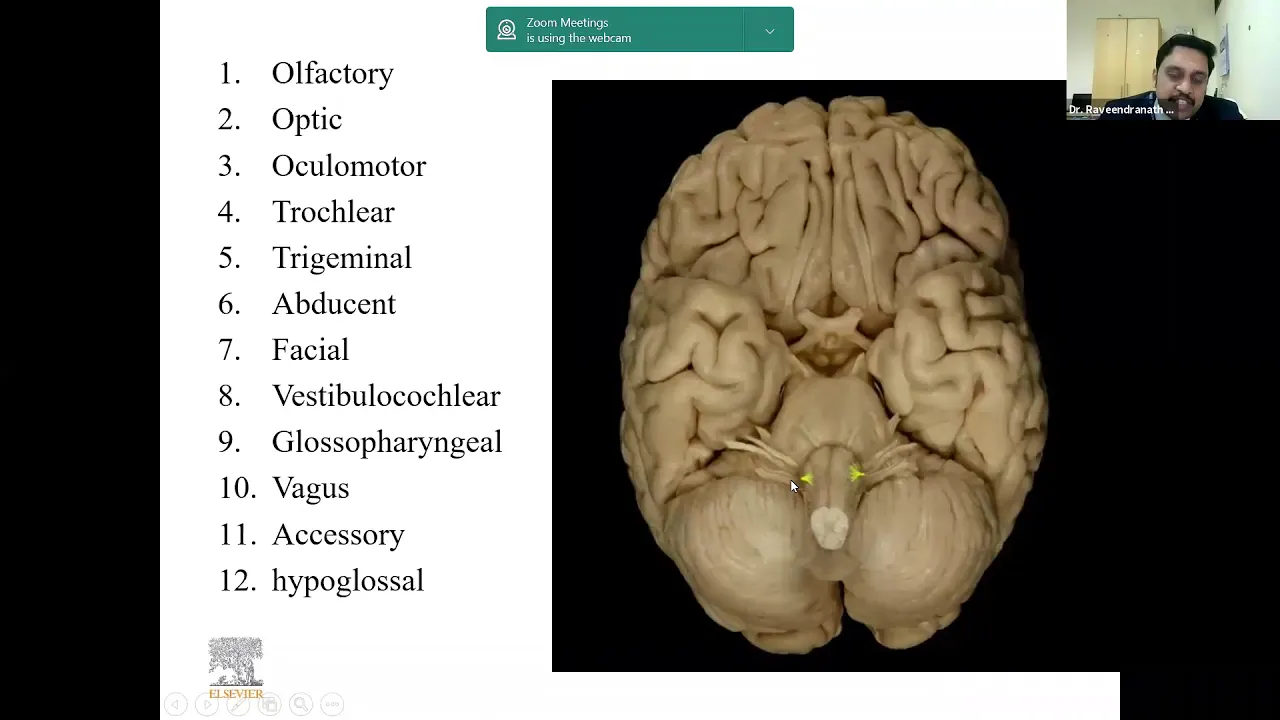
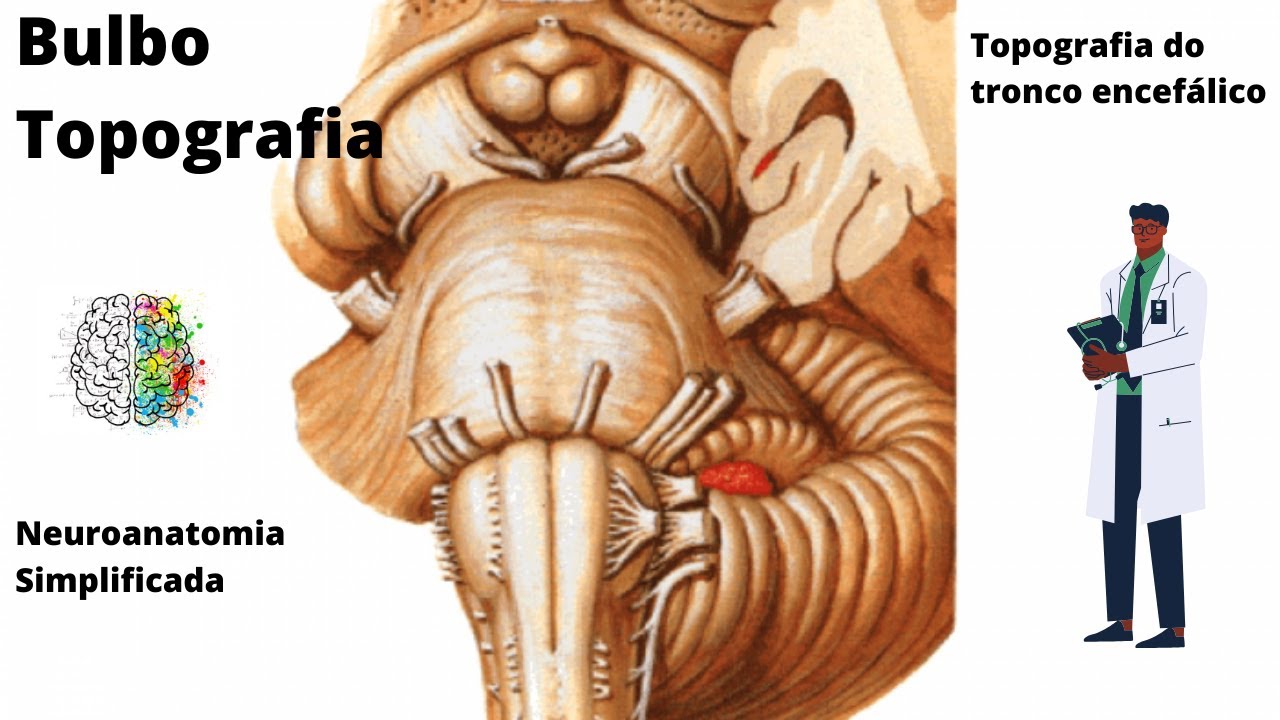
Added:Basic anatomy of the brainstem, specifically the structure and landmarks of the medulla oblongata where the vagus nerve exits.

The vagus nerve exits the cranial cavity through the jugular foramen, which is located just posterior to the olive in the medulla oblongata. The jugular foramen is named because the internal jugular vein also exits through this opening. This anatomical landmark is crucial for identifying the vagus nerve's pathway as it transitions from the skull into the neck.

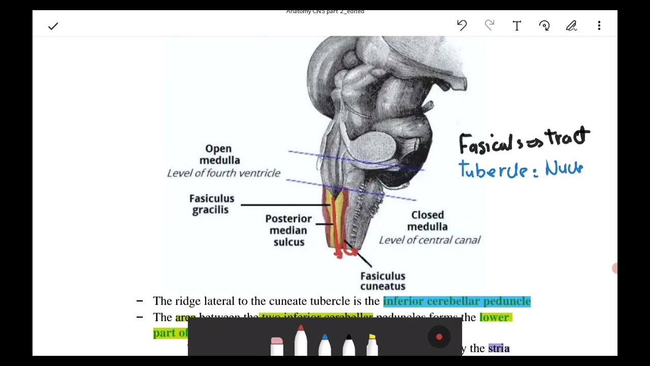
The medulla oblongata is the brainstem segment between the spinal cord and pons, delimited at the foramen magnum. It contains the anterior median fissure, pyramids, and decussation of pyramids where motor fibers cross contralaterally. The anterolateral and posterolateral sulci serve as landmarks, with the olive between them representing the olivary nucleus. The posterior funiculus contains gracile and cuneate fasciculi terminating in tubercles. The trigeminal tubercle is lateral to the cuneate tubercle. Four cranial nerves emerge: glossopharyngeal, vagus, accessory (posterior to olive), and hypoglossal (anterior to olive). The hypoglossal nerve is the only exception to cranial nerve numbering rules.

The vagus nerve (cranial nerve X) is the most extensive cranial nerve, serving as a mixed nerve that carries general somatic afferent (GSA) fibers for touch/pain/temperature sensation from the ear and neck, special visceral afferent (SVA) fibers for taste from the epiglottis, special visceral efferent (SVE) fibers for motor control of pharyngeal and laryngeal muscles (including the pharyngeal constrictors, levator veli palatini, palatoglossus, palatopharyngeus, and intrinsic laryngeal muscles), general visceral afferent (GVA) fibers for visceral sensation from thoracic and abdominal viscera, and general visceral efferent (GVE) fibers for parasympathetic innervation of the heart, lungs, esophagus, stomach, intestines, liver, pancreas, and gallbladder; it originates from the medulla oblongata, exits through the jugular foramen, and forms important plexuses including the pharyngeal plexus, pulmonary plexus, cardiac plexus, esophageal plexus, celiac plexus, and hepatic plexus.

The skull consists of a front portion (containing eyeballs and jaw) and a posterior portion called the occiput. The brain sits inside the skull, with the spinal cord exiting through the foramen magnum. The jugular foramen is a critical opening where cranial nerves 9, 10, and 11 (glossopharyngeal, vagus, and spinal accessory nerves) exit, along with the jugular vein and lymphatic drainage. The space behind the angle of the jaw, in front of the mastoid process, is where the vagus nerve exits and where the largest neck lymph node is located. The back of the head can become tight and restricted, clamping blood flow and proper vagus nerve function. Most cervical spine motion should occur from the occiput and atlas, accounting for up to 50% of head flexion and extension ability.

The medulla oblongata, the lower part of the brainstem continuous with the spinal cord, exhibits distinct external features including the closed part (containing the central canal) and open part (forming the floor of the fourth ventricle); key landmarks include the pyramids (containing corticospinal fibers), olives (housing the inferior olivary nucleus), gracile and cuneate fasciculi (continuations from the spinal cord), and tubercles marking the gracile and cuneate nuclei; the floor of the fourth ventricle displays the rhomboid fossa with the median sulcus, obex, hypoglossal trigone, vagal trigone, vestibular area, and striae medullares, while cranial nerves emerge at specific sites: hypoglossal nerve between pyramid and olive, abducent nerve between medulla and pons, and facial and vestibulocochlear nerves in the cerebellopontine angle.
Fundamental concepts of the Autonomic Nervous System (ANS), particularly the structural and functional differences between the sympathetic and parasympathetic divisions.
![[#1] SISTEMA NERVOSO AUTÔNOMO PARASSIMPÁTICO E SIMPÁTICO: Diferenças anatômicas | MK Fisiologia](https://i.ytimg.com/vi/7OIIYv2oXJw/maxresdefault.jpg)
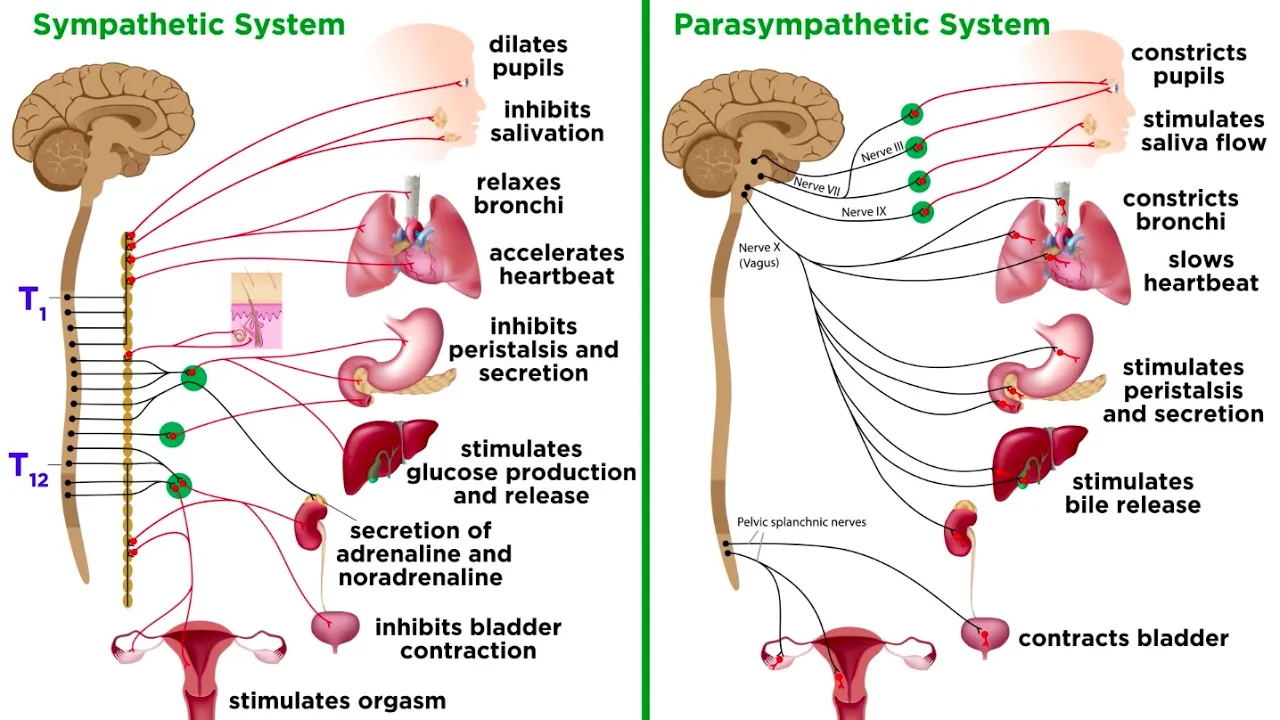
The autonomic nervous system (ANS), the efferent division of the visceral nervous system, controls internal organs like the heart, lungs, gastrointestinal tract, and glands to maintain homeostasis. The ANS is divided into three main systems: parasympathetic (craniosacral), sympathetic (thoracolumbar), and enteric. The key anatomical differences are: (1) Preganglionic neuron origin - parasympathetic neurons arise from brainstem nuclei and sacral spinal cord segments S2-S4, while sympathetic neurons originate only from thoracic and lumbar spinal cord segments T1-L2; (2) Ganglion location - parasympathetic ganglia are located close to or within target organs, whereas sympathetic ganglia are arranged in paravertebral chains along the spine; (3) Axon length - parasympathetic preganglionic axons are long and postganglionic axons are short, while sympathetic preganglionic axons are short and postganglionic axons are long; (4) Adrenal gland exception - the adrenal medulla is directly innervated by preganglionic sympathetic neurons without synaptic relay.

The autonomic nervous system has two opposing divisions that work in opposition to maintain balance: (1) Sympathetic division - activates during stress, danger, or emergencies (fight or flight response); increases heart rate, dilates pupils, and redirects blood flow to muscles; prepares the body for rapid action; (2) Parasympathetic division - activates during rest and digestion; decreases heart rate, stimulates digestion, and conserves energy; promotes relaxation and recovery. These two divisions work together to maintain homeostasis by adjusting the body's readiness for activity versus rest.

The Autonomic Nervous System (ANS) is the motor division of the peripheral nervous system that controls involuntary organs through two opposing divisions: the sympathetic (thoracolumbar distribution, ganglia near CNS, post-ganglionic neurotransmitter norepinephrine) and parasympathetic (craniosacral distribution, ganglia near organs, post-ganglionic neurotransmitter acetylcholine) systems. Both divisions use two neurons (preganglionic and post-ganglionic) with acetylcholine at the preganglionic level, but differ in their post-ganglionic neurotransmitters and ganglion positions. In most organs, these systems have opposite actions (e.g., pupil constriction vs dilation, increased vs decreased heart rate), except in genital organs where they have complementary actions (erection vs ejaculation).

The autonomic nervous system controls involuntary bodily functions like heartbeat and digestion through two opposing divisions: the sympathetic division triggers fight-or-flight responses during emergencies by increasing heart rate, constricting blood vessels, and releasing glucose, while the parasympathetic division manages rest-and-digest functions including digestion and waste elimination; these divisions work together through dual innervation, with the sympathetic originating from thoracic and lumbar spinal cord regions and the parasympathetic from the brain and sacral region, using a two-neuron chain where preganglionic neurons release acetylcholine and postganglionic neurons release either acetylcholine or norepinephrine.

The autonomic nervous system, a subdivision of the peripheral nervous system, controls unconscious bodily functions through two opposing divisions: the sympathetic nervous system (fight or flight response) and the parasympathetic nervous system (rest and digest response). Both divisions use a two-neuron chain with preganglionic neurons releasing acetylcholine, but differ in their postganglionic neurotransmitters and receptor types—sympathetic uses noradrenaline acting on adrenergic receptors (alpha and beta), while parasympathetic uses acetylcholine acting on muscarinic receptors. The sympathetic division originates from the thoracolumbar region of the spinal cord and activates organs like the heart, airways, and blood vessels to prepare the body for stress, whereas the parasympathetic division originates from cranial nerves III, VII, IX, X and the sacral region, promoting relaxation and digestion through effects on the heart, digestive system, and other organs.
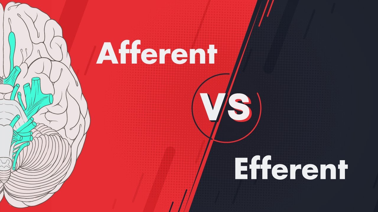
General classification of cranial nerves (CN I-XII), including the concepts of sensory (afferent) versus motor (efferent) pathways.

Cranial nerves are classified into sensory (afferent) and motor (efferent) modalities, with further subdivisions: sensory includes general somatic afferent (pain/temperature/touch via CNs V, VII, IX, X), general visceral afferent (visceral sensations from heart/lungs/GI tract via CNs IX, X), and special visceral afferent (smell/taste/vision/hearing via CNs I, II, VII, VIII, IX, X); motor includes general somatic efferent (skeletal muscles via CNs III, IV, VI, XII), general visceral efferent (parasympathetic control via CNs III, VII, IX, X), and special visceral efferent (branchial arch muscles via CNs V, VII, IX, X).

There are 12 pairs of cranial nerves. Sensory cranial nerves include: Olfactory (I), Optic (II), Oculomotor (III), Trochlear (IV), Trigeminal (V), Abducens (VI), Facial (VII), Vestibulocochlear (VIII), Glossopharyngeal (IX), Vagus (X), Spinal Accessory (XI), and Hypoglossal (XII). Motor cranial nerves include: Oculomotor (III), Trochlear (IV), Trigeminal (V), Abducens (VI), Facial (VII), Glossopharyngeal (IX), Vagus (X), Spinal Accessory (XI), and Hypoglossal (XII). Mixed cranial nerves include: Trigeminal (V), Facial (VII), Vagus (X), and Hypoglossal (XII).

There are 12 pairs of cranial nerves. Sensory cranial nerves include: (1) Olfactory (I), (2) Optic (II), (8) Oculomotor (III), (4) Trochlear (IV), (5) Trigeminal (V), (6) Abducens (VI), (7) Facial (VII), (8) Vestibulocochlear (VIII), (9) Glossopharyngeal (IX), (10) Vagus (X), (11) Accessory (XI), (12) Hypoglossal (XII). Motor cranial nerves include: (3) Oculomotor (III), (4) Trochlear (IV), (6) Abducens (VI), (11) Accessory (XI), (12) Hypoglossal (XII). Mixed cranial nerves include: (5) Trigeminal (V), (7) Facial (VII), (9) Glossopharyngeal (IX), (10) Vagus (X).

Cranial nerves are classified into two main types: (1) Motor nerves (موتور نيرف) - responsible for muscle movement, and (2) Sensory nerves (سنسوري نيرف) - responsible for sensory information. Each cranial nerve has both motor and sensory components. The classification system includes: (1) General somatic efferent (GSE) - controls voluntary skeletal muscle movement, (2) General visceral efferent (GVE) - controls involuntary functions of smooth muscles and glands, (3) Special visceral efferent (SVE) - controls branchial arch muscles, (4) General somatic afferent (GSA) - conveys touch, pain, temperature from body surface, (5) Special somatic afferent (SSA) - conveys vision, hearing, balance, (6) General visceral afferent (GVA) - conveys visceral sensation, (7) Special visceral afferent (SVA) - conveys taste and smell.

This comprehensive section covers afferent pathways and provides a complete classification summary of cranial nerves. Visceral general afferent pathways: glossopharyngeal (IX) → carotid sinus and body → nucleus of solitary tract (blood pressure and oxygenation); vagus (X) → thoracic and abdominal viscera (up to splenic flexure of colon) → nucleus of solitary tract (visceral distension and function). Visceral special afferent pathways (taste): facial (VII) → anterior two-thirds of tongue → nucleus of solitary tract; glossopharyngeal (IX) → posterior one-third of tongue → nucleus of solitary tract; vagus (X) → epiglottis and vallecula → nucleus of solitary tract. Somatic general afferent pathways: trigeminal (V) → all three divisions (V1, V2, V3) → nucleus of trigeminal (principal, mesencephalic, and spinal) → sensory information from face (touch, temperature, pain, proprioception). Additional somatic general afferents: facial, glossopharyngeal, and vagus nerves → external ear, auditory canal, and tympanic membrane → nucleus of trigeminal. Somatic special afferents (hearing, balance) travel via vestibulocochlear (VIII) nerve to vestibular and cochlear nuclei. The final classification summary: purely sensory nerves (I, II, VIII); purely motor nerves (III, IV, VI, XI, XII); mixed nerves (V, VII, IX, X). Each nerve's classification is based on the types of fibers it contains: somatic general efferents (skeletal muscle innervation), visceral general efferents (parasympathetic to glands and smooth muscle), visceral special efferents (branchial motor to branchial arch muscles), somatic general afferents (sensory from body surface), visceral general afferents (sensory from viscera), visceral special afferents (taste), and somatic special afferents (hearing, balance, vision).
Familiarity with cranial nerve functional components and fiber types, such as General Visceral Efferent (GVE) and Special Visceral Efferent (SVE).

Cranial nerves contain four main functional components: general somatic afferent (sensory from skin and muscles), general visceral afferent (internal organ sensations), special visceral efferent (motor to pharyngeal arch muscles), and special visceral afferent (taste sensation); these components explain how cranial nerves differ from spinal nerves in controlling specialized head and neck functions including taste and pharyngeal muscle movement.

Cranial nerves are classified into functional columns based on their function: General Somatic Efferent (GSE) supplies extraocular muscles (CN III, IV, VI) and tongue muscles (CN XII); General Visceral Efferent (GVE) represents parasympathetic fibers (CN III, VII, IX, X); Special Visceral Efferent (SVE) supplies branchial arch muscles (CN V, VII, IX, X, XI); General Somatic Afferent (GSA) carries general sensation from the face (CN V); Special Somatic Afferent (SSA) carries special senses like hearing and vision (CN II, VIII); General Visceral Afferent (GVA) and Special Visceral Afferent (SVA) both carry visceral sensation to the nucleus tractus solitarius (NTS), with SVA specifically carrying taste sensation (CN VII, IX, X).

The cranial nerves containing the General Somatic Efferent (GSE) component are: CN III (Oculomotor) - supplies extraocular muscles except superior oblique, CN IV (Trochlear) - supplies superior oblique muscle only, and CN VI (Abducens) - supplies lateral rectus muscle only. These nerves are purely motor and supply skeletal muscles derived from somites. The cranial nerves containing the Special Visceral Efferent (SVE) component are: CN V (Trigeminal) - supplies muscles of mastication, CN VII (Facial) - supplies muscles of facial expression and stapedius, CN IX (Glossopharyngeal) - supplies stylopharyngeus muscle, CN X (Vagus) - supplies muscles of pharynx and larynx, and CN XI (Accessory) - supplies sternocleidomastoid and trapezius muscles. These nerves supply branchial arch muscles derived from pharyngeal arches.

The cranial nerves are organized into functional columns based on embryological development: the basal plate (anterior, motor/efferent) and LR plate (posterior, sensory/afferent). The seven functional columns are: General Somatic Efferent (GSE) - motor to skeletal muscles (CN 3, 4, 6, 12); General Somatic Afferent (GSA) - general sensation from face (CN 5, 7, 10); Special Somatic Afferent (SSA) - special senses like hearing (CN 8); General Visceral Efferent (GVE) - parasympathetic motor to viscera (CN 3, 7, 9, 10); Special Visceral Efferent (SVE) - motor to branchial arch muscles (CN 5, 7, 9, 10, 11); General Visceral Afferent (GVA) - general visceral sensation (CN 7, 9, 10); Special Visceral Afferent (SVA) - taste sensation (CN 7, 9, 10). A key mnemonic is that somatic columns contain even-numbered cranial nerves (except 10), while visceral columns contain odd-numbered cranial nerves plus 10.

The vagus nerve contains four fiber types: GVE (parasympathetic motor), GVA (visceral sensory), GSA (somatic sensory), and SVE (motor to pharyngeal arch-derived muscles). GSA fibers supply the ear structures and synapse at the superior ganglion, projecting to the trigeminal spinal nucleus. SVA fibers carry taste from the epiglottis and pharynx, synapsing at the nodose ganglion and projecting to the nucleus tractus solitarius. SVE fibers originate from the nucleus ambiguus and supply pharyngeal and laryngeal muscles essential for swallowing and speech. These fibers integrate with cranial nerves IX and XI to form the pharyngeal plexus.
Prerequisite Knowledge
- Concept 01Basic anatomy of the brainstem, specifically the structure and landmarks of the medulla oblongata where the vagus nerve exits.
- Concept 02Fundamental concepts of the Autonomic Nervous System (ANS), particularly the structural and functional differences between the sympathetic and parasympathetic divisions.
- Concept 03General classification of cranial nerves (CN I-XII), including the concepts of sensory (afferent) versus motor (efferent) pathways.
- Concept 04Familiarity with cranial nerve functional components and fiber types, such as General Visceral Efferent (GVE) and Special Visceral Efferent (SVE).
Subsequent Learning
- Step 01Clinical neurological examinations for assessing cranial nerves IX (Glossopharyngeal) and X (Vagus), including the gag reflex, vocal assessment, and palatal elevation.
- Step 02Pathological conditions resulting from vagus nerve damage or dysfunction, such as dysphagia, vocal cord paralysis, gastroparesis, and vasovagal syncope.
- Step 03The neurobiology of the Gut-Brain Axis and how the vagus nerve mediates bidirectional communication between the central nervous system and the gastrointestinal microbiome.
- Step 04Therapeutic applications of Vagus Nerve Stimulation (VNS) in modern medicine for treating epilepsy, clinical depression, and autoimmune inflammatory diseases.
- Step 05Stephen Porges' Polyvagal Theory and its applications in understanding trauma, emotional regulation, and social engagement systems.
Anatomy & Functions
0:03- 1
Vagus nerve has motor, sensory, and parasympathetic fibers.
- 2
It controls swallowing, speech, and heart/gut muscles.
- 3
Branches include pharyngeal, laryngeal, and cardiac nerves.
The Polyvagal Theory and its Neuroanatomical Critiques
While standard neuroanatomy teaches that the vagus nerve (CN X) operates within a simple, antagonistic sympathetic-parasympathetic balance, the 'Polyvagal Theory' offers an alternative, evolutionary framework. Proposed by Stephen Porges, it suggests the vagus nerve consists of two distinct pathways: an older 'dorsal' pathway responsible for immobilization (freeze responses) and a newer 'ventral' pathway regulating social engagement and self-soothing. However, this theory is highly controversial. Mainstream neurophysiologists criticize it for oversimplifying brainstem anatomy and lacking empirical support. They argue that both the dorsal motor nucleus and nucleus ambiguus work in tandem rather than representing separate evolutionary behavioral systems. Introducing this debate encourages students to critically examine how physiological concepts are applied to psychological models.
Clinical neurological examinations for assessing cranial nerves IX (Glossopharyngeal) and X (Vagus), including the gag reflex, vocal assessment, and palatal elevation.

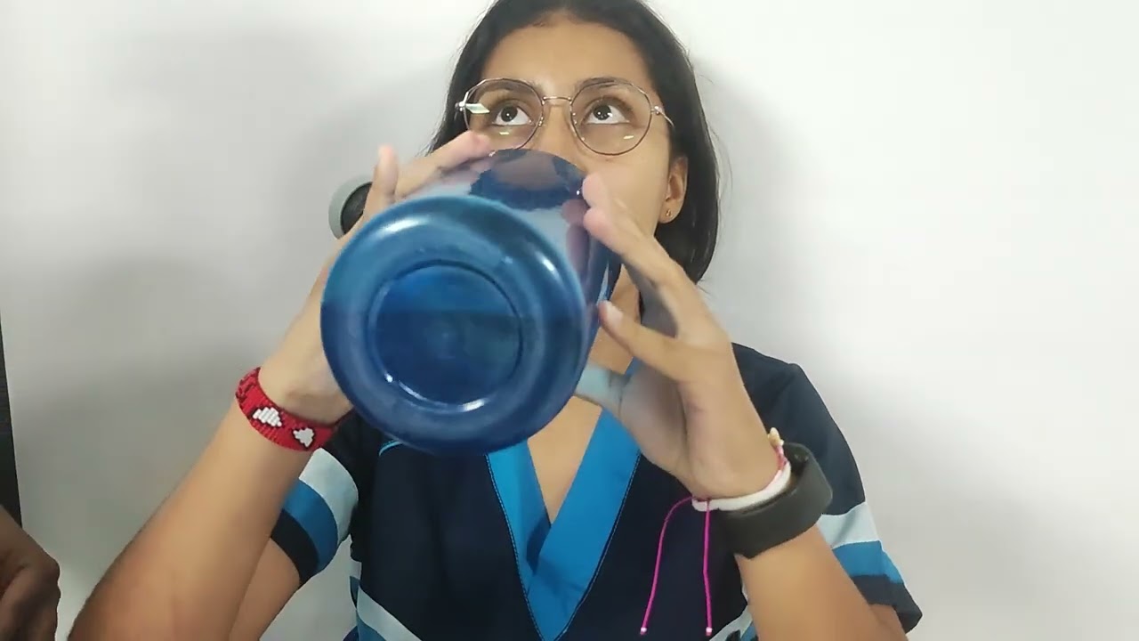
This video demonstrates the clinical examination of cranial nerves IX (glossopharyngeal) and X (vagus), covering four key assessment aspects: tongue protrusion to evaluate tongue movement, water swallowing to assess pharyngeal muscle symmetry, palatal elevation during speech sounds (a/e) to check palatal function, and the gag reflex to test the swallowing reflex. The presenter shows a normal examination with symmetrical tongue protrusion, symmetrical pharyngeal muscle contraction during swallowing, symmetrical palatal elevation during speech, and an intact gag reflex, indicating no neurological alteration in these cranial nerves.

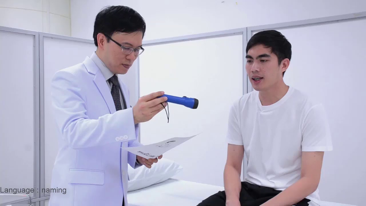
The glossopharyngeal (CN IX) and vagus (CN X) nerves are evaluated together due to shared motor and parasympathetic functions. Testing includes: (1) Voice assessment by having the patient repeat words to evaluate vocal quality; (2) Palate elevation by having the patient open their mouth and say 'ah' to observe symmetrical elevation of the soft palate; (3) Pharyngeal reflex by touching the posterior pharyngeal wall to elicit gag reflex. The presenter demonstrates these techniques.

The glossopharyngeal (CN IX) and vagus (CN X) nerves examination tests gag reflex, palate elevation, and voice quality. The examiner asks the patient to say 'ah' and observes for palate elevation and uvular deviation. The gag reflex is tested by touching the posterior pharyngeal wall. This examination can detect cranial nerve palsies and assess for signs of brainstem dysfunction.

Glossopharyngeal (CN IX) and vagus (CN X) nerve testing evaluates the gag reflex. The patient opens their mouth and says 'ah' while the examiner observes the soft palate and uvula. The examiner may use a tongue depressor to stimulate the posterior pharyngeal wall. The gag reflex involves sensory input from CN IX and motor output from CN X. Asymmetry or absence may indicate nerve pathology.

Cranial nerve IX (glossopharyngeal) is assessed via the gag reflex, though this is not demonstrated in this exam. Cranial nerve X (vagus) is assessed by having the patient open their mouth, stick out their tongue, and say 'ah.' The uvula and soft palate should rise symmetrically with phonation.
Pathological conditions resulting from vagus nerve damage or dysfunction, such as dysphagia, vocal cord paralysis, gastroparesis, and vasovagal syncope.

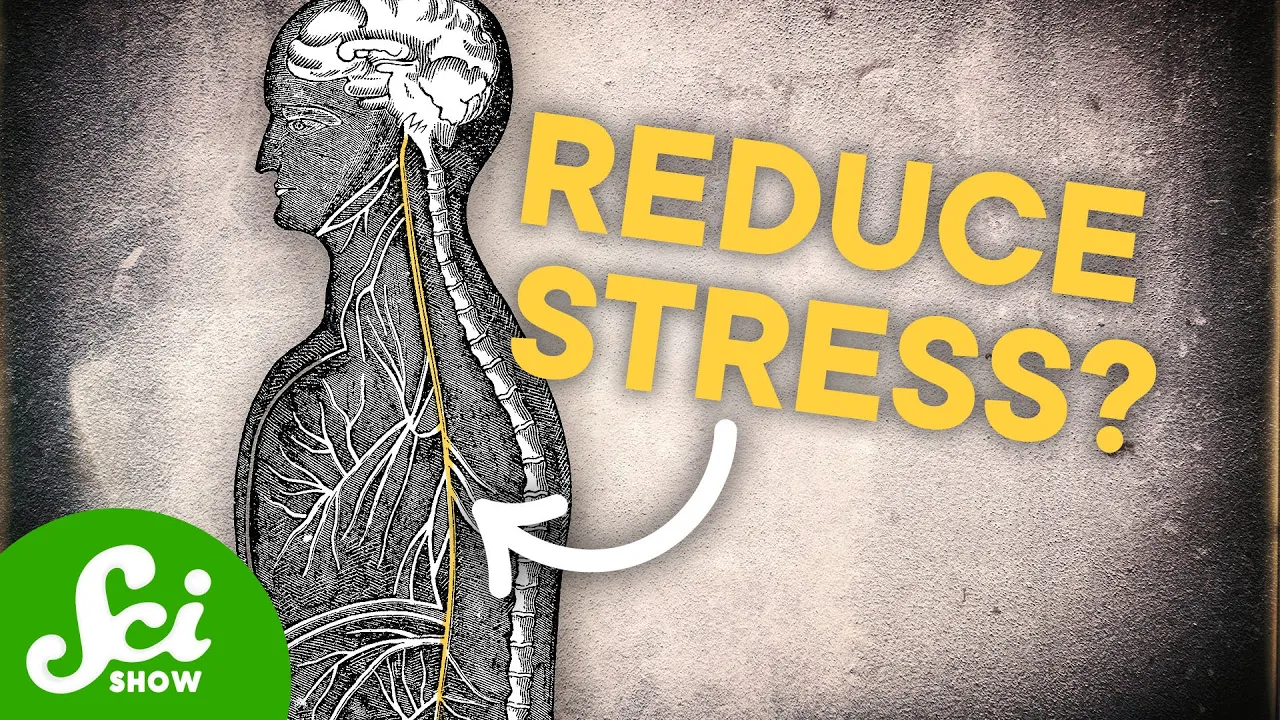
Damage to the vagus nerve can cause several medical conditions. Vasovagal syncope occurs when a vagus nerve near the heart causes a dramatic drop in blood pressure leading to fainting. Gastroparesis results from damaged vagus nerves that prevent gut muscles from moving food out of the stomach. Vagus nerve damage can also disrupt heart rate regulation, cause speech difficulties, or impair swallowing. Importantly, these disorders only occur when there is physical damage to the nerve. Therefore, claims about 'resetting' the vagus nerve for treatment purposes, as promoted by some wellness influencers, are not medically valid for treating these conditions.

Damage to the vagus nerve produces two primary clinical symptoms frequently tested in medical examinations. First, hoarseness (かれ声) results from recurrent laryngeal nerve paralysis, which controls laryngeal muscles and vocal cord movement. The term 'recurrent' describes the nerve's U-shaped path as it descends from the brainstem and loops back up to the larynx. Second, dysphagia (エゲ障害) or difficulty swallowing occurs when the posterior pharynx becomes impaired, as this area is innervated by the vagus nerve. These symptoms represent the most clinically significant manifestations of vagus nerve dysfunction.

The vagus nerve controls taste, pharyngeal activity, epiglottic sensation, gastrointestinal tract innervation, trachea control, and cardiac muscles. Damage causes dysphagia (difficulty swallowing), vocal cord weakness, aphonia (inability to speak), hoarseness (raspy voice), and pain. Damage can affect the nucleus ambiguus and create problems with other cranial nerves.

Dysfunction of the vagus nerve manifests through several clinical signs: diminished or fatiguing gag reflex (where the reflex weakens after repeated stimulation), difficulty swallowing due to impaired pharyngeal and laryngeal function, gastroparesis (delayed stomach emptying), GI problems including constipation, anxiety (due to lack of parasympathetic function leading to chronic fight-or-flight mode), and uvula deviation away from the damaged side when the mouth is opened.

The vagus nerve controls critical functions including swallowing, voice production, heart rate, breathing rate, and the gag reflex. Dysfunction causes difficulty swallowing, voice changes, abnormal heart rate (too fast or slow), breathing irregularities, and loss of gag reflex. Digestive symptoms include early satiety (feeling full after small meals), which may be misdiagnosed as irritable bowel syndrome. The nerve also regulates blood pressure and can cause vasovagal syncope (fainting in crowded or hot environments), sometimes with muscle spasms mistaken for seizures. Understanding these symptoms helps distinguish vagus nerve dysfunction from other conditions like epilepsy.
The neurobiology of the Gut-Brain Axis and how the vagus nerve mediates bidirectional communication between the central nervous system and the gastrointestinal microbiome.

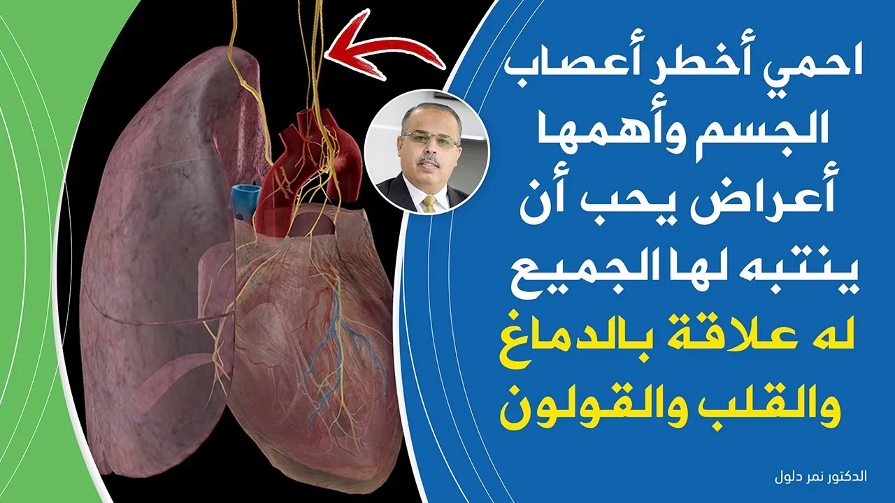
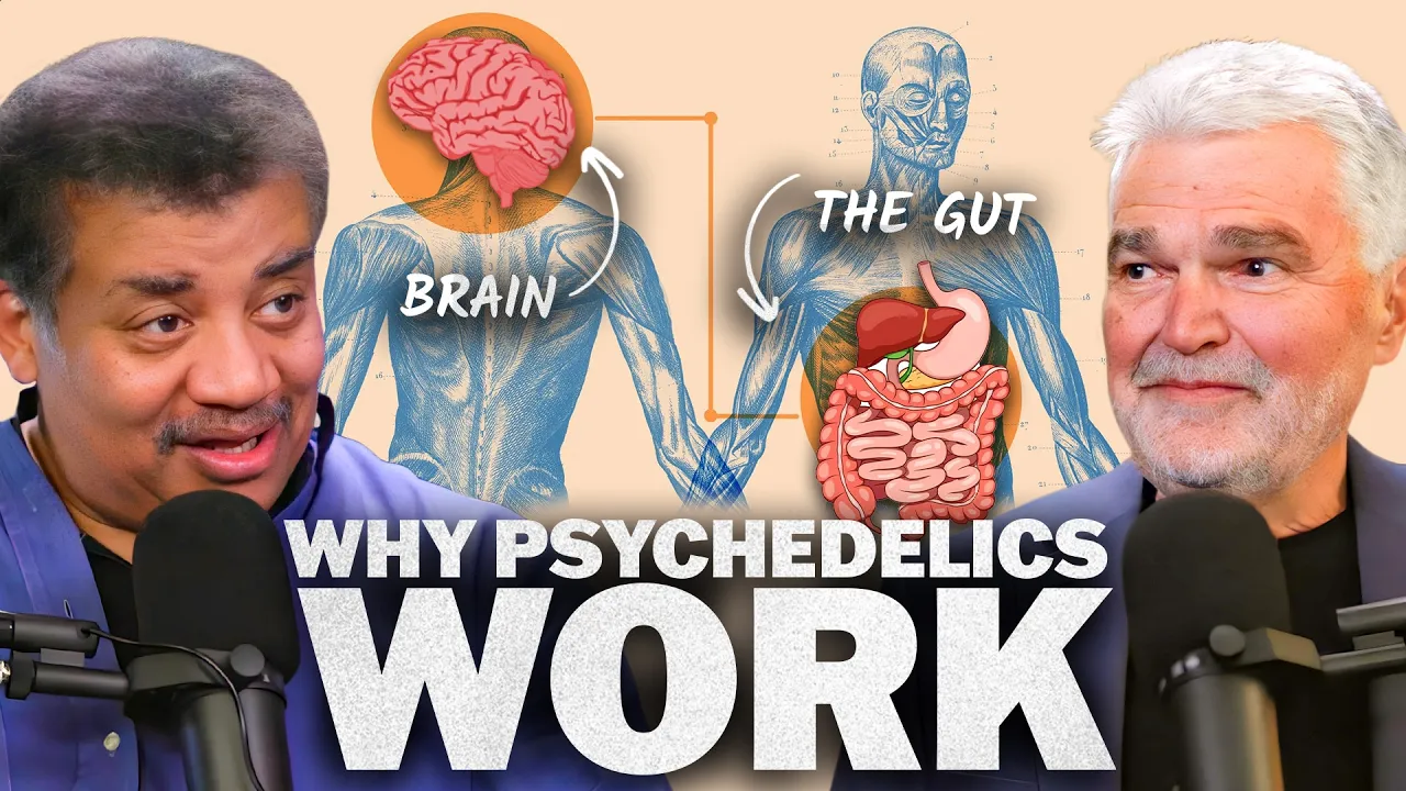
This segment explains the vagus nerve and gut-brain communication in detail. The vagus nerve provides direct bidirectional communication between the gut and brain, primarily carrying sensory information from the gut to the brain. This nerve innervates all organs in the thorax and abdomen. The gut-brain axis involves not just reading signals but also sending commands from the brain to the gut, allowing for active regulation of gut function based on emotional and cognitive states. Thoughts and emotions directly influence gut function through this pathway, with positive thoughts stimulating production of beneficial neurotransmitters and negative thoughts potentially causing gut dysfunction.

The gut-brain axis is a bidirectional communication system between the gastrointestinal tract and the central nervous system, mediated by the vagus nerve and the enteric nervous system (containing 200-600 million neurons), which enables gut bacteria to influence brain function and mood; research demonstrates that germ-free mice exposed to stress show no anxiety or depression symptoms, while mice with normal gut microbiomes do exhibit these symptoms, and introducing gut bacteria to germ-free mice recreates these effects, suggesting that probiotics may help regulate mood by increasing GABA (gamma-aminobutyric acid) production and enhancing brain receptiveness to this calming neurotransmitter.

The vagus nerve serves as a two-way communication highway between the gut and brain, with 80-90% of signals traveling upward from gut to brain. This connection explains how gut health influences cognition and brain function. Contrary to historical medical beliefs, many body parts previously considered sterile actually harbor healthy microbiomes: mother's milk transfers beneficial bacteria to newborns, urine contains a healthy bladder microbiome, semen contains normal bacteria, and the brain itself hosts its own healthy microbiome. These discoveries challenge traditional views of human biology and highlight the importance of maintaining healthy microbial communities throughout the body.

The vagus nerve serves as the primary bidirectional communication pathway between gut and brain, carrying sensory signals including serotonin, GLP-1, and cytokines. Gut microbes are 3.5 billion-year-old organisms that have shaped human evolution—one centimeter of the large intestine contains more microbes than all humans who ever lived. Through lateral gene transfer, microbial genetic material contributed to developing the gut's 'little brain' and eventually influenced brain evolution. This explains why the gut produces neurotransmitters like serotonin that influence mood and behavior, creating a fundamental connection between digestion and mental health.

The gut-brain axis involves bidirectional communication between the gut and brain through direct neural pathways (via the vagus nerve) and indirect signaling through gut microbiota that produce neurotransmitters like dopamine, serotonin, and GABA, which can influence mood, appetite, and overall brain function; maintaining a healthy gut microbiome through diet (particularly fermented foods) supports optimal gut-brain signaling and overall wellbeing.
Therapeutic applications of Vagus Nerve Stimulation (VNS) in modern medicine for treating epilepsy, clinical depression, and autoimmune inflammatory diseases.

Vagus nerve stimulation (VNS) is a medical treatment that exploits the nerve's influence on the brain. It is used to treat epilepsy and depression by sending regular, mild pulses of electrical energy to the brain via the vagus nerve through a device similar to a pacemaker placed under the skin on the chest wall with a wire running to the vagus nerve in the neck. A 2016 study published in PNAS showed that VNS significantly improved measures of disease activity in patients with rheumatoid arthritis, a chronic inflammatory disease affecting 1.3 million people in the United States.

The FDA has approved vagus nerve stimulation for therapeutic use in patients over 12 years old with drug-resistant epilepsy and depression. For epilepsy, VNS helps prevent chronic seizures by modulating neural activity. For depression, VNS increases mood by influencing neural circuits. Both conditions may respond well to VNS because it helps decrease chronic neural inflammation, demonstrating the anti-inflammatory properties of vagus nerve stimulation.

Vagus nerve stimulation (VNS) involves implantable devices that send electrical pulses to the brainstem via the left vagus nerve, functioning similarly to a pacemaker for the brain. The left vagus nerve is typically targeted because the right vagus nerve has a more direct connection to the heart, increasing the risk of interfering with heart rhythms. FDA-approved uses include treatment-resistant epilepsy, certain types of depression, and aiding physical rehabilitation after strokes. For epilepsy, VNS disrupts synchronized neuronal firing during seizures; studies show about 50% of patients experience reduced seizure frequency. For depression, mechanisms may involve affecting noradrenaline neurotransmission or reducing inflammation by slowing production of inflammatory cytokines.

Vagus nerve stimulation (VNS) involves sending mild electrical pulses to the nerve to harness its regulatory powers. It is an approved treatment for seizures and epilepsy, depression unresponsive to other treatments, and cluster headaches. Non-invasive devices can stimulate the nerve through the skin, making treatment accessible without surgery.

Vagus Nerve Stimulation (VNS) is a non-invasive neuromodulation therapy approved for treating drug-resistant epilepsy and depression, which works by stimulating the vagus nerve to modulate brain activity through neurotransmitter changes (increasing GABA and decreasing glutamate), thereby reducing seizure frequency and severity; the therapy involves implanting a device under the clavicle connected to the vagus nerve, with programming parameters including output current (therapeutic range 1.5-2.25 mA), frequency, pulse width, and duty cycle (typically 10%), requiring gradual titration from an initial low dose of 0.8 mA over several weeks to achieve optimal therapeutic response while minimizing side effects.
Stephen Porges' Polyvagal Theory and its applications in understanding trauma, emotional regulation, and social engagement systems.

Stephen Porges' polyvagal theory explains that the vagus nerve has three branches: the dorsal vagal system (shared with reptiles, responsible for freezing responses), the ventral vagal system (social engagement system, right-lateralized, enabling face-to-face communication and emotional regulation), and the sympathetic nervous system. The social engagement system is essential for creating feelings of safety and enabling interactive emotional regulation between individuals.

Polyvagal theory, developed by Stephen Porges, expands the understanding of the autonomic nervous system to include a third branch: the social engagement system. This system, controlled by the vagus nerve, enables social connection, communication, and cooperation. It is the most evolved part of the autonomic nervous system and allows humans to respond to stress through social means rather than fight or flight. The social engagement system develops from early childhood experiences, particularly attachment relationships with caregivers. Children who experience secure attachment and responsive caregiving develop strong social engagement capacities. Those who experience neglect, abuse, or inconsistent caregiving may not develop this system fully. When faced with stress, individuals may default to fight or flight responses rather than using social skills to navigate difficulties. This developmental pattern can perpetuate across generations, explaining why trauma survivors often struggle with social functioning and why healing trauma is essential for healthy relationships.

Polyvagal Theory explains that trauma causes the nervous system to shut down through the dorsal vagal response, which immobilizes the body and prevents healing by interfering with the social engagement system; this system, regulated by the ventral vagal pathway, enables healthy social interactions and co-regulation with others, and when it becomes dysregulated, individuals lose the ability to perceive safety cues and engage in relationships, requiring interventions that stimulate the social engagement system to restore nervous system balance and facilitate trauma recovery.

Trauma causes the nervous system to become 'retuned' for survival, creating chronic defensive states that make clients resistant to treatment; clinicians can help by reducing threat cues, increasing safety cues, and using co-regulation techniques like breath work and physical presence to negotiate with the nervous system and help clients access regulated states.

According to Stephen Porges' polyvagal theory, humans have three primary nervous system states: the sympathetic fight-or-flight system, the shutdown system (where the vagus nerve becomes overactive), and the social engagement system. The social engagement system should be our default state as mammals and primates. This state involves natural impulses to connect with others through eye contact, sharing meals, movies, and cooperative activities. Healing trauma involves returning to this state of openness and connection rather than remaining stuck in fight, flight, or shutdown responses.
Anatomy & Functions
0:03- 1
Vagus nerve has motor, sensory, and parasympathetic fibers.
- 2
It controls swallowing, speech, and heart/gut muscles.
- 3
Branches include pharyngeal, laryngeal, and cardiac nerves.
The Polyvagal Theory and its Neuroanatomical Critiques
While standard neuroanatomy teaches that the vagus nerve (CN X) operates within a simple, antagonistic sympathetic-parasympathetic balance, the 'Polyvagal Theory' offers an alternative, evolutionary framework. Proposed by Stephen Porges, it suggests the vagus nerve consists of two distinct pathways: an older 'dorsal' pathway responsible for immobilization (freeze responses) and a newer 'ventral' pathway regulating social engagement and self-soothing. However, this theory is highly controversial. Mainstream neurophysiologists criticize it for oversimplifying brainstem anatomy and lacking empirical support. They argue that both the dorsal motor nucleus and nucleus ambiguus work in tandem rather than representing separate evolutionary behavioral systems. Introducing this debate encourages students to critically examine how physiological concepts are applied to psychological models.
The vagus nerve, or cranial nerve X, is the longest cranial nerve with diverse functions, many of which are critical. It consists of both sensory, or afferent; and motor, or efferent, fibers; and involves 4 nuclei in the medulla.
It passes through two ganglia – superior and inferior - shortly after exiting the skull via the jugular foramen. - Motor neurons that originate from the nucleus ambiguus contain special visceral efferent fibers. They control most muscles of the pharynx, larynx, and some muscles of the soft palate and tongue, and thus play an important role in swallowing and speech; - Fibers from the dorsal vagal motor nucleus are general visceral efferent fibers. They provide parasympathetic innervation to cardiac, pulmonary, and esophageal muscles; as well as the glands of the gastrointestinal tract.
- Sensory neurons that end in the spinal trigeminal nucleus have their cell bodies in the superior ganglion. They contain general somatic afferent fibers that convey sensation from the outer ear and tympanic membrane. - Neurons that terminate in the solitary nucleus have their cell bodies in the inferior ganglion. They provide general visceral afferent fibers that conduct sensory impulses from the carotid and aortic bodies. There are also special visceral afferent fibers that convey taste sensation from the pharynx, palate, and epiglottis.
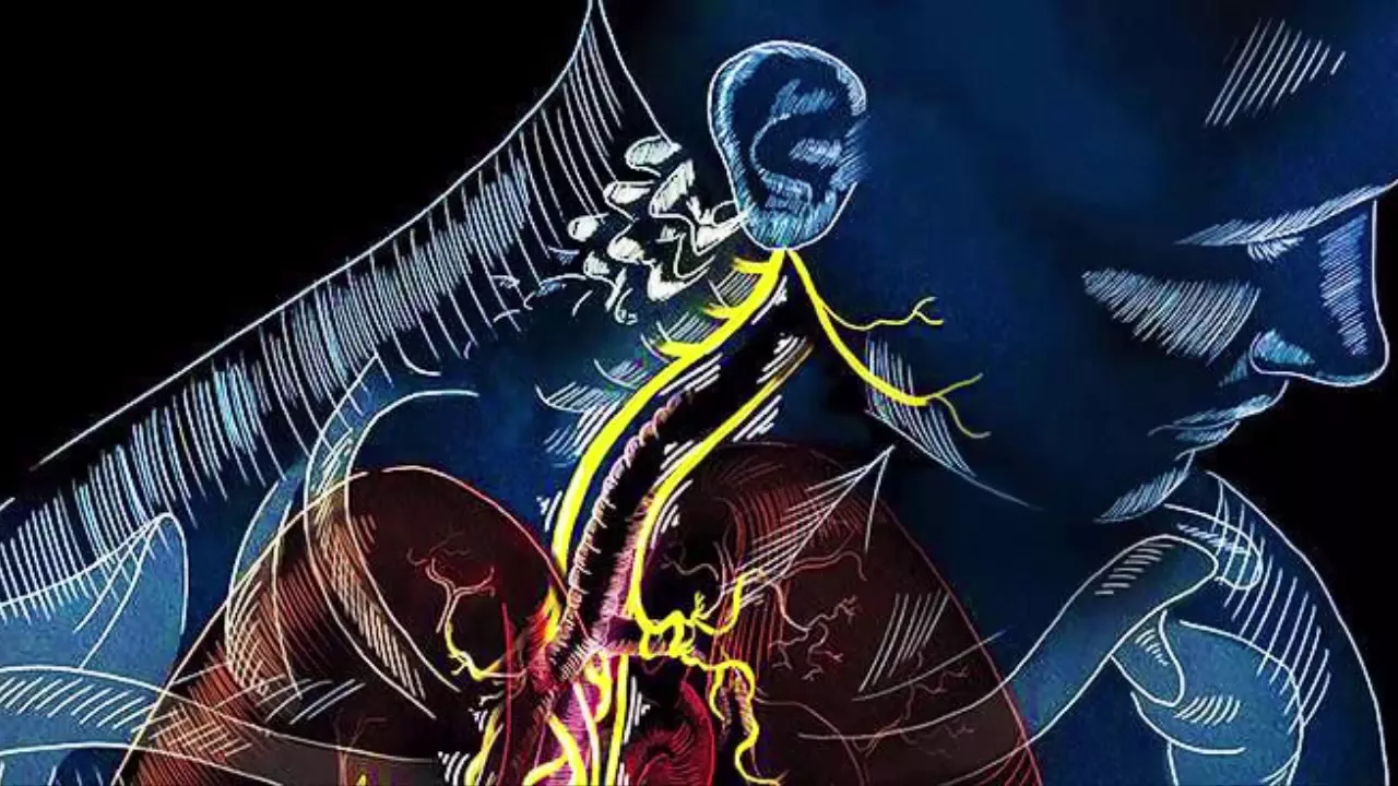
The vagus nerve descends within the carotid sheath together with carotid arteries and internal jugular vein. In the neck, it gives out several branches: - The pharyngeal branch carries both motor and sensory fibers to most muscles of the pharynx and palate, as well as the palatoglossus muscle of the tongue; and conveys sensory information from the carotid body. - Next is the superior laryngeal nerve which shortly divides into internal and external branches. The internal branch supplies sensation to the mucosa - from the epiglottis to the level just above the vocal folds. The external branch controls the cricothyroid muscle of the larynx. - There are also superior cardiac branches which descend and merge with other cardiac branches to form cardiac plexuses.
The right vagus nerve then continues downward, anteriorly to the subclavian artery, at which point, the right recurrent laryngeal nerve branches off and loops upward to enter the larynx.
On the other side, left recurrent laryngeal nerve loops around the aortic arch to ascend to the larynx. These recurrent laryngeal nerves control all muscles of the larynx except for the cricothyroid muscle. They also carry sensory information from the level of the vocal folds and below. As it continues to the thorax and abdomen, the vagus nerve gives rise to more branches, contributing to the formation of cardiac, pulmonary, esophageal, gastric and celiac plexuses.
Damage to vagus nerve results in hoarseness or loss of voice, difficulty swallowing/speaking; impaired gag reflex, reduced gastrointestinal motility, increased heart rate and other parasympathetic problems. The effect is fatal if both nerves are damaged.
Vagus nerve is usually evaluated together with glossopharyngeal nerve. In addition to observing any speech or swallowing problems, patients are tested for symmetry of the gag reflex and symmetry of uvula elevation when saying "ah". The uvula often deviates away from the affected side.
Hoarseness or loss of voice in combination with a normal uvula elevation and normal gag reflex typically indicates a lesion of the recurrent laryngeal nerve.
Up Next

Neuromarketing: How Brands Exploit Your Brain's Subconscious
@DWPlanetA
1.8M views•2021-06-18

Bessel van der Kolk on How Trauma Affects the Body and Brain
@bigthink
226.3K views•2025-10-03

Action Potential in Neurons, Animation: Phases Explained
@Alilamedicalmedia
1.6M views•2016-04-25

How Exercise Benefits Your Brain: Science Explained
@TED
11.4M views•2018-03-21
Related Study Plans & Knowledge Roadmaps
Structured learning paths in Neuroscience