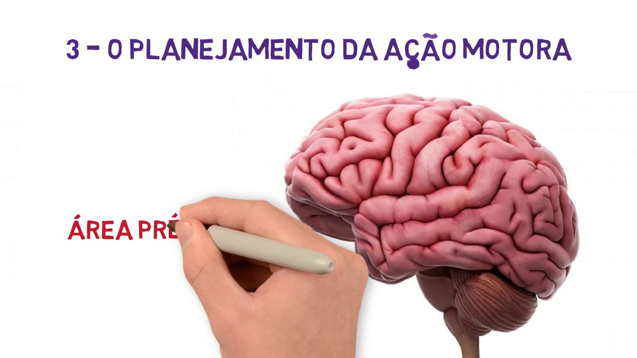


The motor cortex, located on the top center of the brain, controls voluntary movements through five interconnected regions: the primary motor cortex (generating neural impulses for movement), premotor cortex (preparing and guiding movements), supplementary motor area (planning and coordinating bilateral movements), posterior parietal cortex (transforming sensory information into motor commands), and primary somatosensory cortex (functionally part of motor circuitry). Movement planning is collected in area 6 and delivered to area 4, which then sends commands to specific body areas. The cerebellum regulates the timing and sequence of these movements, ensuring smooth execution.
Motor Cortex Explained: Brain Anatomy & Movement Control
Added:Basic neuroanatomy, specifically the major lobes of the brain (frontal, parietal, occipital, and temporal) and their general functions.

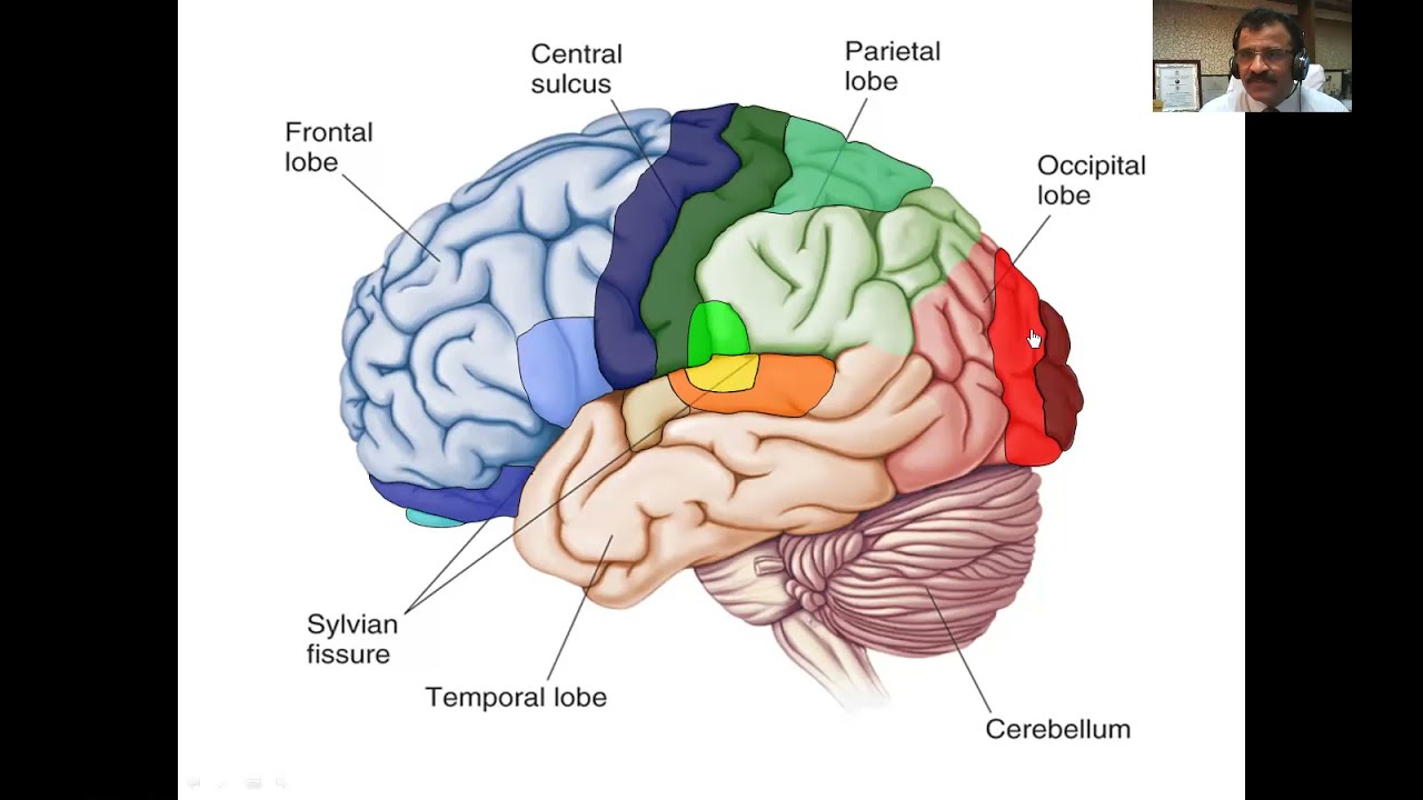
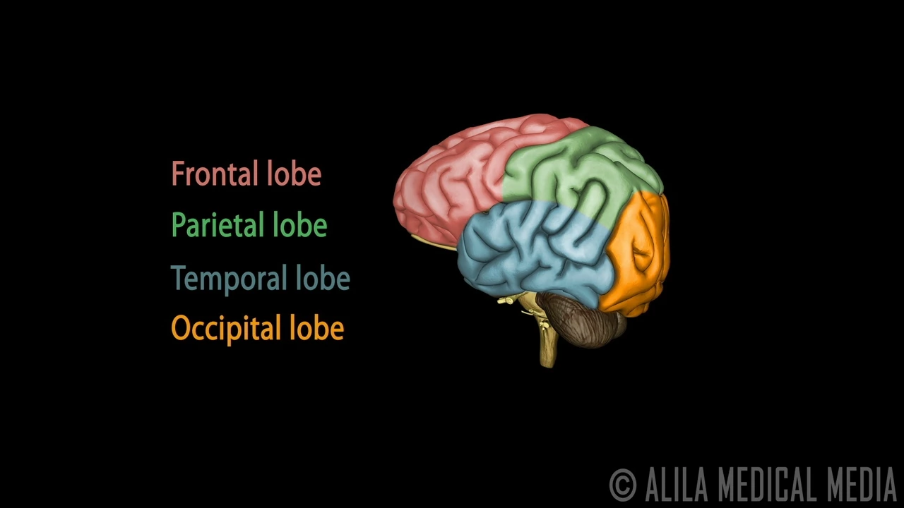
The brain has four main cerebral lobes with distinct functions. The frontal lobe houses the primary motor cortex and regulates reward, motivation, behavior, decision-making, and executive functioning. It is the last lobe to mature, with final maturation around ages 20-25. The temporal lobe is key for auditory processing, memory formation, and language recognition. The parietal lobe handles sensory perception, spatial reasoning, and body movement coordination. The occipital lobe contains all visual cortex and processes visual information. Each lobe has disproportionate representation for certain body parts, such as hands and lips, indicating their functional importance.

The frontal lobe (anterior to central sulcus, superior to lateral sulcus) controls voluntary motor functions, decision-making, logical thinking, motivation, emotions, aggression, and self-awareness. The parietal lobe (posterior to central sulcus, superior to lateral sulcus) processes sensory information including touch, pain, and pressure. The occipital lobe (posterior to parieto-occipital sulcus) processes visual input. The temporal lobe (inferior to lateral sulcus, anterior to occipital lobe) handles hearing, smell, and memory functions.

The cerebrum consists of four major lobes, each with distinct functions: the frontal lobe (motor lobe) controls motor functions, memory formation, emotions, decision-making, and personality; the parietal lobe (sensory lobe) processes tactile sensations, proprioception, and spatial awareness; the occipital lobe (visual lobe) handles vision and visual processing; and the temporal lobe (auditory lobe) is responsible for hearing, language comprehension, and memory formation. Each lobe contains specific functional areas: the frontal lobe includes the primary motor cortex (area 4), Broca's area (areas 44-45), and frontal eye field (area 8); the parietal lobe contains the primary somatosensory cortex (areas 3,1,2) and gustatory cortex (area 43); the occipital lobe has the primary visual cortex (area 17) and visual association areas (areas 18-19); and the temporal lobe includes the primary auditory cortex (areas 41-42) and Wernicke's area (areas 20-22). The white matter of the cerebrum includes the corpus callosum (connecting hemispheres) and internal capsule (connecting cortex to brainstem and spinal cord).

The cerebrum has four major lobes with distinct functions: (1) The frontal lobe, situated anterior to the central sulcus, is associated mainly with voluntary motor functions, planning, motivation, emotion, and social judgment; (2) The parietal lobe, located posterior to the central sulcus, is mainly concerned with sensory functions of the somatosensory category such as touch, stretch, movement, temperature, and pain; (3) The temporal lobe, separated from the frontal and parietal lobes by the lateral sulcus, is associated with hearing, learning, visual memory, and language; (4) The occipital lobe, located at the rear of the cerebrum, serves as the visual processing center of the brain.

The cerebrum, the largest part of the brain located superiorly and anteriorly, consists of four lobes (frontal, parietal, temporal, and occipital) named after corresponding cranial bones, each containing association areas responsible for specific functions: the frontal lobe handles higher intellect, personality, mood, social conduct, and language (dominant hemisphere); the parietal lobe manages language, calculation, and visual-spatial functions (dominant hemisphere) or discrimination (non-dominant hemisphere); the temporal lobe is associated with memory and language; and the occipital lobe contains the primary visual cortex responsible for vision.
The fundamentals of neuronal communication, including how action potentials and neurotransmitters transmit signals throughout the nervous system.

A synapse is the junction where neurons communicate with target cells, consisting of a presynaptic membrane (axon terminal), a synaptic cleft (small gap), and a postsynaptic membrane (target cell membrane); chemical synapses release neurotransmitters from synaptic vesicles that cross the cleft and bind to receptors on the postsynaptic membrane, while electrical synapses have direct physical connections via gap junctions allowing ion flow between cells.

Electrical forebrain stimulation activates neural pathways transmitting action potentials to the striatum. Macarena injection into the striatum enables real-time mapping of this circuitry. Signal changes occur only in the ipsilateral striatum, demonstrating lateralized connectivity—forebrain communicates exclusively with the same-side striatum. Control experiments confirm specificity: iron oxide nanoparticles without C2AB protein show no response. Electrophysiological recordings validate biological relevance, showing spike rate correlations with MRI signals. This demonstrates the system's capability for mapping neural circuits and understanding how electrical stimulation propagates through connected brain regions.

The nervous system serves as the body's communication network, receiving and processing information to coordinate responses. Neurons are the fundamental functional units, specialized cells with dendrites (signal reception), cell body (processing), and axons (signal transmission). Synaptic transmission is the process by which neurons communicate across synapses through neurotransmitters. Major neurotransmitters include acetylcholine (muscle contraction and memory), norepinephrine and epinephrine (stress response), histamine (immune responses), GABA (inhibitory signaling), glutamate (excitatory signaling), serotonin (mood regulation), and dopamine (reward and movement). Synaptic receptors bind neurotransmitters and can open ion channels, allowing sodium or potassium ions to flow across the membrane, changing electrical potential. This creates membrane potential changes that determine whether neurons will generate action potentials.

The nervous system consists of approximately 100 billion neurons that transmit electrical messages. Each neuron has a cell body, dendrites, and an axon. The nerve message is electrical along the axon, propagating from dendrites to the nerve termination. At rest, the membrane potential is -70 mV. When stimulated above threshold, action potentials are generated with constant amplitude regardless of stimulation intensity (all-or-nothing principle). At synapses, electrical signals become chemical as neurotransmitters are released and bind to postsynaptic receptors. Synapses can be excitatory or inhibitory. Neurons integrate all incoming signals at the cell body and generate action potentials if the net input exceeds threshold. The intensity of stimulation is encoded in the frequency of action potentials.

Neurons communicate with each other using both electrical and chemical signals. Messages begin as electrical signals called action potentials that travel rapidly down the neuron. When these signals reach the gap between two neurons (the synapse), they must be transformed from an electrical signal to a chemical message to cross the gap. This transformation involves the release of neurotransmitters that can trigger new action potentials on other neurons, allowing messages to be relayed throughout the nervous system.
The distinction between the somatic (voluntary) and autonomic (involuntary) nervous systems.

Key differences between somatic and autonomic nervous systems: (1) Somatic system controls voluntary skeletal muscle movements, while autonomic system controls involuntary internal organ functions, (2) Somatic system uses one motor neuron from anterior horn of spinal cord directly to muscle, while autonomic system uses two neurons (preganglionic and postganglionic), (3) Somatic system uses acetylcholine at the neuromuscular junction, while autonomic system uses different neurotransmitters. The somatic system is under conscious control while the autonomic system operates involuntarily.

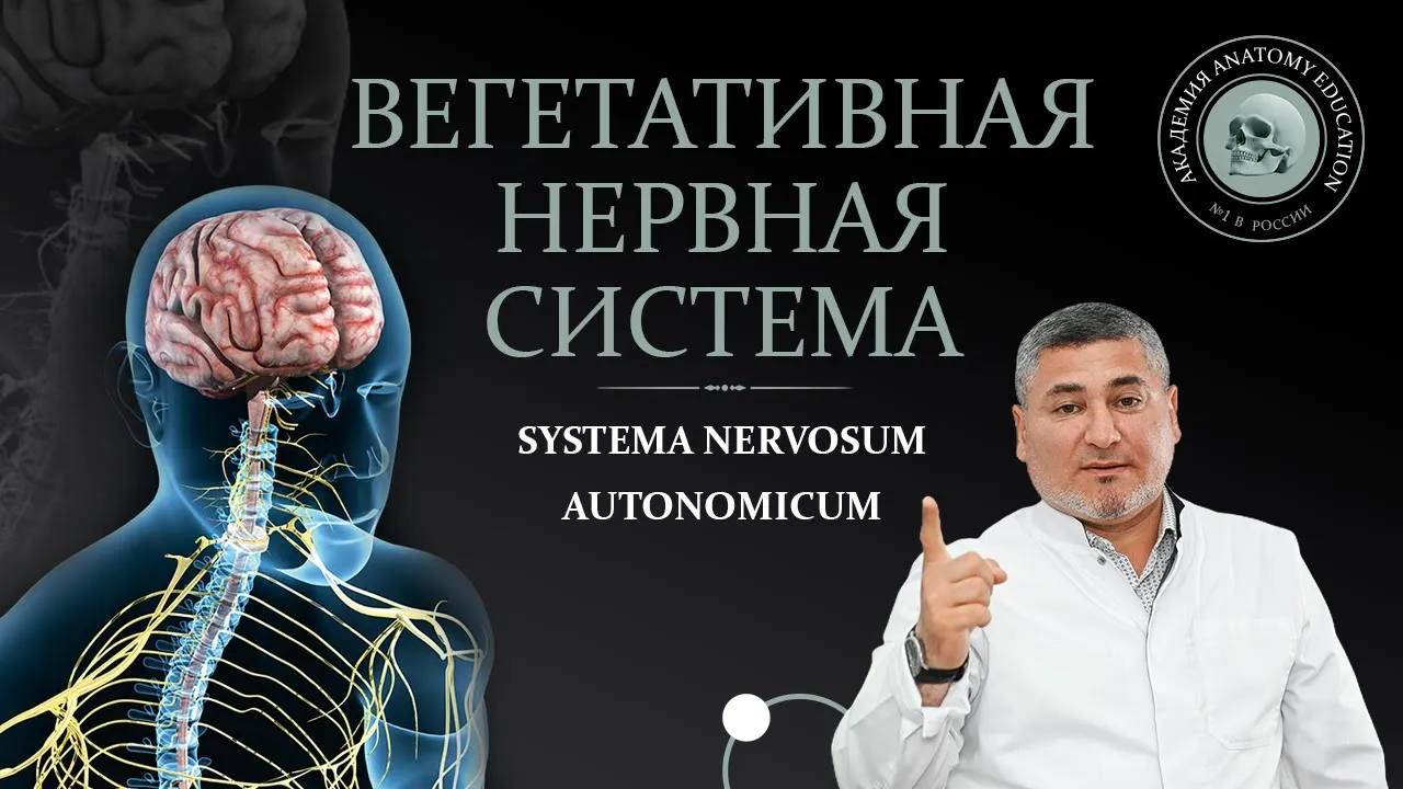
The nervous system is divided into two main parts: the somatic nervous system (voluntary control) and the autonomic nervous system (involuntary control). The somatic system controls skeletal muscles and is under conscious control. The autonomic system controls internal organs (heart, lungs, digestive system) and operates automatically without conscious control. This division allows the body to handle both voluntary movements and involuntary organ functions simultaneously.

The human body has two distinct nervous systems: the somatic nervous system and the autonomic (vegetative) nervous system. When the brain and spinal cord are damaged, the somatic nervous system becomes impaired, and the autonomic nervous system becomes activated, manifesting as pathological signs. The autonomic nervous system controls movement through different laws and reflex arcs compared to the somatic system. The somatic nervous system influences internal organs through motor-visceral reflexes, while the autonomic system works through receptors in internal organs, muscles, and joints. Understanding this distinction is crucial because traditional rehabilitation that only activates the somatic nervous system is ineffective when the autonomic system is dominant. Patients with pain may have either somatic or autonomic nervous system involvement. To differentiate, observe how pain responds to dynamic load changes. If pain decreases when the patient sits down, this indicates somatic involvement. If pain remains unchanged or increases, this suggests autonomic involvement.

The somatic nervous system controls voluntary movements of skeletal muscles, has one-neuron effector chains, and myelinated fibers. The autonomic nervous system controls involuntary processes of smooth muscle, cardiac muscle, and glands, has two-neuron effector chains, and unmyelinated postganglionic fibers. The autonomic nervous system regulates metabolic processes, trophic processes, secretion, and organ function without conscious control. Despite its complexity, the ANS can be influenced through practices like yoga. The ANS may cause false symptoms that complicate diagnosis, and much of its function remains poorly understood.

The nervous system is divided into somatic and autonomic systems. The somatic nervous system controls voluntary movements of skeletal muscles and is under conscious control. The autonomic nervous system controls involuntary functions of internal organs and operates automatically without conscious thought.
The core concept of localization of brain function, which posits that specific regions of the brain are responsible for specific bodily tasks.

Localization of function is the concept that specific brain regions control specific behaviors. The brain has two hemispheres, each controlling the opposite body side, with four lobes per hemisphere: frontal (motor cortex), temporal (auditory), parietal (somatosensory), and occipital (visual). Language centers are in the left hemisphere: Broca's area (speech production) and Wernicke's area (comprehension). Evidence for localization includes brain imaging showing distinct activation patterns and lesion studies. However, evidence against localization includes Lashley's maze experiments showing no single area was critical for learning, and MRI studies revealing multiple damaged areas in Broca's patients, suggesting some functions involve distributed neural networks.

Localization of function is the principle that specific areas of the brain are responsible for performing specific functions. For example, the temporal parietal junction (TPJ) is responsible for theory of mind—the ability to judge what other people are thinking or feeling. The hippocampus is associated with long-term memory and learning capacity, while the amygdala is linked to emotional regulation. These brain structures exist in relation to each other and perform specific functions, with different parts talking to each other to enable better performance.

Localization of function is the concept that specific brain regions are responsible for particular functions, such as the motor cortex controlling movement on the opposite side of the body, Broca's area for speech production, and Wernicke's area for speech comprehension; however, this theory has limitations as it oversimplifies brain function by not accounting for network interactions between regions and brain plasticity, though it is supported by case studies like patient Tan.

Localization of function is the theory that specific cognitive and behavioral functions are located in particular brain regions. Key brain areas include: the motor area in the frontal lobe controlling voluntary movements; the somatosensory area in the parietal lobe processing touch, pressure, and pain; the visual area in the occipital lobe processing visual information; and the auditory area in the temporal lobe processing sound. These four areas span both brain hemispheres, while language centers (Broca's and Wernicke's areas) are exclusively in the left hemisphere. Understanding these fundamental concepts provides the foundation for analyzing how brain structure relates to mental processes.

Localization theory proposes that specific cognitive functions are localized to specific brain regions. This concept emerged from famous case studies: Phineas Gage (frontal lobe injury affecting personality and inhibition), Broca's patient (inferior frontal gyrus lesion causing articulation difficulties, establishing Broca's area), and patient HM (bilateral medial temporal lobe surgery causing inability to form new memories, placing memory function in the temporal lobe). This theory suggests one part of the brain is responsible for one cognitive function.
Prerequisite Knowledge
- Concept 01Basic neuroanatomy, specifically the major lobes of the brain (frontal, parietal, occipital, and temporal) and their general functions.
- Concept 02The fundamentals of neuronal communication, including how action potentials and neurotransmitters transmit signals throughout the nervous system.
- Concept 03The distinction between the somatic (voluntary) and autonomic (involuntary) nervous systems.
- Concept 04The core concept of localization of brain function, which posits that specific regions of the brain are responsible for specific bodily tasks.
Subsequent Learning
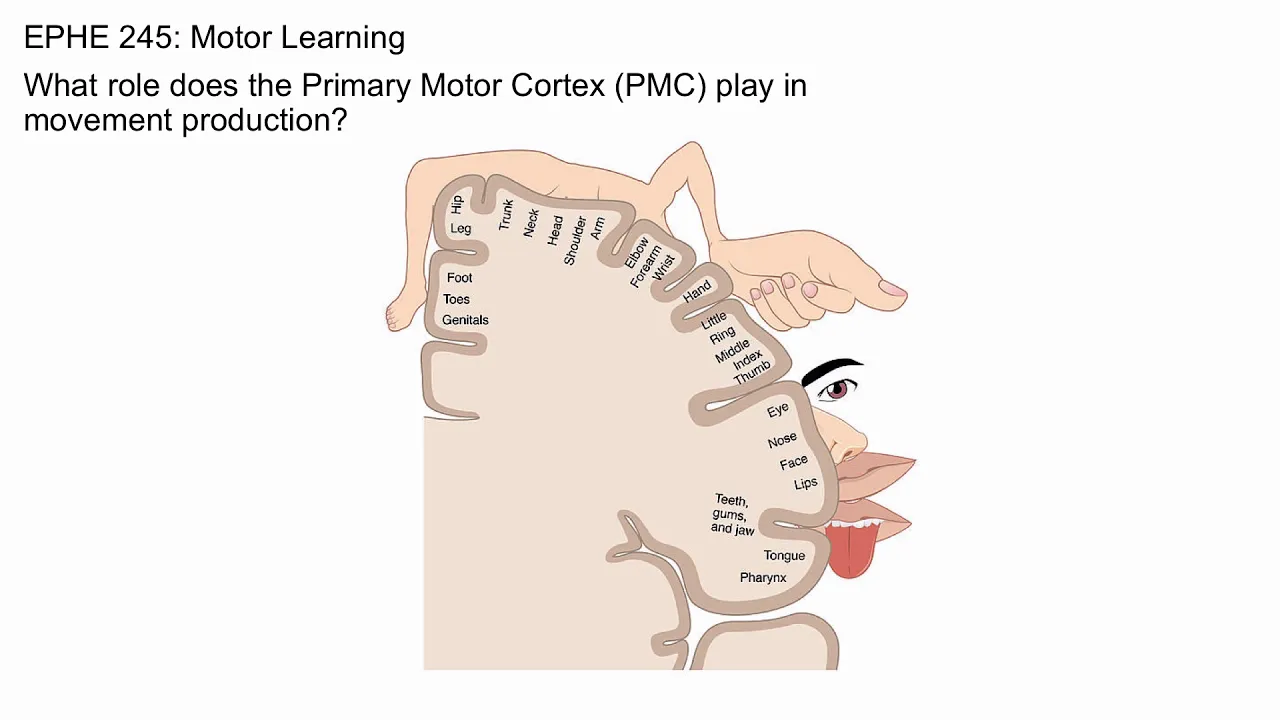
- Step 01The concept of the motor homunculus, exploring how different body parts are somatotopically mapped across the primary motor cortex.
- Step 02The anatomy of descending motor pathways, particularly the corticospinal tract, which transmits signals from the motor cortex to spinal nerves.
- Step 03The modulatory roles of the basal ganglia and cerebellum in planning, coordinating, and refining voluntary movements.
- Step 04Clinical pathologies associated with motor system damage, such as stroke, Parkinson's disease, and Amyotrophic Lateral Sclerosis (ALS).
- Step 05Real-world applications in neurotechnology, such as utilizing motor cortex signals to develop Brain-Computer Interfaces (BCIs) and neuroprosthetics.
Motor Cortex
0:00- 1
Explains the motor cortex's five subparts and their core functions.
- 2
Describes how the brain generates and controls voluntary movements.
- 3
Details the flow from planning in area 6 to execution in area 4.
The Action-Map Theory of Motor Cortex Organization
While traditional neuroscience teaches that the motor cortex is organized as a neat, somatotopic map of individual muscles (the classic homunculus), the 'Action-Map' theory provides a significant counterpoint. Pioneered by researchers like Michael Graziano, this theory suggests that the motor cortex is actually organized to control complex, ethologically relevant behaviors—such as reaching, grasping, or defending—rather than isolated muscle contractions. By applying longer, more naturalistic electrical stimulations to the motor cortex, scientists observed coordinated, multi-joint movements rather than simple muscle twitches. This perspective challenges the rigid, top-down 'keyboard' model of movement, suggesting instead that the motor cortex operates as a highly dynamic network that coordinates behavioral goals and whole-body actions in response to sensory feedback.
The concept of the motor homunculus, exploring how different body parts are somatotopically mapped across the primary motor cortex.
![[IYNA/HKU] The Neuroscience of Movement](https://i.ytimg.com/vi/D26uRJPqumA/maxresdefault.jpg)
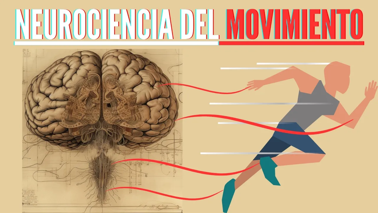
The motor homunculus is a somatotopic map of the body in the primary motor cortex, where different body parts are represented in specific regions. The medial (inner) part of the motor cortex controls the lower body (kneecap, feet), while the lateral (outer) part controls the face, lips, and hands. This organization shows that some body parts have disproportionately large representations in the motor cortex, reflecting the fine motor control needed for those areas. The hands and face receive particularly extensive cortical representation, which explains our ability for precise hand movements and facial expressions.

The motor homunculus is a somatotopic map of the body in the primary motor cortex. Different body parts are represented in different areas, with the face and hand areas being particularly large. This explains why some movements are more susceptible to damage.

The motor homunculus is a somatotopic map representing the entire body in the motor cortex, similar to the sensory homunculus in the somatosensory cortex. Each body part has a proportional representation based on its functional importance. Areas requiring more precise control have larger representations in the motor homunculus.

The motor homunculus is a representation of the body in the primary motor cortex, where different body parts are mapped to specific cortical areas. The representation is not proportional to actual body size but reflects the amount of motor control required for each body part. Body parts requiring fine motor control, such as the hands and lips, have disproportionately large representations. This topographic organization explains why certain body parts are more sensitive to motor control and why damage to specific cortical areas affects specific body functions.

The motor homunculus represents the mapping of body areas onto the primary motor cortex. Different body parts have varying sizes of cortical representation based on their motor demands. Areas requiring fine motor control occupy larger portions of the motor cortex, while areas involved in gross motor movements occupy smaller portions.
The anatomy of descending motor pathways, particularly the corticospinal tract, which transmits signals from the motor cortex to spinal nerves.

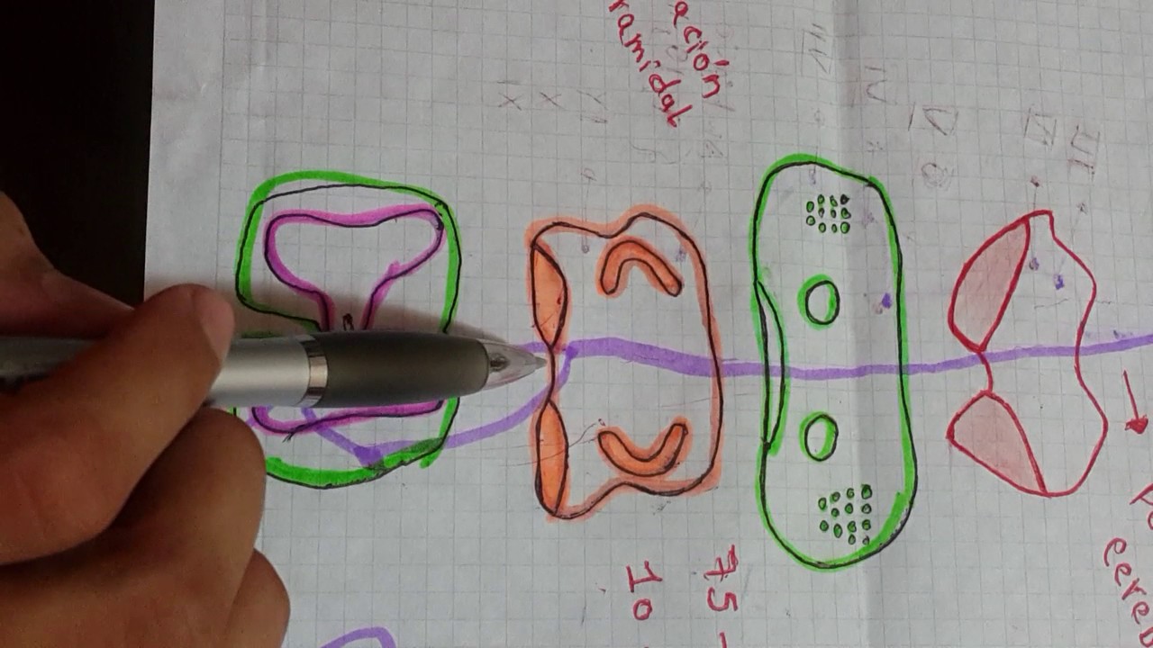
The corticospinal tract is the primary descending motor pathway originating from the primary motor cortex (precentral gyrus), premotor cortex, and supplementary motor areas in the frontal lobe. Upper motor neuron axons travel through the corona radiata, internal capsule, cerebral peduncles, and pyramids, where approximately 85-90% decussate at the pyramidal decussation to form the lateral corticospinal tract (controlling fine, skilled movements of distal limbs) and 10-15% remain uncrossed as the anterior corticospinal tract (controlling axial and proximal muscles). These fibers synapse on lower motor neurons in the spinal cord's ventral horn, which then innervate skeletal muscles; lesions at different points along this pathway produce characteristic upper motor neuron signs including spasticity, hyperreflexia, and the Babinski sign.

The corticospinal tract is a descending motor pathway controlling voluntary movements of the extremities. It begins in the frontal lobe (areas 4, 6, 8, precentral gyrus) and terminates in the spinal cord. The pathway passes through the internal capsule (genu), cerebral peduncles, and brainstem. At the pyramidal decussation in the medulla, 75-90% of fibers remain ipsilateral to the anterior funiculus, while 10-15% cross contralaterally to the lateral funiculus. The tract synapses with lower motor neurons in the anterior horn of the spinal cord, which then transmit signals via acetylcholine and adrenaline to effectors for voluntary muscle contraction.

The corticospinal tract is the primary motor pathway descending from the motor cortex. Upper motor neurons travel from the cortex through the brainstem to the lower medulla, where they decussate. Lower motor neurons then exit the spinal cord at the anterior horn and synapse directly on target muscles. The lateral corticospinal tract (larger) controls limb musculature (arms and legs), while the anterior corticospinal tract controls axial musculature (abdominal muscles, trapezius, back muscles, paraspinal muscles). This pathway enables voluntary movement by transmitting motor commands from the brain to muscles throughout the body.

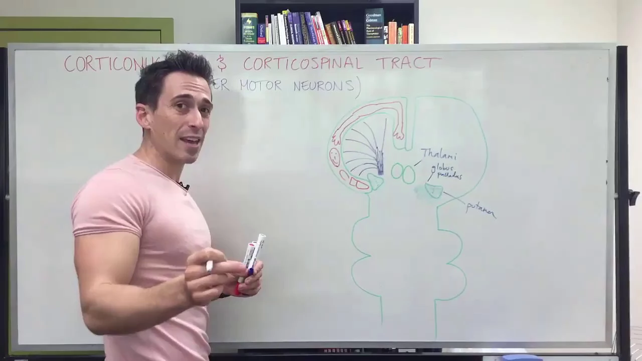
The motor cortex generates conscious voluntary movements through two main descending motor pathways: the corticonuclear tract (which controls cranial nerves V, VII, IX, X, XI, and XII for head and neck movements) and the corticospinal tract (which controls skeletal muscles of the limbs and trunk). Both pathways originate from the primary motor cortex in the precentral gyrus, travel through the corona radiata and internal capsule, and descend through the brainstem. The corticonuclear tract terminates at cranial nerve nuclei in the pons and medulla, while the corticospinal tract crosses at the medullary spinal cord junction (decussation), with approximately 90% crossing over to innervate the opposite side of the body. The lateral corticospinal tracts control fine motor movements of the hands and feet, while the anterior corticospinal tracts control trunk and axial muscle movements.

Descending tracts are neural pathways from higher CNS centers to lower centers, following a three-neuron chain: upper motor neurons (first-order), interneurons (second-order), and lower motor neurons (third-order). The corticospinal tract is the primary pathway for voluntary motor control, with fibers originating from pyramidal cells in the fifth layer of the cerebral cortex—two-thirds from the precentral gyrus (motor areas 4 and 6) and one-third from the postcentral gyrus (parietal areas 3, 1, 2). The tract passes through the posterior limb of the internal capsule, corona radiata, and midbrain, maintaining somatotopic organization. At the medulla-spinal cord junction, most fibers decussate to form the lateral corticospinal tract (descending in lateral white column), while uncrossed fibers form the anterior corticospinal tract. The tract terminates in the anterior gray column, synapsing with interneurons that control alpha and gamma motor neurons. Functionally, it confers speed and agility to voluntary movements, particularly skilled fine movements like writing and playing instruments.
The modulatory roles of the basal ganglia and cerebellum in planning, coordinating, and refining voluntary movements.

Two critical brain structures collaborate with the motor cortex for movement planning. The cerebellum provides information from previous movement attempts, helping the brain identify useful versus incorrect strategies. It modulates planned movement information by filtering out errors and reinforcing successful approaches. The basal ganglia, located deep in the brain, regulate information flow and energy distribution. They synthesize multiple fragmented signals into fewer, more powerful commands, consolidating information to improve energy efficiency. They also route only essential information to planning areas while filtering out irrelevant signals.

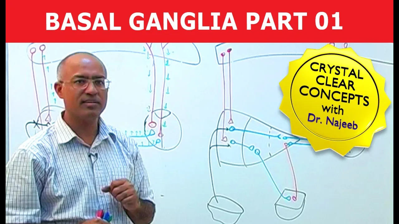
Motor plans are stored in basal ganglia and cerebellum. When initiating movement, the idea must first consult basal ganglia for planning and refinement before descending to lower motor neurons. Basal ganglia electrical activity begins even before movement execution. A cortical-basal ganglia-thalamic loop allows consultation, refinement, and execution coordination.

The basal ganglia and cerebellum are involved in movement planning and coordination. The basal ganglia are involved in initiating and regulating movements, while the cerebellum is involved in fine-tuning and coordinating movements.

The corticospinal tract is modulated by the basal ganglia and cerebellum. The cerebellum ensures movements are smooth and coordinated. The basal ganglia inhibit involuntary movements. Dysfunction can cause Parkinson's (increased inhibition) or Huntington's (decreased inhibition).

The basal ganglia (striatum and globus pallidus) modulate motor control through a pathway where cortex stimulates striatum, which inhibits globus pallidus, releasing thalamic inhibition and facilitating movement. Dysfunction causes Parkinson's disease with rigidity, akinesia, and tremor. The cerebellum (10% of brain volume, half of brain neurons) integrates sensory information about ongoing movements and provides corrective feedback to the motor cortex and basal ganglia. This allows smooth, coordinated movements and rapid adjustments. Damage causes ataxia, dysmetria, and intention tremor.
Clinical pathologies associated with motor system damage, such as stroke, Parkinson's disease, and Amyotrophic Lateral Sclerosis (ALS).
![C5N - LOS ENIGMAS DEL CEREBRO: CEREBRO MOTOR [BLOQUE 2]](https://i.ytimg.com/vi/xv0yXesMwYI/hqdefault.jpg?sqp=-oaymwEmCOADEOgC8quKqQMa8AEB-AHWBIAC4AOKAgwIABABGFQgZShjMA8=&rs=AOn4CLBsfxEVUGPN-uC6IdZeYi76Xyo6DQ)
Stroke interrupts blood flow to the brain, causing neuronal damage and motor paralysis. Functional imaging shows recovery prediction circuits activate within 1-1.5 months, with rehabilitation windows extending to 3 months. Parkinson's disease involves progressive neuron death in the substantia nigra, depleting dopamine and causing bradykinesia and reduced movement amplitude. ALS affects spinal motor neurons, causing muscle atrophy through loss of both motor control and nutritional support. These conditions demonstrate how specific brain regions control movement, and how their dysfunction leads to progressive motor impairment.

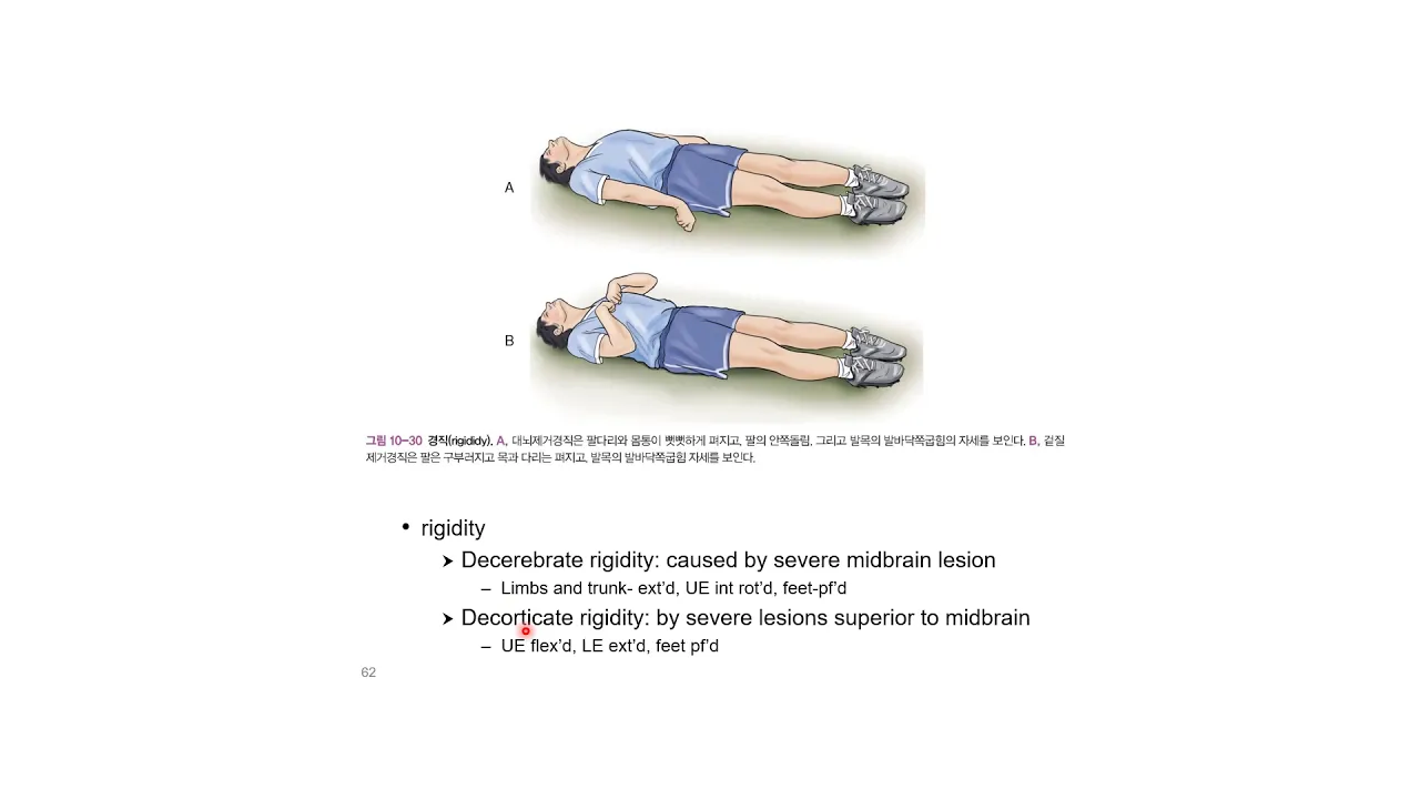
Damage to different motor system components produces distinct clinical syndromes. Pyramidal pathway damage causes muscle weakness (paralysis), classified as monoplegia, hemiplegia, paraplegia, or tetraplegia. Basal ganglia damage results in hyperkinesia (excessive involuntary movements) or hypokinesia (reduced movements). Cerebellar damage causes ataxia, characterized by loss of movement precision and coordination. These patterns help clinicians localize neurological lesions.

Multiple diseases cause motor tract damage including stroke, brain injury, cerebral palsy, multiple sclerosis, traumatic brain injury, and ALS. Stroke particularly affects the motor cortex due to its vascular supply. ALS specifically targets motor neurons, causing progressive muscle weakness. The specific disease determines the pattern of motor impairment, with some conditions causing spasticity while others cause flaccidity. Understanding these disease-specific patterns aids in diagnosis and treatment planning.

The motor system consists of pathways from the primary motor cortex traveling down the spinal cord through pyramidal tracts, crossing at the decussation and terminating in the ventral horn where they synapse with alpha motor neurons. The basal ganglia (caudate, putamen, globus pallidus, subthalamic nucleus, substantia nigra) help determine movement amplitude, direction, and initiation. Dopamine, synthesized in the substantia nigra and projecting to the striatum, is critical for motor control. The cerebellum contains Purkinje cells, among the largest neurons in the brain, and is essential for control of skilled, automatic movements. All Purkinje cells are inhibitory. Motor system diseases include spinal cord injuries (causing flaccid paralysis), polio (viral destruction of motor neurons), ALS (progressive death of motor neurons), and myasthenia gravis (autoimmune destruction of acetylcholine receptors). Parkinson's disease involves disruption of the basal ganglia circuit, specifically death of dopaminergic neurons in the substantia nigra. Symptoms include resting tremor, rigidity, bradykinesia, and difficulty initiating movements. Treatments include levodopa (a dopamine precursor), deep brain stimulation (electrical impulses in the subthalamic nucleus), and stem cell transplantation.

Clinical differentiation between upper motor neuron (UMN) and lower motor neuron (LMN) lesions is essential. UMN lesions cause spasticity, hyperreflexia, and Babinski sign. LMN lesions cause flaccidity, hyporeflexia, and fasciculations. The pyramidal tract originates from Betz cells in the motor cortex, travels through the corticospinal tract, and synapses at the anterior horn cells. Stroke is the most common cause of UMN motor dysfunction, with 80% being ischemic and 20% hemorrhagic. Amyotrophic Lateral Sclerosis (ALS) is a neurodegenerative disease affecting both UMN and LMN, characterized by progressive weakness, spasticity, and fasciculations. ALS predominantly affects women over 50-60 years old, with an incidence of 1-7 per 100,000.
Real-world applications in neurotechnology, such as utilizing motor cortex signals to develop Brain-Computer Interfaces (BCIs) and neuroprosthetics.

BCIs can already control devices like tablets through brain signals. Paralyzed individuals can use their brains to operate technology. This represents the practical application of neural interfaces for restoring lost motor functions and enabling new forms of human-machine interaction.

Brain-computer interfaces (BCIs) enable direct communication between the brain and external devices. In 2008, researchers demonstrated that monkeys could control robotic arms using motor cortex signals, treating the robotic arm as a natural limb extension. The technology uses biofeedback and closed-loop learning, where systems adapt to neural signals. Microelectrodes implanted in the motor cortex read neural activity patterns, which algorithms decode to determine intended movements. Clinical applications show patients with paralysis or multiple sclerosis can control prosthetic limbs within days of training, achieving precision comparable to natural limb movement. This technology represents a breakthrough in treating neurological conditions and restoring function to those with paralysis.

Brain-computer interfaces (BCIs) are implantable devices that use electrodes to detect electrical signals from specific brain regions, particularly the motor cortex, and translate these signals into commands for computers or external devices using machine learning algorithms; they primarily help individuals with paralysis, spinal cord injuries, or neurodegenerative diseases like ALS regain communication and interaction abilities, with current applications focusing on reading neural signals rather than writing to the brain, requiring hundreds to thousands of electrodes for effective operation.
![L’Homme connecté [LA SEMAINE DU CERVEAU]](https://i.ytimg.com/vi_webp/_ej2bPNIEl4/maxresdefault.webp)

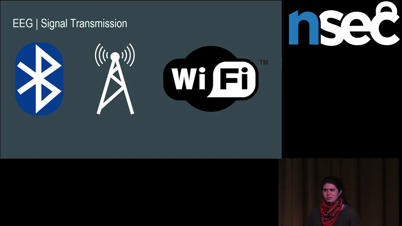
Brain-computer interfaces measure brain activity through three systems: EEG (scalp sensors), ECoG (brain surface sensors), and cortical implants (100 electrodes in specific brain regions). The motor cortex contains a homunculus mapping body parts to brain regions, with hands and face having disproportionate representation. Training patients to imagine specific actions generates consistent brain signals (biomarkers) that computers can recognize. Applications include: controlling mental switches for ALS patients (late 1990s), brain-controlled mouse for tetraplegic patients (2006), mental typing for communication restoration (40 characters/minute), and mental writing achieving 80 characters/minute after 3 years of training.

Brain-computer interfaces (BCIs) are neurotechnological systems that measure brain activity through electrodes and translate these signals into control commands for external devices like exoskeletons, enabling paralyzed individuals to perform voluntary movements such as grasping objects; this technology combines real-time brain signal analysis with machine learning algorithms and context-aware sensors to interpret user intentions and execute corresponding actions, with applications extending from motor rehabilitation to potential treatments for psychiatric conditions like depression and addiction.
Motor Cortex
0:00- 1
Explains the motor cortex's five subparts and their core functions.
- 2
Describes how the brain generates and controls voluntary movements.
- 3
Details the flow from planning in area 6 to execution in area 4.
The Action-Map Theory of Motor Cortex Organization
While traditional neuroscience teaches that the motor cortex is organized as a neat, somatotopic map of individual muscles (the classic homunculus), the 'Action-Map' theory provides a significant counterpoint. Pioneered by researchers like Michael Graziano, this theory suggests that the motor cortex is actually organized to control complex, ethologically relevant behaviors—such as reaching, grasping, or defending—rather than isolated muscle contractions. By applying longer, more naturalistic electrical stimulations to the motor cortex, scientists observed coordinated, multi-joint movements rather than simple muscle twitches. This perspective challenges the rigid, top-down 'keyboard' model of movement, suggesting instead that the motor cortex operates as a highly dynamic network that coordinates behavioral goals and whole-body actions in response to sensory feedback.
on this edition of grey matters we're going to discuss the function and operation of the brains motor cortex the motor cortex is a band of the cerebral cortex involved in the planning control and execution of voluntary movements it stretches over the top center of the brain from here to here like you might imagine the Madam's hair back but made of a squishy great noodle-like substance the motor cortex can be subdivided into five basic parts the primary motor cortex which makes up area for the premotor cortex the supplementary motor area the posterior parietal cortex and the primary somatosensory cortex all of which make up area 6 the primary motor cortex is responsible for generating neural impulses that pass down the spinal cord and control movement the premotor areas responsible for motor control including the preparation for movement the sensory guidance of movement the spatial guidance of reaching and the direct control of some proximal and trunk muscles of the body the supplementary motor area is responsible for internally generating planning of movement and the coordination of the two sides of the body such as in by manual coordination Mysteria parietal cortex is responsible for transforming multi-sensory information into motive fans the primary somatosensory cortex is considered to be functionally part of the motor control circuitry simply put the motor cortex controls the body's movement as your context clues may have allowed you to gather so let us say that we want to make been here move his arm with simple stimulation of the motor cortex thusly we will see that Ben's arm reacts as desired but of course movement isn't quite as simple as all this even in performing the simplest movements your brain doesn't merely send a letter or assembly culturally a text directly to the body parts as a command to move rather each movement is actually a series of movements the motion is simple is lifting your leg may be generated at one part of the motor cortex but travel through various other parts including other parts of the brain entirely before the series of movements that comprises the lifting of your leg is coherently assembled into a command for this to function properly and smoothly you need a sort of internal cloth to regulate the sequence of movements this is the responsibility of the cerebellum now if we deactivate the cerebellum and try to stimulate the same area again the reaction may not manifest because the cerebellum controls both timing and speed of the movement of course the reaction may also certainly be delayed you see the motor cortex as mentioned above is divided into two larger areas these are known as area 4 and area 6 a planning for any given movement is collected from various parts of the brain and concentrated into area 6 the cohesive set of instructions as then delivered to area 4 or the primary motor cortex which then disperses motion can to the appropriate area of the voting according to those instructions provided nothing interferes such as a seventh spinal column or ruptured columns this essential process of the brain performs accurately and efficiently allowing humans to engage in wonders such as the Russian ballet team athletics and although cannibalism join us next time for another exciting episode of grey max this is Rupert Worchester Shaya saying good
Up Next
Brain Implants and Mind Reading: Neurotech Security Implications
@NorthSecIo
19.4K views•2018-10-10

Bessel van der Kolk on How Trauma Affects the Body and Brain
@bigthink
226.3K views•2025-10-03

Vagus Nerve (CN X): Anatomy, Nuclei & Functions Explained
@Alilamedicalmedia
305.2K views•2022-10-31

How Exercise Benefits Your Brain: Science Explained
@TED
11.4M views•2018-03-21
Related Study Plans & Knowledge Roadmaps
Structured learning paths in Neuroscience