














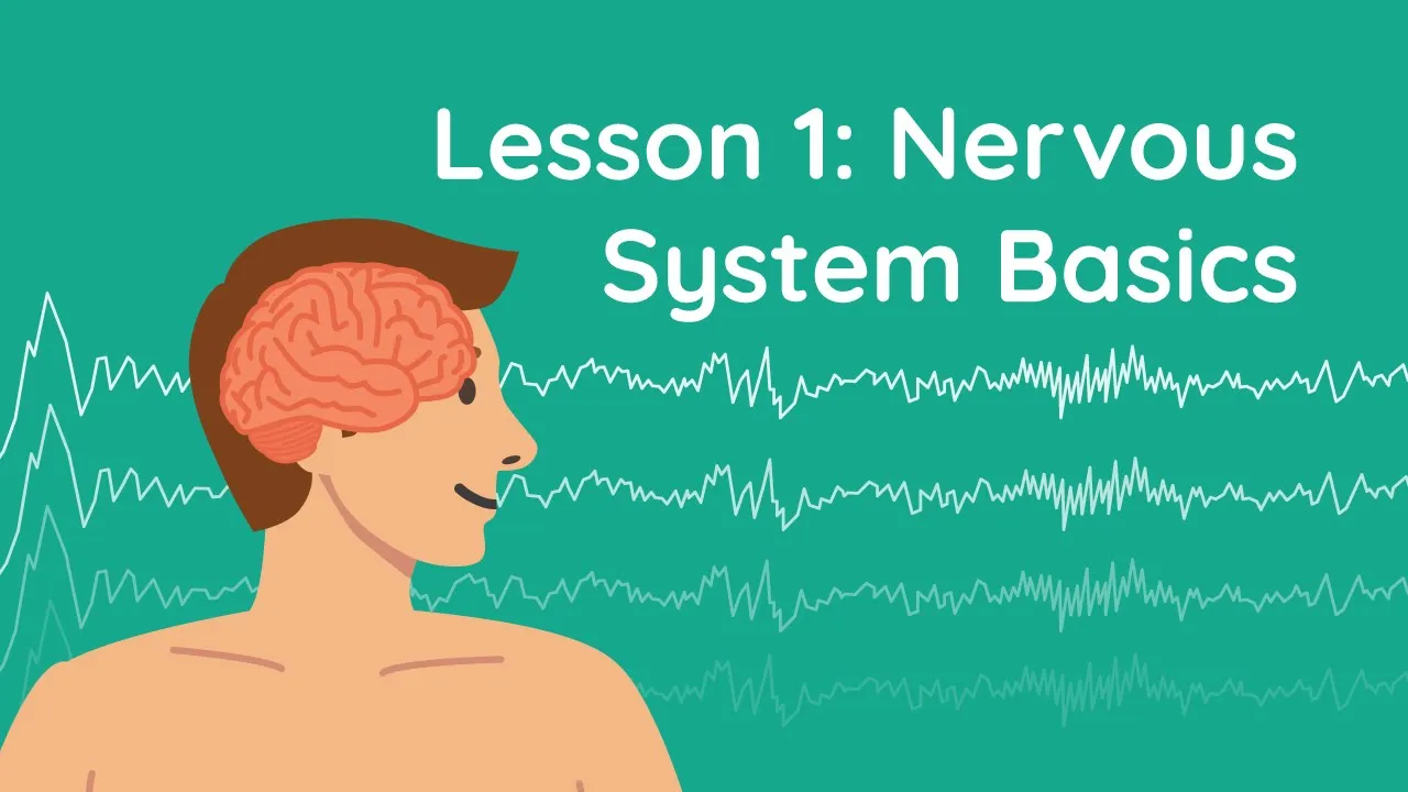







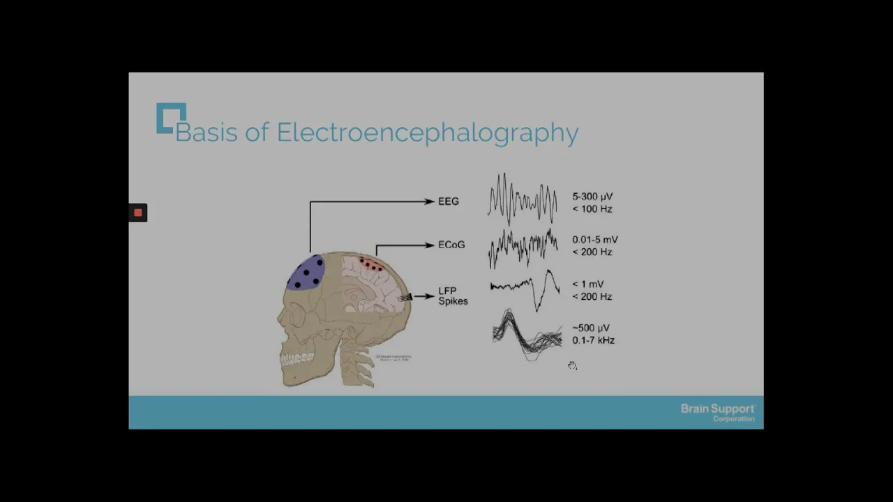






EEG (Electroencephalography) measures electrical changes in the brain produced by neuronal firing using scalp electrodes, detecting the summation of excitatory postsynaptic potentials from densely packed pyramidal neurons near the cortical surface; action potentials are electrical signals generated by sodium and potassium ion movements across neuronal membranes, traveling unidirectionally due to refractory periods, and are the fundamental basis for the electrical signals that EEG captures to diagnose conditions like epilepsy, where abnormal waveforms such as spike waves, sharp waves, or unusually slow waves indicate excessive or synchronous neuronal activity.
EEG Explained: How Brain Activity & Action Potentials Create Electroencephalograms | Neurological Diagnostics
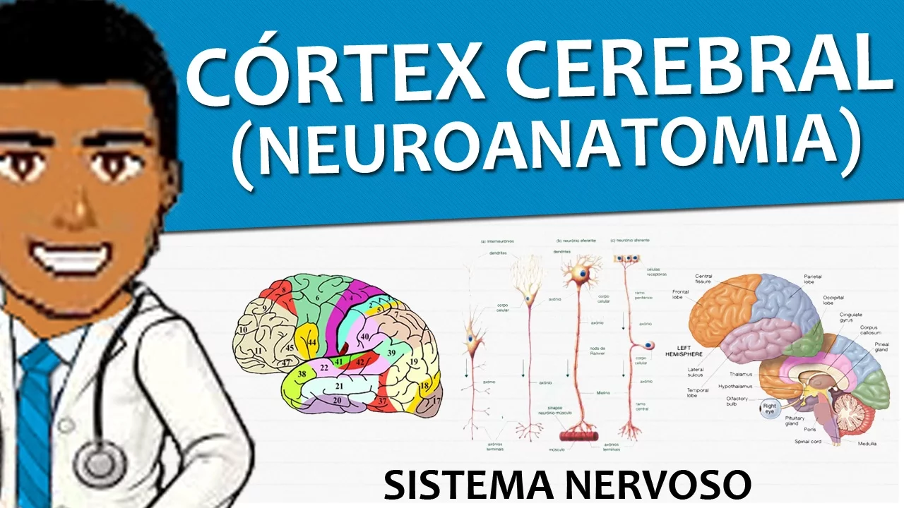
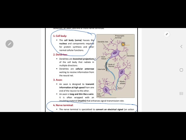
Added:Basic anatomy of a neuron, specifically the structure of pyramidal cells, dendrites, and axons in the cerebral cortex.
The biophysics of resting membrane potentials and action potentials, including the movement of sodium and potassium ions.
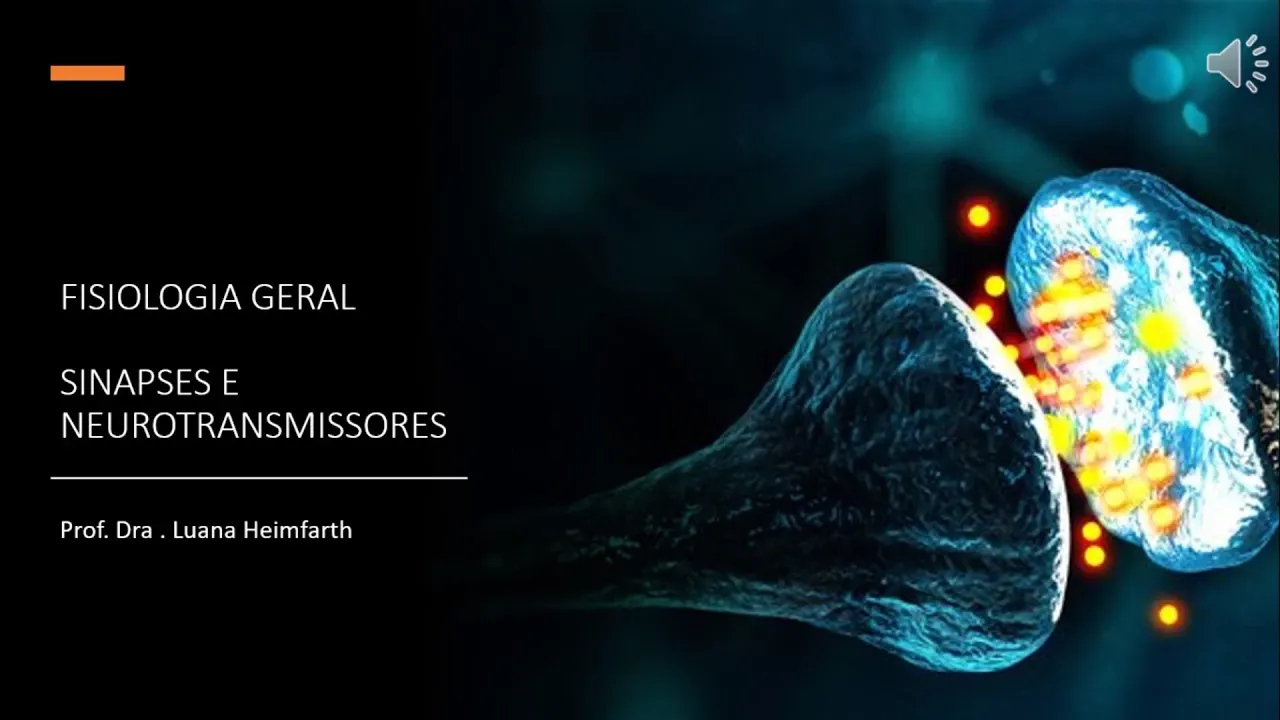
The mechanisms of chemical synaptic transmission, particularly how Excitatory and Inhibitory Postsynaptic Potentials (EPSPs and IPSPs) are generated.
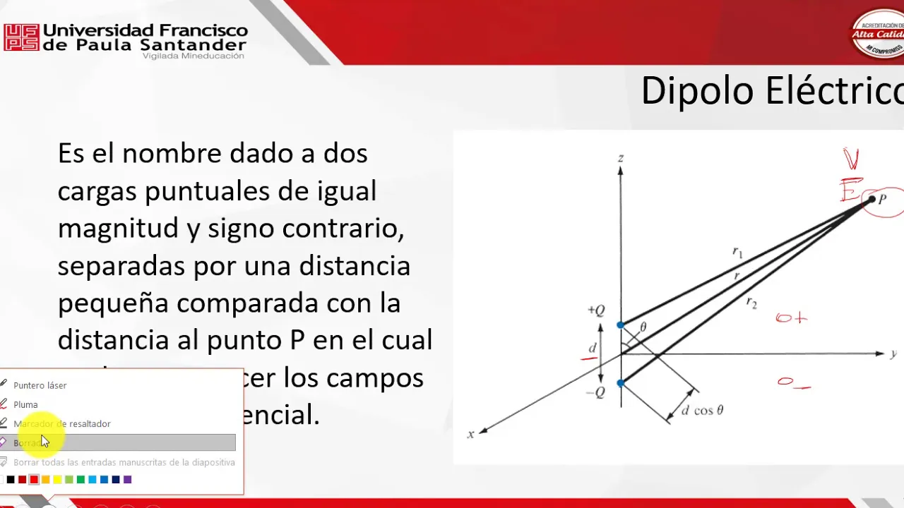
Basic electrical principles, including voltage, current, conductance, and how electrical fields propagate through physical media (volume conduction).
Classification and interpretation of EEG frequency bands (Alpha, Beta, Theta, Delta, and Gamma waves) across different states of consciousness and sleep stages.
Clinical diagnostics of epilepsy, specifically recognizing epileptiform patterns like spike-and-wave discharges and localized vs. generalized seizures.
EEG artifact identification and signal processing, including how to filter out physiological noise from eye blinks, muscle tension, and cardiac activity.
Comparison of EEG with other neuroimaging modalities, such as MEG (magnetoencephalography) and fMRI, analyzing their trade-offs in spatial and temporal resolution.
2.1K views12likes6:03Original Release: 2012-05-01
EEG and action potentials.
What is an EEG? EEG stands for electronraph.
This is a method by which the electrical changes in the brain produced when neurons fire are measured using scalp electrodes. Traces are produced of the electrical activity and can be viewed for abnormalities.
How does an EEG work? First, let us consider what an action potential is.
Brain activity is regulated by neuronal firing with different neurons sinapsing with others to elicit responses. Neurons consist of a cell body send dendritic projections into it which other neurons can signapse with as well as an axon which has a selectively permeable membrane. An action potential is the conduction of an electrical signal down the axon.
Action potentials are generated by sodium and potassium ion movements. At rest, the inside of the axon is at minus70 molts compared to the extracellular matrix. This is maintained by the activity of sodium potassium ATPAS pumps that actively deposit three sodium ions out of the cell for every two potassium ions into the cell. When the nerve fires, the axon rapidly deolarizes and its membrane potential rises to plus 40 molts. This happens because the sodium potassium pumps stop and sodium channels in the membrane open allowing sodium to enter the axon. If the stimulus does not cause the membrane voltage to rise over the threshold value of - 55 molts, the action potential will not be conducted. This is the all or nothing principle. Following depolarization, the sodium channels shut and potassium channels in the membrane open allowing rapid repolarization of the axon back to aroundus 70 molts.
Following repolarization, there is a refractory period when the nerve cannot fire again for a short time.
The action potential travels down the axon by depolarization of one area of membrane causing a flow in a current down the axon triggering deolarization of the next section. Due to the refractory periods, this means that the action potential can only flow in one direction. In my nerves, there is saltatory conduction where the axon is only depolarized at the nodes of rovier, thus allowing faster conduction.
How does this relate to an EEG? Near the cortical surfaces of the brain are pyramidal nerve cells. When an apherent neuron activates one of these cells by conducting an action potential via a signapse, the dendrite deolarizes. It therefore becomes positively charged due to ion influx leaving the extracellular space negatively charged. As the action potential flows down the dendrite towards the soma, this creates a current running through the neuron which exits it in a brain area away from the sinapse area creating an area of positively charged extracellular space. There is therefore a dipole in the extracellular space around the dendrite. When a scalp electrode is placed, a negative charge can be detected above the synaptic end of the dendrite. What is measured is this excitatory postsinaptic potential.
The detective voltages are tiny for each dendrite. However, due to the densely packed nature of these pyramidal neurons in the brain, each electrode will detect a summation of the electrical signals from many different dendrites in the same area. This occurs because multiple neurons will be activated at the same time and they will usually have the same orientation.
Around 20 plus electrodes are placed on the head using the international 1020 electrode placement system as a basis.
An EEG will produce an individual trace for each electrode placed. The normal electrical wave traces produced by an EEG include alpha, beta, theta, and delta waves. Different stimuli will produce different wave traces from different brave areas. For example, alpha waves should be detected at the back of the head while you wake with your eyes shut.
One of the main uses of an EEG is to look for epileptic brain activity which can manifest as spiken waves, sharp waves, or as unusually slow waves on the EEG trace. An epileptic seizure is characterized as abnormal, excessive, or synchronous neuronal activity in the brain. EEGs can be used to help confirm an epilepsy diagnosis as up to 90% of epileptics will show abnormal waveforms in their EEGs between seizures known as subclinical seizures. However, not all epileptics will have these shown in every EEG. These abnormal waveforms can be induced in a patient by asking them to hyperventilate to reduce blood carbon dioxide levels or to look at a flashing stimulus or else the patient may be asked not to sleep beforehand as being drowsy can inre increase the chances of seeing some interrectal activity.
Nonpileptic seizures can also occur due to psychological rather neurological conditions and EEGs can be used to identify these by their lack of epileptic activity in the traces.
If an EEG is recorded during an epileptic seizure, the brain will often show large amplitude spike in waves.
These waves may occur locally for a few electrode traces or widespread over. The pattern of waves produced will vary depending on the type of seizure and whether the seizure is a general one affecting the entire brain or a partial one localized to one cortical lobe. The epicenter of seizure activity may be located deeper in the brain and if so then the EEG may not be able to detect any abnormal activity as the electrodes measure the voltages near the scalp surface. An EEG can also be used to determine the focal center of a partial seizure, allowing that area of the brain to be localized for surgical treatment of epilepsy. Status epilepticus occurs when there is a recurrent seizing state for over 30 minutes. EEGs can be used to confirm that the seizures are occurring and as status epilepticus is a life-threatening condition, EEGs are therefore able to help confirm diagnosis before permanent neurological damage occurs.
In conclusion, EEG is a method by which the electrical activity of the brain can be measured and imaged in traces. This has proved medically useful in the diagnosis of epilepsy, a neurological disorder.
Up Next

12:08
Motor Cortex: Primary, Premotor, & Supplementary Areas Explained | CNS Physiology
@PhysiologyLearning
7.6K views•2022-05-27

21:55
Deciphering Neural Circuits Controlling Anorexia in Mice | Richard Palmiter
@AllenInstitute
2.3K views•2011-10-10

4:10
Vagus Nerve (CN X): Anatomy, Nuclei & Functions Explained
@Alilamedicalmedia
305.2K views•2022-10-31

13:03
How Exercise Benefits Your Brain: Science Explained
@TED
11.4M views•2018-03-21
Related Study Plans & Knowledge Roadmaps
Structured learning paths in Neuroscience
neuroscience
Appetite Regulation: Brain Circuits & Hormones
01Foundations of Endocrinology and Brain Anatomy
02Introduction to Energy Homeostasis
03Peripheral Signals: Leptin, Ghrelin, and Insulin
+ 2 more learning modules
14h5 modules
Explore Roadmap
neuroscience
Neuroeconomics: Value, Risk & Brain Mechanisms
01Foundations of Neuroscience and Brain Anatomy
02Introduction to Neuroeconomics and Subjective Value
03Dopaminergic Prediction Errors & Reinforcement Learning
+ 3 more learning modules
18h6 modules
Explore Roadmap
neuroscience
Spatial Navigation: Cells, Grids & Brain Maps
01Foundations of Brain Anatomy and Memory
02Place Cells and Head-Direction Cells
03Grid Cells: The Brain's Coordinate System
+ 2 more learning modules
12h5 modules
Explore Roadmap
neuroscience
BCIs: Neural Decoding & Prosthetic Control
01Neuroscience Foundations: How the Brain Signals Intent
02Signal Acquisition: Recording the Brain's Activity
03Neural Decoding: Translating Brain Waves into Data
+ 2 more learning modules
25h5 modules
Explore Roadmap