Non-homologous end joining (NHEJ) is a cellular repair mechanism for double-strand DNA breaks where the Ku protein binds to broken ends, recruits DNA PKcs, Artemis trims single-stranded tails, and the XRCC4-XLF complex ligates the ends together; however, this process often results in deletions or imprecise joins because any nucleolytic digestion that occurred during the break is not restored.
Non-Homologous End Joining (NHEJ) | DNA Double-Strand Break Repair Animation
Added:Deep Dive
Basic structure of DNA, including the double helix, phosphodiester backbone, and complementary base pairing.

DNA और RNA वंशागतिकी का आण्विक आधार Molecular Basis of Inheritance CH 6 L 2 ✔ 12th/NEET Biology
217.5K
DNA double helix structure consists of two strands connected by phosphodiester bonds between nucleotides. The sugar-phosphate backbone forms the outer structure, while nitrogenous bases project inward. Base pairing follows specific rules: Adenine (A) always pairs with Thymine (T) through 2 hydrogen bonds, and Guanine (G) always pairs with Cytosine (C) through 3 hydrogen bonds. This complementary base pairing is fundamental to DNA structure and function.
Click to watch →
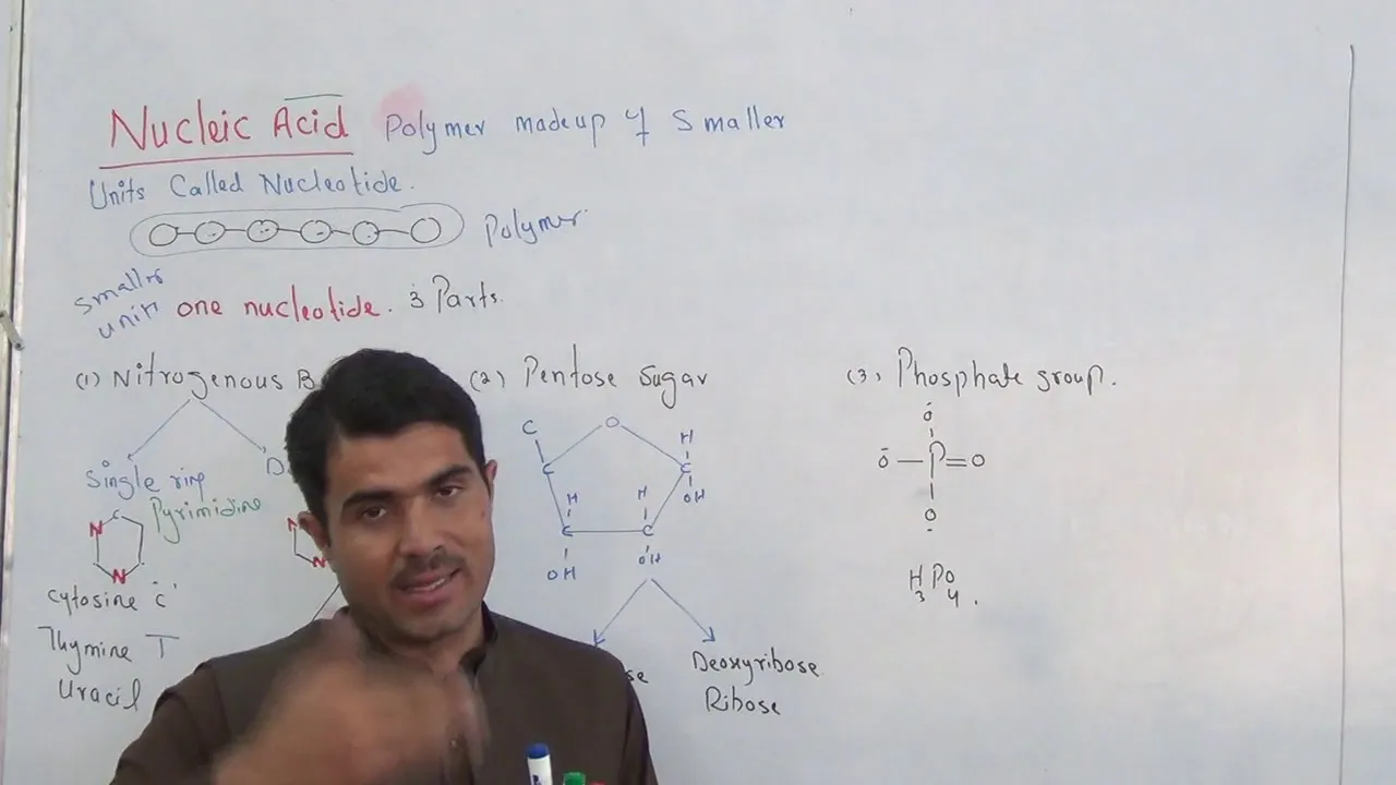
Nucleic Acid full detail along with 14 questions DNA,RNA in english part 1 by Dr Hadi Khan
72.1K
The DNA double helix consists of two anti-parallel strands held together by hydrogen bonds between complementary bases. Each strand is composed of deoxyribonucleotides linked by phosphodiester bonds, which form when the phosphate group of one nucleotide bonds to the hydroxyl group at carbon 5 of the adjacent sugar. This bond formation releases a water molecule. The 5' end has a free phosphate group, while the 3' end has a free hydroxyl group, giving DNA directionality. The DNA double helix has a diameter of 2 nanometers, with base pairs spaced 0.34 nanometers apart. One complete rotation contains 10 base pairs spanning 3.4 nanometers. The helix has major and minor grooves that allow proteins to access genetic information. Base pairing follows specific rules: adenine always pairs with thymine (2 hydrogen bonds), and guanine always pairs with cytosine (3 hydrogen bonds).
Click to watch →

BIOLOGÍA SELECTIVIDAD. ÁCIDOS NUCLEICOS
78.1K
DNA is a double-stranded polymer (bi-catenar) with nucleotides containing deoxyribose, phosphate, and bases (adenine, thymine, guanine, cytosine). The primary structure is the nucleotide sequence connected by phosphodiester bonds. The secondary structure is the double helix discovered by Watson and Crick in 1953. The two strands are antiparallel (one 5' to 3', the other 3' to 5'). Base pairing follows specific rules: adenine always pairs with thymine through two hydrogen bonds, while guanine always pairs with cytosine through three hydrogen bonds. This complementary base pairing is fundamental to DNA structure and function.
Click to watch →
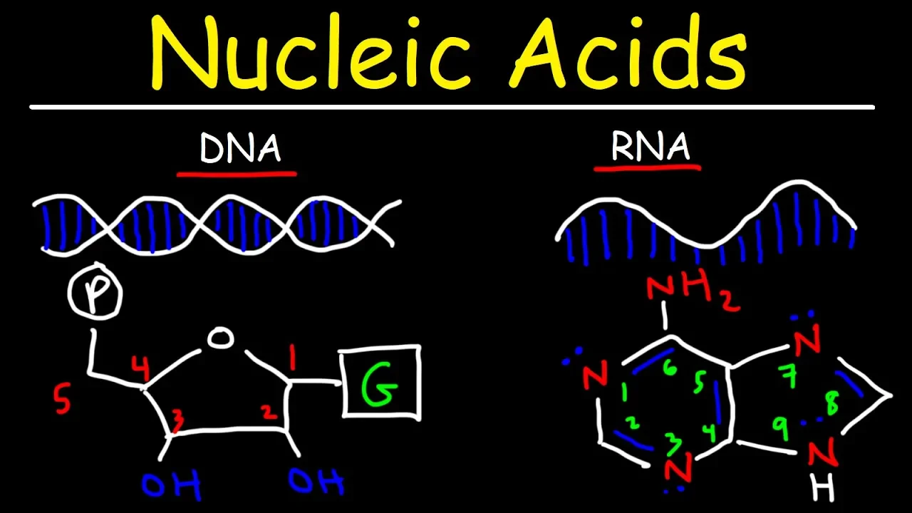
Nucleic Acids - RNA and DNA Structure - Biochemistry
828.8K
DNA forms a double helix with two antiparallel strands running in opposite directions (5' to 3' and 3' to 5'). The sugar-phosphate backbones connect through phosphodiester linkages between the 3' carbon of one sugar and the 5' carbon of the next. Nitrogenous bases pair specifically: adenine (A) pairs with thymine (T) via two hydrogen bonds, and cytosine (C) pairs with guanine (G) via three hydrogen bonds. To write complementary sequences, apply base-pairing rules and reverse the direction. For example, 5'-ATCG-3' becomes 5'-CGTA-3'. This complementary base pairing is fundamental to DNA replication, transcription, and genetic inheritance, ensuring accurate transmission of genetic information across generations.
Click to watch →

المحاضرة (30) | تركيب الـDNA.
62.1K
DNA consists of two antiparallel strands forming a double helix. The strands run in opposite directions (5' to 3' and 3' to 5'). Nitrogenous bases pair specifically: adenine with thymine, guanine with cytosine, through hydrogen bonds. The sugar-phosphate backbone connects nucleotides via covalent bonds. This complementary base pairing ensures genetic information can be accurately copied during replication.
Click to watch →
Deep Dive
The concept of DNA damage, specifically distinguishing between single-strand breaks and double-strand breaks.
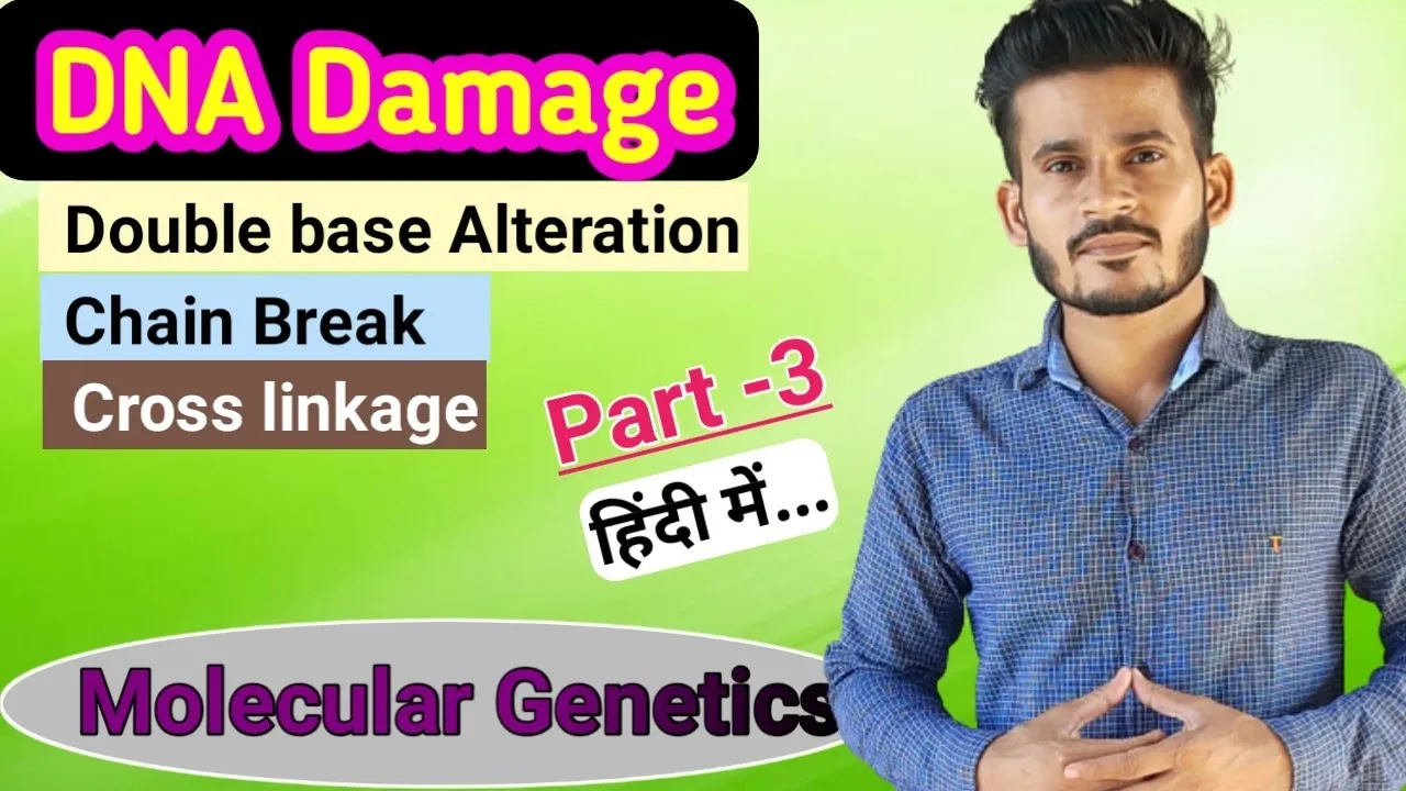
DNA Damage and Repair | Part 3 | MSc | CSIR NET | GATE
18.3K
DNA strand breaks are categorized into single-strand breaks and double-strand breaks based on the extent of damage. Single-strand breaks occur when only one DNA strand is broken, while double-strand breaks compromise both strands simultaneously. These breaks can be caused by ionizing radiation, free radicals, and chemical reactions. Single-strand breaks are repaired through specific repair mechanisms that use the intact complementary strand as a template. Double-strand breaks are more dangerous and are repaired through homologous recombination repair and non-homologous end joining repair mechanisms.
Click to watch →
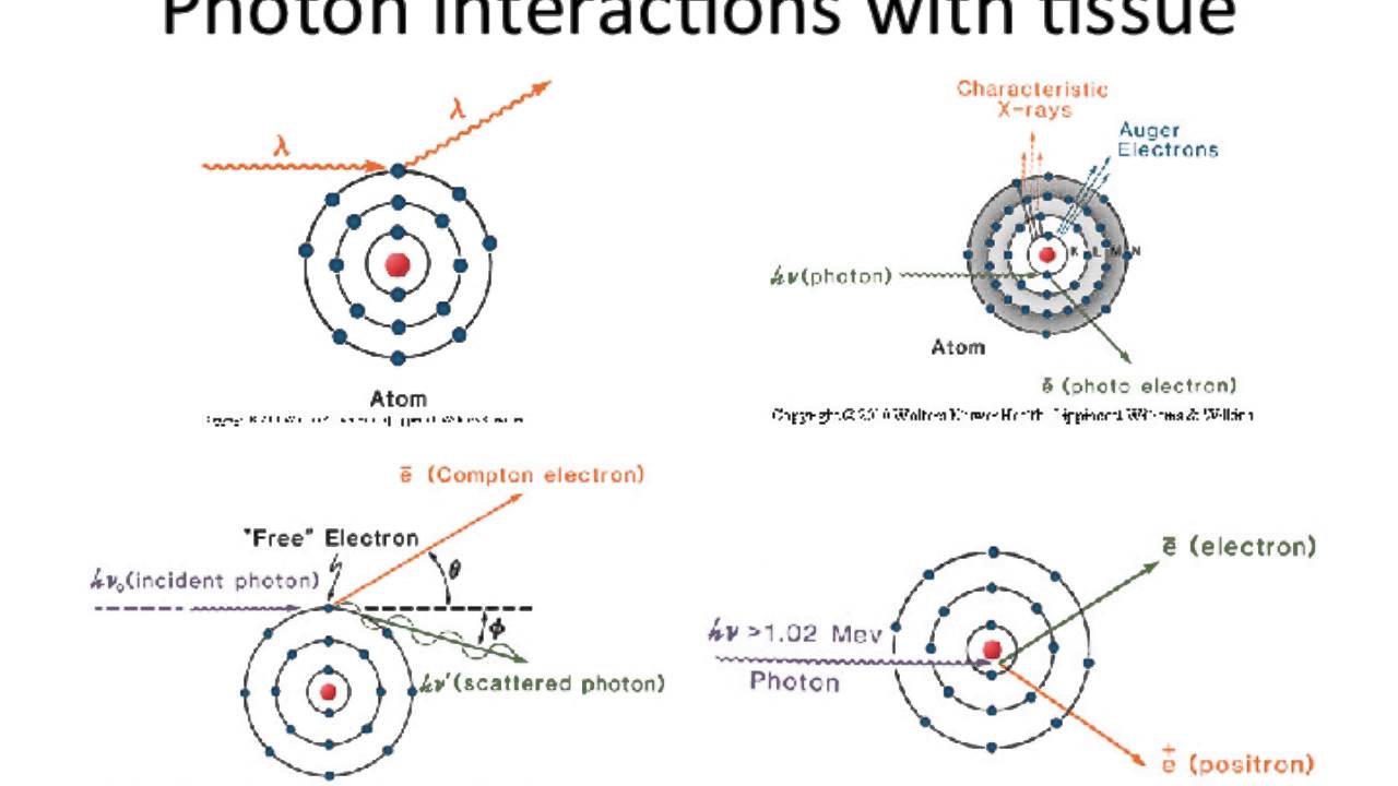
Lecture 2 - Introduction to Radiation Biology and Physics
91K
Radiation causes two types of DNA damage with different biological consequences. Single strand breaks are generally not lethal because cells have efficient repair mechanisms (base excision repair, nucleotide excision repair, mismatch repair). One gray of radiation causes approximately 1,000 single strand breaks, which cells can repair effectively. Double strand breaks are lethal and lead to mitotic catastrophe or apoptosis. One gray causes about 40 double strand breaks on average. Cells repair double strand breaks through non-homologous end joining (error-prone) or homologous recombination (high-fidelity).
Click to watch →

DNA Damage Response
4.2M
This video explains how DNA damage response mechanisms work in healthy cells versus cancer cells. In healthy cells, single-strand DNA breaks are repaired by PARP proteins, while double-strand breaks are repaired through homologous recombination using BRCA1 protein. In hereditary breast cancer patients with BRCA1 defects, double-strand breaks cannot be repaired, potentially transforming cells into cancer cells. PARP inhibitors exploit this vulnerability by blocking single-strand break repair in tumor cells, causing them to accumulate lethal double-strand breaks, while healthy cells with functional BRCA1 can still repair the damage and survive.
Click to watch →
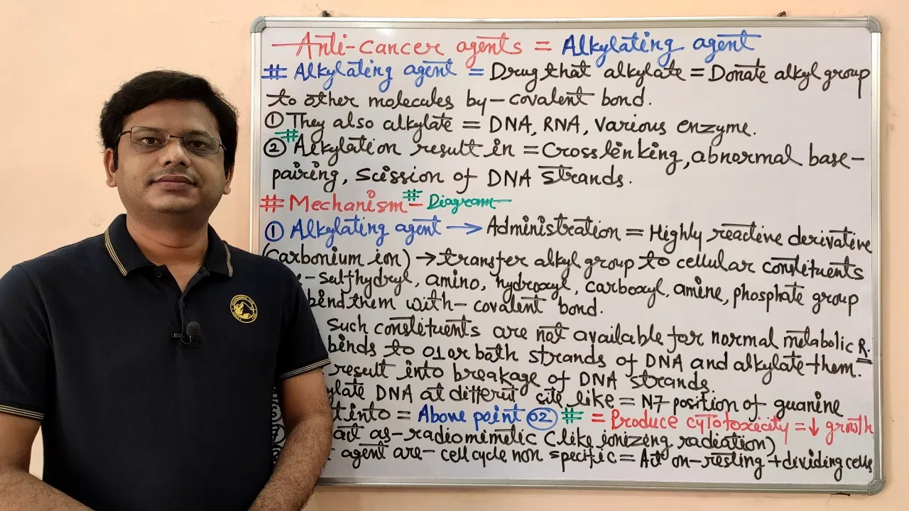
Anticancer Drugs (Part-04) Alkylating Agents| Mechanism of Action of Alkylating Agents | Anticancer
68.7K
Alkylating agents can cause both single-strand breaks and double-strand breaks in DNA. Single-strand breaks occur when the agent modifies one strand of the DNA double helix, while double-strand breaks involve damage to both strands simultaneously. Both types of damage prevent proper DNA replication and transcription, triggering cellular repair mechanisms that may lead to apoptosis (programmed cell death) if the damage is too extensive to repair.
Click to watch →

Molecular Biology-DNA Repair, NHEJ, by RAGHVENDRA Sir #CSIR, #JRF, #GATE, #DBT, #ICMR #JNU PhD
8.7K
DNA repair is essential for maintaining genetic integrity, with two main categories of damage: double-strand breaks (requiring homologous recombination or non-homologous end joining) and single-strand damage (repaired via base excision repair, nucleotide excision repair, or direct repair). Double-strand breaks are repaired through homologous recombination (using a sister chromatid as template for accurate repair) or non-homologous end joining (direct ligation without template, which can introduce errors). Single-strand damage is addressed through three strategies: excision repair (removing damaged bases), bypass repair (continuing replication past lesions), and direct repair (reversing specific damages without removing nucleotides). Key proteins involved include Ku, XRCC4, Ligase IV, and DNA polymerase with proofreading activity.
Click to watch →
Deep Dive
Fundamentals of the eukaryotic cell cycle, as repair pathway choices are often cell-cycle dependent.
![[TALK 3] NGBS2025: The Mre11-Rad50-Nbs1 complex: repairing DNA strand breaks - Karl-Peter Hopfer](https://i.ytimg.com/vi/ZuMwXSa6YWY/maxresdefault.jpg)
[TALK 3] NGBS2025: The Mre11-Rad50-Nbs1 complex: repairing DNA strand breaks - Karl-Peter Hopfer
127
DNA repair strategy depends critically on cell cycle phase. During G2 and S phases, sister chromatids provide templates for homologous recombination repair, which is more accurate than non-homologous end joining. A logistic switch controlled by cell cycle-dependent kinases regulates pathway choice, involving factors like BRCA1, RACK1B complex, and CTIP. These proteins undergo phosphorylation and priming during normal cell cycle progression. In response to DNA damage, additional phosphorylations occur, directing assembly of repair factors around break sites. This generates chromatin structures believed to be inaccessible for chromatin remodeling, creating a specialized environment conducive to homologous recombination. The MRN complex remains in an inactive state until activated by CTIP, which functions as both an activator and scaffolding protein to coordinate repair factor assembly.
Click to watch →
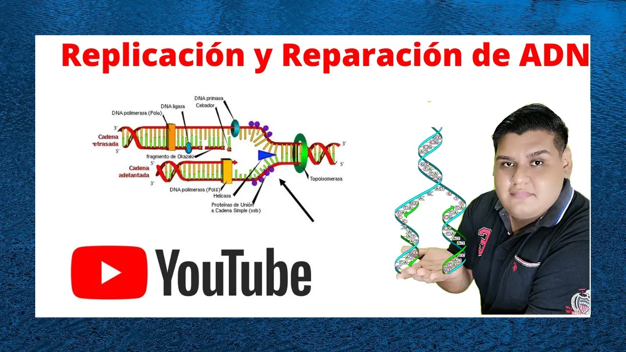
Replicación y Reparación del ADN Cap 13 Biologia de Karp
5.6K
This section covers eukaryotic DNA replication within the cell cycle and repair mechanisms. Replication occurs during S phase, with only 10-15% of cells actively replicating at any time. Origins contain conserved sequences bound by the origin recognition complex (ORC). The pre-replication complex (pre-RC) assembles with six MCM proteins during late mitosis/early G1. Activation occurs in S phase, with MCM proteins moving with the replication fork. After replication, MCM proteins are displaced and cannot be reloaded until the next cycle, ensuring each origin fires only once. Eukaryotic Okazaki fragments average 150 nucleotides. Major repair pathways include nucleotide excision repair (NER) for bulky lesions, base excision repair (BER) for damaged bases, and non-homologous end joining (NHEJ) for double-strand breaks. Defects in these pathways cause human diseases including xeroderma pigmentosum and neurodegenerative disorders.
Click to watch →
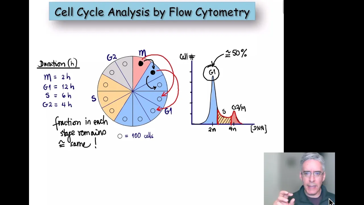
Cell cycle analysis by flow cytometry
26.9K
The eukaryotic cell cycle comprises four phases: M phase (mitosis, chromosome segregation producing two daughter cells with diploid chromosomes), G1 phase (synthesis of cellular components for S phase entry), S phase (DNA replication increasing chromosome number from 23 to 46 pairs, transitioning from diploid to tetraploid state), and G2 phase (preparation for mitosis). Progression is regulated by growth factors (stimulatory signals) and checkpoints (repressive signals). The Restriction Point commits cells to completing the cycle. Four checkpoints verify DNA integrity and proper chromosome alignment. Unresolved defects trigger apoptosis, preventing damaged cells from proliferating and potentially causing cancer.
Click to watch →
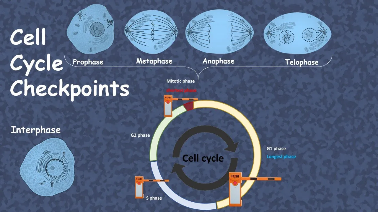
cell cycle checkpoint (advanced)
39.3K
Cell cycle progression depends on cyclin-Cdk complexes and their downstream effectors. Cyclin D-CDK4 phosphorylates pRb to release E2F, enabling cyclin E synthesis for S phase entry. Cyclin E-CDK2 activates CDC45 for DNA replication. When DNA damage occurs, ATM/ATR sensors activate p53, which induces p21 CIP to inhibit cyclin D-CDK4 in G1, or Chk1 to activate CDC25 and suppress cyclin E-CDK2 in S phase. Both responses halt replication until damage is repaired. If repair fails, p53 initiates apoptosis through Bax-mediated mitochondrial disruption and caspase activation, eliminating damaged cells.
Click to watch →

BIOLOGIA - Lezione 6 - Il Ciclo Cellulare
380.5K
The cell cycle encompasses all events between one cell division and the next in eukaryotic cells. Its duration varies by species, cell type, and environmental conditions. The cycle comprises two main phases: interphase (growth and preparation) and the M phase (cell division). Interphase includes G1 (cell growth and metabolic activities), S (DNA synthesis and duplication), and G2 (preparation for mitosis). The M phase involves mitosis (nuclear division) and cytokinesis (cytoplasmic division). While most cells undergo mitosis, specialized gamete-producing cells perform meiosis for sexual reproduction.
Click to watch →
Deep Dive
An introduction to basic enzymes involved in DNA metabolism, such as polymerases, ligases, and nucleases.
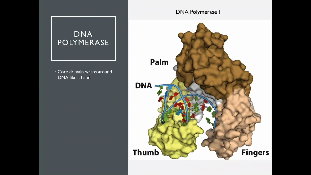
OSSM Biochem Chapter 25 - DNA Polymerase
263
DNA metabolism involves two major classes of enzymes: polymerases that synthesize nucleic acids and nucleases that degrade them. DNA polymerase adds nucleotides to the 3' end of growing strands in the 5' to 3' direction, using parental DNA as a template. Nucleases include exonucleases, which remove nucleotides from ends (3' to 5' or 5' to 3'), and endonucleases, which cut internally at specific recognition sites. These enzymes work together to build, modify, and repair genetic material throughout the cell cycle.
Click to watch →

Recombinant DNA Technology | RDT | Biotechnology | Methods in Biology | Unit - 13
4.1K
Three essential enzyme categories are used in RDT: nucleases for cutting DNA, polymerases for synthesis, and ligases for joining fragments. DNA polymerase requires a template strand and primer to initiate synthesis. DNA Polymerase I from E. coli has three activities: 5'→3' polymerase, 3'→5' proofreading, and 5'→3' exonuclease. The Klenow fragment is a modified version with exonuclease activity removed, ideal for RDT applications. DNA ligase catalyzes phosphodiester bond formation between DNA fragments, with T4 DNA ligase being most effective for in vitro applications. DNA end chemistry is fundamental to cloning efficiency: blunt ends require more efficient ligation than sticky ends. Modification enzymes include Alkaline Phosphatase (removes 5' phosphate to prevent self-ligation), Polynucleotide Kinase (adds 5' phosphate using ATP), and Terminal Deoxynucleotidyl Transferase (adds nucleotides to 3' ends without template). These enzymes enable precise DNA manipulation for cloning and genetic engineering applications.
Click to watch →

Enzyme manipulation - 4A
211
DNA manipulation (genetic engineering) involves direct alteration of an organism's genome using biotechnology to introduce, edit, or remove genes. Three key enzymes enable this process: restriction enzymes (endonucleases) act as 'genetic scissors' that cut DNA at specific recognition sites, producing either blunt ends (straight cuts) or sticky ends (staggered cuts with overhanging nucleotides); DNA ligases function as 'molecular glue' that joins DNA fragments by reforming phosphodiester bonds in the sugar-phosphate backbone; and polymerases add nucleotides to build DNA or RNA strands. These enzymes, all sharing the 'ase' suffix, work together to cut, join, and replicate DNA for scientific and medical applications.
Click to watch →

44.Restriction Enzymes, DNA ligases and End modification enzymes| Introduction to Biotechnology
496
This video explains the three major categories of DNA manipulation enzymes: nucleases (which break down nucleic acids, including exonucleases that remove nucleotides from ends and endonucleases that cut within DNA strands), restriction enzymes (specific endonucleases that recognize and cut specific DNA sequences, classified into types I, II, and III based on their structure and mechanism), and DNA ligases (which join DNA fragments by forming phosphodiester bonds). The video also covers practical applications including gel electrophoresis for analyzing DNA fragments, Southern blotting for detecting specific DNA sequences, and restriction mapping for determining DNA fragment structures. Key factors affecting enzyme activity include DNA purity, buffer conditions, temperature, and the presence of methyl groups that can block restriction enzyme activity.
Click to watch →

Sinh học phân tử - Buổi 6 - Phương pháp phân tích ADN
787
This section covers three essential enzymes used in molecular biology. Restriction enzymes (endonucleases) cut DNA at specific recognition sequences (4-8 nucleotides, often palindromic). Type 1 and Type 3 enzymes have restriction and methylation activities, require ATP, and cut distant from recognition sites. Type 2 enzymes only have restriction activity, don't require ATP, and cut at or near recognition sites. DNA polymerases synthesize DNA from nucleotides, classified by template and product (DNA-dependent DNA polymerase for replication, RNA-dependent DNA polymerase for reverse transcription). Key activities include 5' to 3' polymerization, 3' to 5' proofreading exonuclease, and 5' to 3' exonuclease. DNA ligase forms phosphodiester bonds between 3' hydroxyl and 5' phosphate groups. T4 DNA ligase joins both double and single-stranded DNA using ATP, while E. coli DNA ligase primarily joins double-stranded DNA using NAD+. These enzymes are fundamental for DNA replication, repair, and molecular cloning techniques.
Click to watch →
Prerequisite Knowledge
- Concept 01Basic structure of DNA, including the double helix, phosphodiester backbone, and complementary base pairing.
- Concept 02The concept of DNA damage, specifically distinguishing between single-strand breaks and double-strand breaks.
- Concept 03Fundamentals of the eukaryotic cell cycle, as repair pathway choices are often cell-cycle dependent.
- Concept 04An introduction to basic enzymes involved in DNA metabolism, such as polymerases, ligases, and nucleases.
Subsequent Learning
- Step 01Homologous Recombination (HR), the alternative high-fidelity pathway for repairing double-strand breaks, and the mechanisms of pathway choice.
- Step 02The role of NHEJ in physiological processes like V(D)J recombination, which generates antibody and T-cell receptor diversity in the immune system.
- Step 03How CRISPR-Cas9 genome editing technology exploits the error-prone nature of NHEJ to achieve gene knockouts.
- Step 04Human pathologies linked to NHEJ deficiencies, including severe combined immunodeficiency (SCID), radiation sensitivity, and cancer predisposition.
NHEJ repair
NHEJ repair
0:05Playing Section
- 1
Explains non-homologous end joining for DNA double-strand break repair.
- 2
Details protein steps: Ku binding, DNA-PKcs, Artemis trimming, and ligation.
- 3
Notes potential sequence loss due to nuclease degradation at break site.
Homologous Recombination (HR) and the Error-Prone Nature of NHEJ
While Non-Homologous End Joining (NHEJ) is a rapid pathway for repairing DNA double-strand breaks, it is inherently error-prone, often introducing insertions, deletions, or causing chromosomal translocations because it ligates broken ends without a template. The primary alternative is Homologous Recombination (HR). Unlike NHEJ, HR is a high-fidelity, error-free repair mechanism restricted to the S and G2 phases of the cell cycle. HR utilizes an undamaged sister chromatid as a homologous template to precisely restore the original genetic sequence. Understanding HR provides a crucial counterpoint to NHEJ, illustrating the evolutionary trade-off cells make between the speed of repair (NHEJ) and the absolute preservation of genetic fidelity (HR).
Deep Dive
Homologous Recombination (HR), the alternative high-fidelity pathway for repairing double-strand breaks, and the mechanisms of pathway choice.
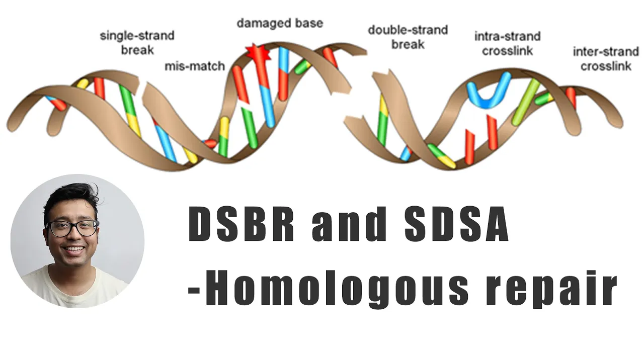
Double stranded break repair using homologous chromosome
30.5K
The complete homologous recombination pathway follows a sequential process: (1) Double strand break occurs; (2) MRN complex binds and trims 5' ends to create 3' overhangs; (3) RPA binds to protect single-stranded DNA; (4) Rad51 facilitates strand invasion; (5) D-loop formation occurs between the invading strand and sister chromatid; (6) DNA polymerase extends the invading strand to form a holiday junction; (7) Resolution occurs through either DSBR (crossover) or SDSA (non-crossover) pathways. This template-directed repair ensures accurate restoration of the original genetic information.
Click to watch →
Lec 6: Homologous and non homologous recombination
8.7K
DNA double-strand breaks (DSBs) are repaired through three main pathways: homologous recombination (HR), which uses a sister chromatid as a template for high-fidelity repair; non-homologous end joining (NHEJ), which directly rejoins broken DNA ends regardless of sequence and is error-prone; and alternative NHEJ (A-EJ), which serves as a backup pathway when conventional repair fails. The choice between pathways depends on protein factors: MRN complex binding promotes HR, while Ku70/Ku80 binding promotes NHEJ. HR involves strand invasion, D-loop formation, and resolution of Holliday junctions, serving both genome protection and evolutionary diversification through gene rearrangements.
Click to watch →

Homologous Recombination for Double Strand Breaks Part 1
83.7K
Double strand breaks (DSBs) are critical DNA lesions where both strands of the double helix are severed, commonly caused by ionizing radiation. These breaks rarely occur cleanly with blunt ends; instead, they produce messy ends with overhangs and potential nucleotide loss. Cells employ two primary repair mechanisms: homologous recombination (HR), which replaces damaged DNA with nearly identical copies from homologous chromosomes, and nonhomologous end joining (NHEJ), which directly ligates processed ends. While NHEJ is the predominant repair pathway, HR provides more accurate restoration of the original genetic sequence. Understanding these mechanisms is fundamental to comprehending how cells maintain genomic stability.
Click to watch →
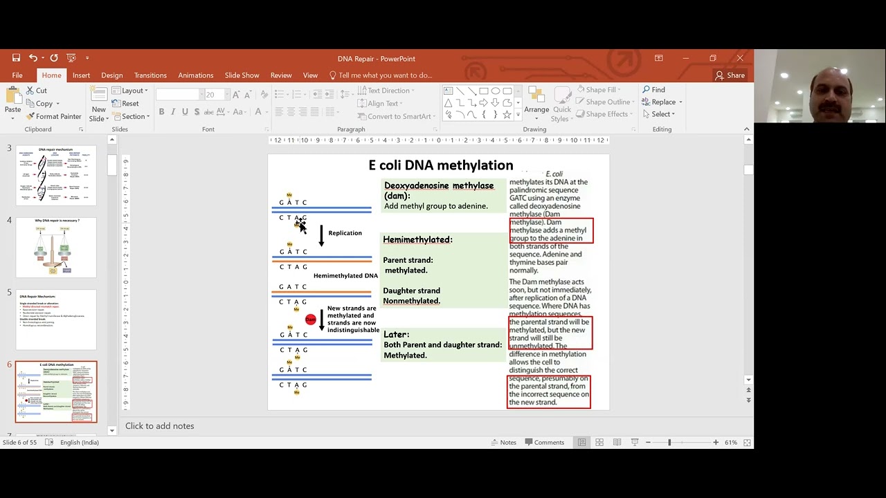
DNA damage and molecular mechanism for DNA repair
194
Double-strand breaks (DSBs) are repaired by three pathways: homologous recombination (HR), non-homologous end joining (NHEJ), and microhomology-mediated end joining (MMEJ). NHEJ repairs DSBs by directly joining broken ends without a template, occurring during any cell cycle phase but being error-prone with small insertions or deletions. Key NHEJ proteins include Ku (recognizes DNA ends), DNA-PK (recruits and phosphorylates), Artemis (trims ends), XRCC4 and DNA ligase IV (join ends), and XLF (facilitates joining). HR uses a homologous template, restricted to S and G2 phases, and is high-fidelity with no genetic material loss. HR involves MRN complex recognition, Rad51-mediated strand invasion, DNA synthesis using the template, and resolution of resulting structures. Species-specific differences exist in HR proteins between yeast and humans.
Click to watch →

DNA mutation and repair, Bio(molecular biology), Tuturial 3
7.7K
Homologous recombination (HR) and non-homologous end joining (NHEJ) are the two main pathways for repairing double-strand breaks. HR uses a homologous template for accurate repair but requires a sister chromatid or homologous chromosome, so it is primarily active in S and G2 phases. NHEJ directly ligates broken ends without a template and can operate throughout the cell cycle, but is error-prone and can cause small insertions or deletions. The choice between pathways depends on cell cycle phase and template availability. Defects in these pathways cause genetic disorders and cancer, demonstrating the critical importance of DNA repair for genomic stability.
Click to watch →
Deep Dive
The role of NHEJ in physiological processes like V(D)J recombination, which generates antibody and T-cell receptor diversity in the immune system.

Non-Homologous End Joining: Key Proteins in DNA Repair
83.5K
Non-homologous end joining (NHEJ) is a critical DNA repair mechanism that fixes double-stranded DNA breaks without requiring a homologous template, making it essential during G1 and early S phases when no sister chromatid is available for homologous recombination; the process involves Ku70/Ku80 heterodimers binding to broken ends, recruiting DNA-PK catalytic subunits and Artemis for end processing, and finally using Ligase 4 along with XRCC4 and XLF to join the DNA ends, and this mechanism is particularly vital for V(D)J recombination in B cells to generate antibody diversity.
Click to watch →
T and B Cell Development: V(D)J Recombination
87.1K
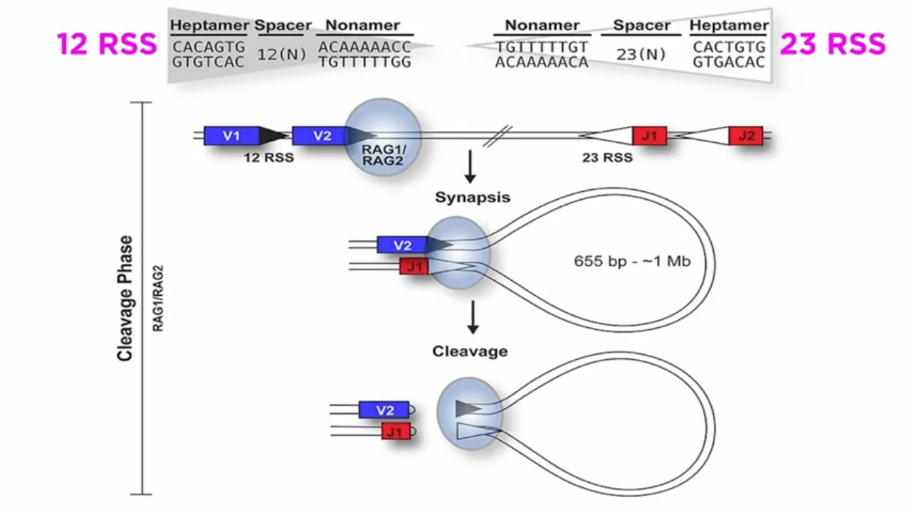
V(D)J recombination is the fundamental process by which T and B cells generate their diverse antigen receptors, creating millions of unique receptor variants through the recombination of variable (V), diversity (D), and joining (J) gene segments; this process involves RAG1 and RAG2 enzymes cleaving DNA at recombination signal sequences (RSSs) with conserved heptamer and nonamer elements separated by 12 or 23 nucleotide spacers, followed by non-homologous end-joining (NHEJ) that repairs coding ends imprecisely to increase diversity while precisely joining signal ends, ultimately producing functional immunoglobulin and T-cell receptor proteins essential for adaptive immunity.
Click to watch →
VDJ recombination overview | Generation of antibody diversity | Antibody diversity mechanism | VDJ
161.4K
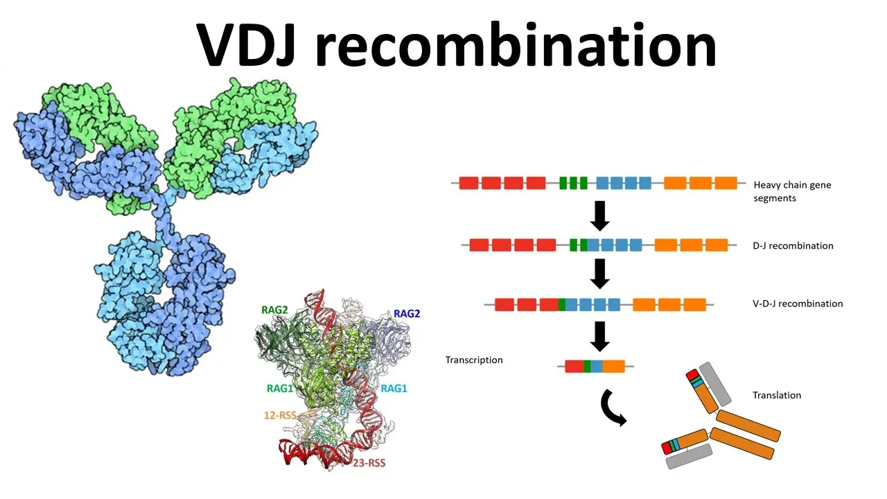
VDJ recombination proceeds through sequential molecular events: RAG proteins recognize RSS, DNA looping brings RSS together, single-strand cleavage creates hairpin intermediates, and double-stranded breaks are repaired by NHEJ machinery (Ku70/80, Artemis, DNA PK, XRCC4). Antibody diversity emerges from three mechanisms: combinatorial joining of multiple gene segments (at least 51 V, 27 D, and 6 J segments yielding ~8,000 combinations), junctional flexibility during repair, and somatic hypermutation post-antigen exposure. These mechanisms collectively enable the immune system to generate an enormous antibody repertoire capable of recognizing virtually any pathogen.
Click to watch →

Lecture 15: Generation of diversity ( GOD ) of lymphocyte antigen receptors ( Contd. )
153
V(D)J recombination utilizes the non-homologous end joining (NHEJ) DNA repair pathway. Ku70/Ku80 heterodimer binds to DNA breaks and recruits DNA-PKcs and Artemis. DNA-PKcs activates Artemis, which processes the DNA hairpins by opening them to create single-stranded ends. This processing is essential for generating the diversity of the final receptor. The NHEJ machinery, while originally evolved for DNA repair, has been co-opted for the specific purpose of generating receptor diversity in lymphocytes.
Click to watch →
How Our Body Creates Millions of Antibodies! | V(D)J Recombination Made Easy | CSIR NET Life Science
388
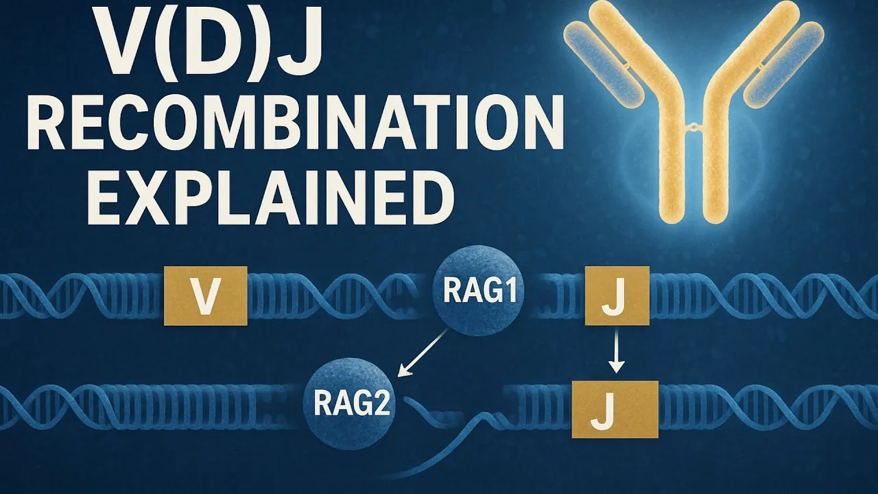
The complete VDJ recombination mechanism involves several coordinated steps: (1) RAG1/2 recognizes RSS and brings gene segments into synapsis; (2) RAG1/2 creates single-strand nicks, forming hairpin coding ends; (3) DNA ligase 4 joins RSS regions to form signal joints; (4) Artemis opens hairpins, generating overhangs; (5) NHEJ proteins repair coding joints with potential nucleotide loss; (6) TdT adds non-templated nucleotides at heavy chain joints. This process generates antibody diversity through combinatorial selection of V, D, and J segments plus junctional diversity from hairpin processing and TdT activity.
Click to watch →
Deep Dive
How CRISPR-Cas9 genome editing technology exploits the error-prone nature of NHEJ to achieve gene knockouts.

Understanding CRISPR-Cas9
292.5K
NHEJ is an error-prone DNA repair pathway that joins broken DNA ends without a template. The Ku70/Ku80 heterodimer binds to DNA ends and recruits DNA-PKcs, which attracts XRCC4 and XLF proteins that form a complex around the ends. DNA ligase IV then joins the ends by making sugar-phosphate bonds. This process often introduces mutations such as nucleotide insertions or deletions, making it useful for gene knockout applications.
Click to watch →
![[講義]【2020年ノーベル化学賞】ゲノム編集のツールとしてのCRISPR-Cas9 (CRISPR-casシステムについての解説)](https://i.ytimg.com/vi/jxYyeZCtQ2s/maxresdefault.jpg)
[講義]【2020年ノーベル化学賞】ゲノム編集のツールとしてのCRISPR-Cas9 (CRISPR-casシステムについての解説)
507
NHEJ (Non-Homologous End Joining) repairs double-strand breaks by directly joining broken ends, but is error-prone and often introduces insertions or deletions (indels). These indels cause frameshift mutations that disrupt gene reading frames, creating premature stop codons and non-functional proteins. This mechanism is commonly used for gene knockouts in CRISPR experiments. By inducing double-strand breaks at specific gene locations, NHEJ can effectively 'knock out' genes by disrupting their function.
Click to watch →

CRISPR | CRISPR - CAS9 | Genome Editing Tool
101.8K
When CRISPR-Cas9 creates a double-strand break in DNA, cells repair it using two main pathways. In organisms without homologous chromosomes (like bacteria), the NHEJ pathway is used. This pathway simply joins the broken DNA ends together, but during this process, 2-4 nucleotides may be lost from each end due to trimming by repair proteins. This imprecise repair often results in gene disruption, making NHEJ particularly useful for knocking out or disabling specific genes in genetic research.
Click to watch →
CRISPR-Cas9 Explained: Genome Editing Technology & Applications
927.8K
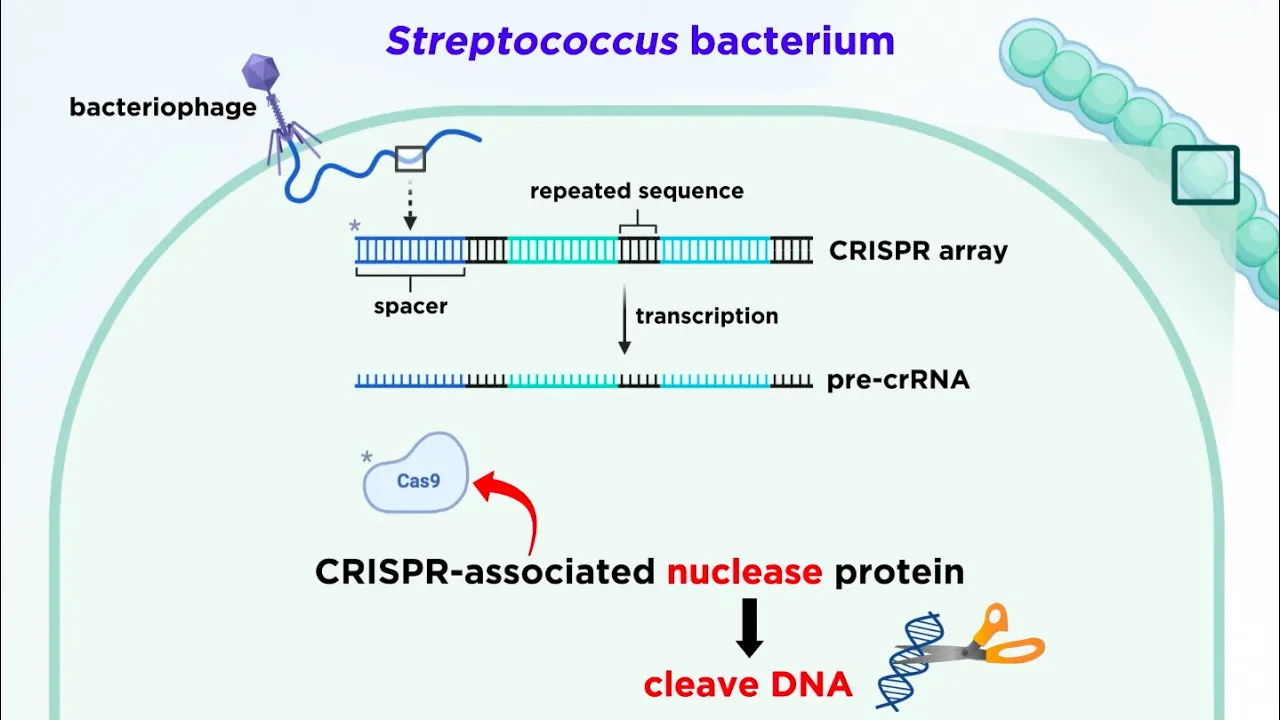
After Cas9 cleaves DNA, the natural repair mechanism is enacted through two pathways. NHEJ repairs double-strand breaks by directly ligating the broken ends without requiring a homologous template. This pathway can introduce insertions or deletions (indels) at the joining sites, creating DNA strands with non-uniform size. NHEJ is more common in eukaryotic cells.
Click to watch →
CRISPR dalam Rekayasa Genetik
280
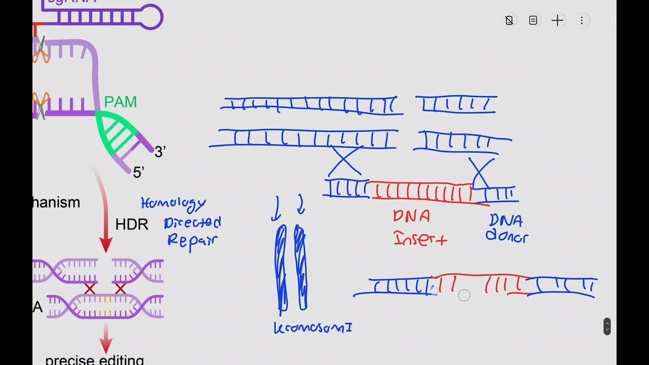
CRISPR-Cas9 creates double-strand breaks in DNA, triggering two repair pathways. Homology Directed Repair (HDR) uses a homologous template (like the other chromosome) to precisely repair DNA, enabling gene insertion or deletion. Non-Homologous End Joining (NHEJ) is error-prone, simply connecting broken ends and often causing deletions or random insertions. HDR requires donor DNA with homologous sequences and an insert, while NHEJ can create gene knockouts without donor DNA. The choice between pathways is largely random, requiring multiple cells to achieve desired modifications.
Click to watch →
Deep Dive
Human pathologies linked to NHEJ deficiencies, including severe combined immunodeficiency (SCID), radiation sensitivity, and cancer predisposition.

Non-homologous end joining
3.3K
The choice between NHEJ and homologous recombination is regulated at the 5' end resection step: unresected breaks undergo NHEJ while resected breaks commit to homologous recombination. NHEJ operates throughout the cell cycle but is most critical during G1 when no homologous template exists. CDK1 regulates this choice by phosphorylating Nbs1 to initiate resection. NHEJ is essential for V(D)J recombination in immune development, where RAG proteins create hairpin-capped breaks opened by Artemis and joined by NHEJ. Terminal deoxynucleotidyl transferase adds non-templated nucleotides to maximize receptor diversity. Defective NHEJ causes severe combined immunodeficiency (SCID). Telomere protection relies on NHEJ proteins like Ku for capping and subtelomeric silencing. Human syndromes include Lig4 syndrome and XLF CID (radiosensitivity, microcephaly, SCID) and Artemis deficiency (SCID without neurological defects). Mice lacking Lig4 or Xrcc4 are embryonic lethal, demonstrating NHEJ essentiality for mammalian viability.
Click to watch →
Recognizing SCID Phenotypes
420
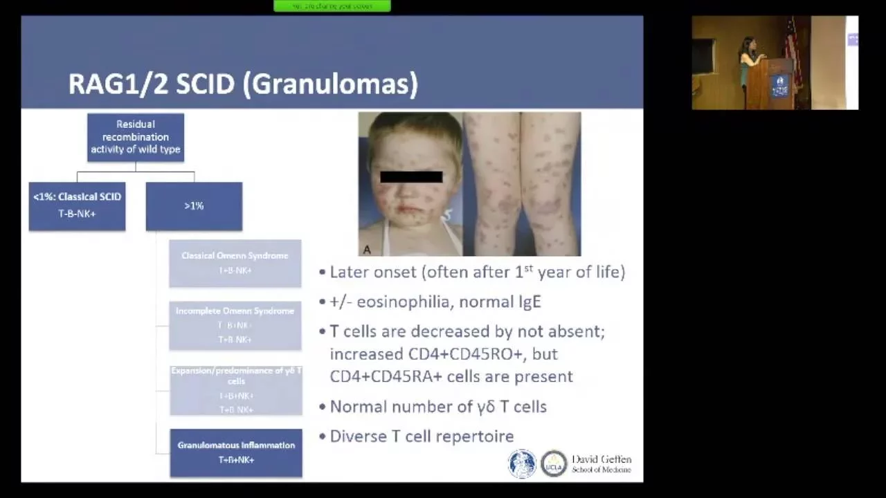
Leaky SCID phenotypes result from partial RAG activity and include Omenn syndrome (skin rash, alopecia, lymphocytosis, elevated IgE, oligoclonal T cell repertoire), incomplete Omenn syndrome, and gamma delta T cell expansion. Radiation-sensitive SCID results from NHEJ pathway defects (DNA-PKcs, Artemis, XLF, Ligase IV) that impair DNA repair for all cell types, causing radiosensitivity critical for transplant conditioning. These represent distinct clinical entities requiring different management approaches despite overlapping immunophenotypes.
Click to watch →
Efectos biológicos de Rad.Ion. a nivel molecular y celular. Curvas Sobrevida- BCP - Parte 1
7.4K
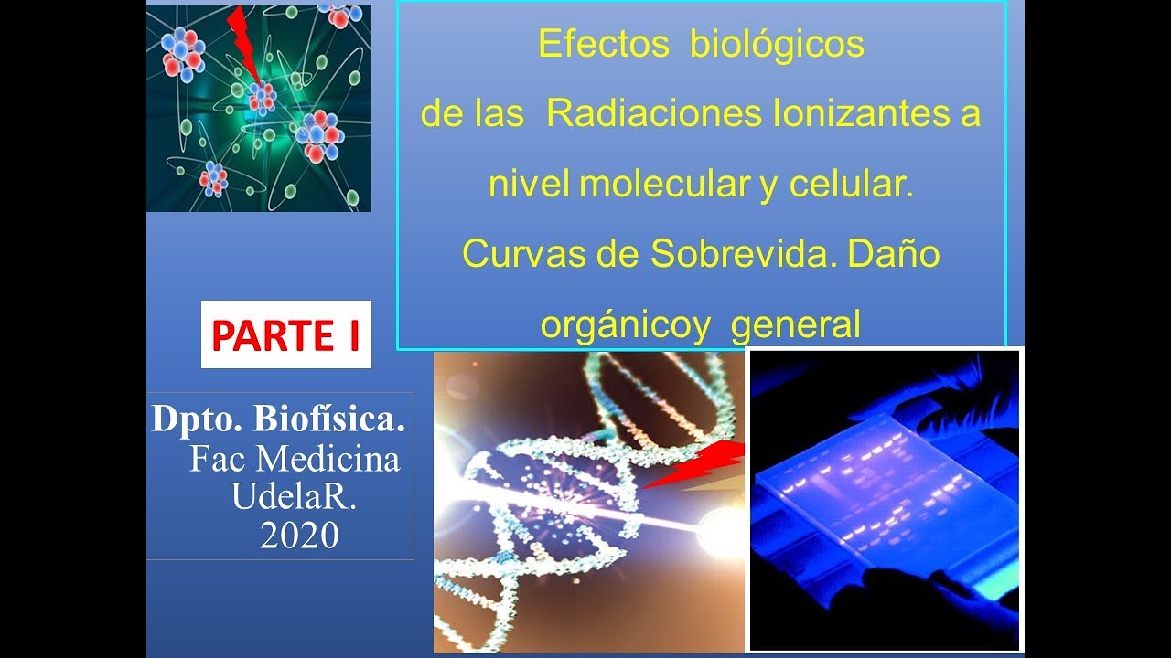
Double-strand breaks are the most critical DNA lesions. Repair mechanisms: Homologous Recombination (HR) uses sister chromatid as template (accurate, S/G2 phases); Non-Homologous End Joining (NHEJ) directly ligates ends (error-prone, all phases). BRCA1 and BRCA2 genes are critical for HR; mutations cause hereditary breast/ovarian cancer (10% of cases). NHEJ defects cause radiosensitivity and cancer predisposition. Improper repair leads to chromosomal aberrations (deletions, translocations, inversions) visible in karyotypes.
Click to watch →
Understanding SCID IN QUIZ FORM in just 2 minutes
3.6K
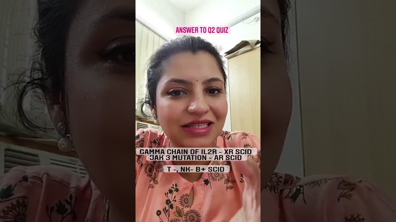
SCID (Severe Combined Immunodeficiency) is caused by defects in the Non-Homologous End Joining (NHEJ) pathway, which is essential for DNA repair and V(D)J recombination in T and B cells; this defect prevents proper immune cell development, leading to severe susceptibility to infections.
Click to watch →
Double Stranded DNA Repair - Homologous Recombination and Nonhomologous End Joining
5.3K
Double-stranded DNA breaks, often caused by ionizing radiation, trigger two distinct repair mechanisms. Non-homologous end joining (NHEJ) directly ligates broken DNA ends without a template, occurring before S phase and being highly error-prone. This mechanism is essential for rapid repair but carries mutation risks. NHEJ defects cause serious diseases including ataxia-telangiectasia (characterized by ataxia, telangiectasias, and cancer predisposition), severe combined immunodeficiency (SCID) with recurrent infections, and Fanconi anemia. Understanding these pathways reveals how DNA repair failures directly translate into human disease states.
Click to watch →
NHEJ repair
NHEJ repair
0:05- 1
Explains non-homologous end joining for DNA double-strand break repair.
- 2
Details protein steps: Ku binding, DNA-PKcs, Artemis trimming, and ligation.
- 3
Notes potential sequence loss due to nuclease degradation at break site.
Homologous Recombination (HR) and the Error-Prone Nature of NHEJ
While Non-Homologous End Joining (NHEJ) is a rapid pathway for repairing DNA double-strand breaks, it is inherently error-prone, often introducing insertions, deletions, or causing chromosomal translocations because it ligates broken ends without a template. The primary alternative is Homologous Recombination (HR). Unlike NHEJ, HR is a high-fidelity, error-free repair mechanism restricted to the S and G2 phases of the cell cycle. HR utilizes an undamaged sister chromatid as a homologous template to precisely restore the original genetic sequence. Understanding HR provides a crucial counterpoint to NHEJ, illustrating the evolutionary trade-off cells make between the speed of repair (NHEJ) and the absolute preservation of genetic fidelity (HR).
Basic structure of DNA, including the double helix, phosphodiester backbone, and complementary base pairing.
The concept of DNA damage, specifically distinguishing between single-strand breaks and double-strand breaks.
Fundamentals of the eukaryotic cell cycle, as repair pathway choices are often cell-cycle dependent.
An introduction to basic enzymes involved in DNA metabolism, such as polymerases, ligases, and nucleases.
Homologous Recombination (HR), the alternative high-fidelity pathway for repairing double-strand breaks, and the mechanisms of pathway choice.
The role of NHEJ in physiological processes like V(D)J recombination, which generates antibody and T-cell receptor diversity in the immune system.
How CRISPR-Cas9 genome editing technology exploits the error-prone nature of NHEJ to achieve gene knockouts.
Human pathologies linked to NHEJ deficiencies, including severe combined immunodeficiency (SCID), radiation sensitivity, and cancer predisposition.
319.8K views2.6Klikes1:44@OUPAcademicOriginal Release: 2014-04-11
to see how a cell repairs a double strand break in DNA by non-homologous enjoining let's imagine that we have a parental DNA duplex containing the sequence a b CDE e we then imagine that the duplex is broken by a double strand break in the C region degradation of regions from each side of the break which are generally less than 10 base pairs long may occur this process is sometimes called resection Q protein binds around the broken ends leaving the actual DNA ends exposed coup also recruits the DNA PK catalytic subunit also called DNA pkcs DNA pkcs recruits the nucleus artamis and phosphates it artmus trims any single stranded tails that are present at the break lias 4 acting in complex with xrcc4 and XLF cunos legates the broken ends together as a result the duplex is rejoined but note that any region that was removed by a nuclease will be missing in the repair duplex in this case joining does not restore the DNA to its pre-break AB CDE sequence because the C region was removed by nuclea digestion we see how a DNA double strand break can be repaired without deletion or imprecise join in another video how to repair a double strand brake by hology dependent double strand brake repair
Up Next
15:53
CRISPR Gene Editing Technology Explained by Jennifer Doudna
@TED
1.9M views•2015-11-12
14:12
Quantitative Real-Time PCR (qPCR): Principle, Method & Data Analysis
@animatedbiologywitharpan
129.2K views•2023-09-06
23:33
Microbial Degradation of Plastics: Biodegradation Pathways & Sustainability
@majeedhammad
2.9K views•2021-04-11
16:04
CRISPR and Genetic Engineering: How Gene Editing Works and Why It Matters
@kurzgesagt
30.5M views•2016-08-10
Related Study Plans & Knowledge Roadmaps
Structured learning paths in Biotechnology
biotechnology
AAV Production: Transfection, Prep & qPCR
01Introduction to Gene Therapy and AAV Biology
02Mammalian Cell Culture: Suspension HEK293 Cells
03Upstream Bioprocessing: Triple-Transfection Optimization
+ 2 more learning modules
18h5 modules
Explore Roadmap
biotechnology
Building Gut-on-a-Chip: PDMS, Cells & TEER
01Foundations of Microfluidics and Gut Anatomy
02Cleanroom Fabrication & PDMS Soft Lithography
03Epithelial Cell Culture and On-Chip Seeding
+ 2 more learning modules
26h5 modules
Explore Roadmap
biotechnology
Optimizing CFPS: E. coli Extract & Yield
01Fundamentals of Molecular Biology and Translation
02Introduction to Cell-Free Protein Synthesis (CFPS)
03Preparing E. coli Crude Lysate (The Extract)
+ 2 more learning modules
19h5 modules
Explore Roadmap
biotechnology
Haematococcus PBR: Design, Stress & Astaxanthin
01Biology and Life Cycle of Haematococcus pluvialis
02Bench-Scale Photobioreactor (PBR) Design and Engineering
03Culture Media Formulation and Inoculation
+ 2 more learning modules
15h5 modules
Explore Roadmap