Building Gut-on-a-Chip: PDMS, Cells & TEER
Learning Goal: Establish a fully functional, physiologically relevant gut-on-a-chip microphysiological system. This curriculum covers designing and fabricating a microfluidic device using polydimethylsiloxane (PDMS) soft lithography, establishing a viable co-culture of human intestinal epithelial cells (Caco-2) and gut microbiota, and validating the biological barrier integrity using on-chip Transepithelial Electrical Resistance (TEER) measurements.
- Prerequisites: Basic college-level biology/histology, introductory fluid mechanics, and standard laboratory biosafety training (BSL-2).
- Estimated Total Study Time: 26 Hours
Module 1: Foundations of Microfluidics and Gut Anatomy
This module introduces the mechanical and biological principles that govern organ-on-a-chip systems. You will study how fluid behaves at micro-scales—specifically, the transitions to low Reynolds number regimes, laminar flow, and wall shear stress. In parallel, you will analyze the histology of the human intestinal epithelium, focusing on its cellular composition, polarized structure, and the physiological significance of the extracellular matrix (ECM).
Recommended Videos

- Why this video: This academic lecture provides an in-depth mathematical and physical foundation of fluid dynamics at micro-scales. It is essential for understanding why fluids behave in a strictly laminar fashion within micrometer-sized channels, a concept crucial to designing microfluidic devices.
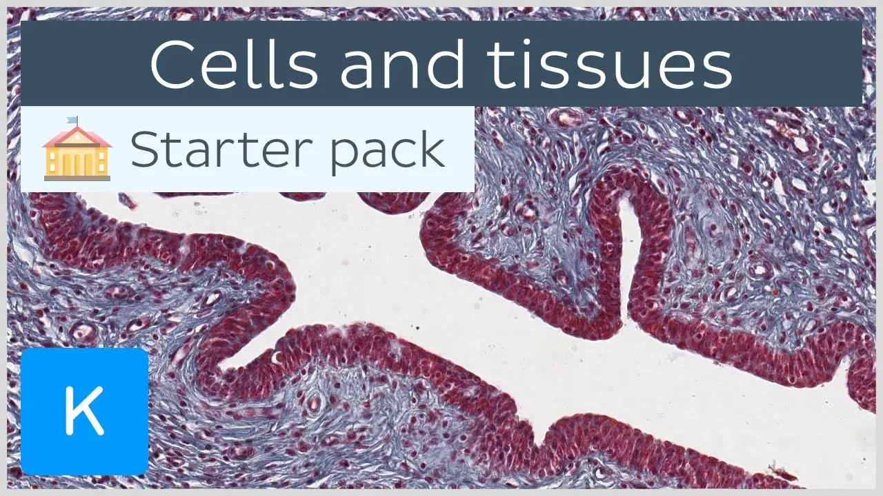
- Why this video: A comprehensive review of human histology is necessary to model the gut barrier. This video covers the primary characteristics of epithelial tissue, explaining cell shapes, polarization, and how these cells interface with the extracellular matrix to form protective, semi-permeable membranes.

- Why this video: Delivered by one of the pioneer researchers in the field, this talk bridges the gap between engineering and biology. It explains the core philosophy of microphysiological systems (MPS)—why static Petri dish cultures fail to replicate human physiology and how dynamic microenvironments mimic organ-level functions.
Knowledge Checkpoint
- Understand the definition of Reynolds number () and why it is typically in microfluidic channels.
- Explain how laminar flow differs from turbulent flow and why mixing in microfluidics depends primarily on diffusion.
- Identify the main structural layers of the intestinal barrier, focusing on the role of the mucosal lining and polarized epithelial cells.
- Articulate how mechanical strain (e.g., cyclic stretching or fluid shear stress) influences tissue differentiation on-chip.
Module 2: Cleanroom Fabrication & PDMS Soft Lithography
This module focuses on the micro-engineering and material fabrication phase. You will follow the step-by-step master mold fabrication using photolithography, learn to handle and cure polydimethylsiloxane (PDMS), and master the chemical principles of atmospheric plasma treatment to covalently bond microfluidic layers.
Recommended Videos

- Why this video: An exceptionally clear, step-by-step walk-through of the photolithography process in a cleanroom. It details wafer preparation, SU-8 spin coating, soft baking, alignment with photomasks, UV exposure, and development to yield a high-fidelity master mold.

- Why this video: This tutorial outlines the practical replica molding process. It demonstrates how to mix PDMS polymer base with curing agent at a 10:1 ratio, degas the mixture in a vacuum chamber, cure it, peel the elastomeric structures, and punch fluidic ports.

- Why this video: Focused intensely on the physical replica molding process, this video provides excellent close-up details on cutting cured PDMS slabs, aligning them, and prepping surfaces for permanent bonding.

- Why this video: This video focuses on atmospheric oxygen plasma treatment, explaining the molecular mechanisms (e.g., exposing silanol groups ) that enable irreversible covalent bonding between PDMS and glass slides.
Knowledge Checkpoint
- Describe the difference between positive and negative photoresists (such as SU-8).
- State the correct mixing ratio for SYLGARD 184 (elastomer base to curing agent) and explain why degassing is required.
- Understand the chemical surface activation process that occurs during oxygen plasma bonding.
- Troubleshoot common bonding failures (e.g., dust contamination, incomplete contact, or over-exposure to plasma).
Module 3: Epithelial Cell Culture and On-Chip Seeding
In this module, you will master biological preparations for gut-on-a-chip culture. This includes maintaining Caco-2 human intestinal epithelial cells, coating the hydrophobic PDMS microchannels with an extracellular matrix (ECM) scaffold, calculating physiologic shear stresses, and introducing cells into microfluidic channels under flow.
Recommended Videos

- Why this video: This masterclass highlights Caco-2 cell line biology. It details why Caco-2 is the standard cell line used for modeling the intestinal barrier, their cancer-derived origin, and their ability to spontaneously differentiate into polarized monolayers.
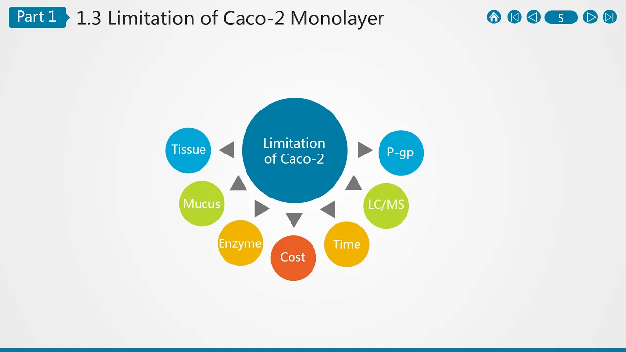
- Why this video: This video demonstrates how to run permeability assays using differentiated Caco-2 cells. It introduces the functional behavior of intestinal monolayers, highlighting brush border formation and cellular tight junctions.

- Why this video: Fluid flow generates shear forces that are biologically critical for gut maturation. This tutorial explains how to calculate fluid flow rates, channel geometry, and viscosity to target physiological wall shear stress levels.
Curriculum Gap Acknowledgment & Self-Study
⚠️ Curriculum Gap: The existing video library has limited coverage of the physical, step-by-step setup of microfluidic tubing, syringe pumps, bubble traps, and cell suspension injection directly inside a microchannel under a cell culture hood.
To close this gap, search YouTube or academic literature for:
- "Caco-2 cell seeding microfluidic channel perfusion protocol"
- "How to connect microfluidic tubing and syringe pumps without bubbles"
Knowledge Checkpoint
- Explain why Caco-2 cells require up to 21 days to fully differentiate in static cultures, and how microfluidic flow accelerates this maturation timeline.
- List common ECM proteins (e.g., Collagen I, Fibronectin, Matrigel) used to promote epithelial attachment inside microfluidic channels.
- Calculate the target flow rate () required to produce a physiological wall shear stress of () inside a microchannel with known dimensions.
- Describe the structural features of polarized epithelial cells (i.e., apical vs. basolateral domains).
Module 4: TEER Measurement & Barrier Function Validation
To ensure the engineered tissue acts as an effective physical barrier, you must validate its junctional integrity. You will learn the electronic and biophysical principles of Transepithelial Electrical Resistance (TEER), construct or integrate electrodes within microchannels, and measure the electrical resistance of the cell layer under flow.
Note: In practical research workflows, you must first validate the base epithelial monolayer's barrier integrity using TEER before exposing it to microbiota (as presented in Module 5).
Recommended Videos

- Why this video: This video introduces the physical and mathematical concepts behind TEER. It explains how applying an AC current across an epithelial monolayer allows researchers to quantify the resistance of paracellular pathways (tight junctions).
- Why this video: Transitioning from Transwells to microfluidic chips introduces unique challenges like electrode positioning and temperature fluctuations. This video shows how to integrate stable, planar electrodes directly within microfluidic systems to achieve reproducible measurements.

- Why this video: This protocol preview shows how researchers physically align metal wire or sputtered planar electrodes directly with PDMS microchannels to enable real-time electronic monitoring without taking the chip out of the incubator.
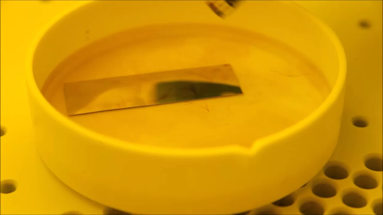
- Why this video: A highly technical fabrication guide showing how microfluidic channels are aligned and permanently bonded over patterned glass electrodes or electrical components to produce integrated bio-sensors.
Curriculum Gap Acknowledgment & Self-Study
⚠️ Curriculum Gap: The existing video pool demonstrates general TEER theory and commercial Transwell insert measurements (such as Chopstick electrodes), but lacks hands-on lab footage showing a researcher manually threading platinum wire electrodes into microfluidic PDMS ports or soldering leads under flow.
To close this gap, search YouTube or academic literature for:
- "How to integrate electrodes in PDMS microfluidic chip for TEER"
- "On-chip TEER measurement tutorial microfluidics"
Knowledge Checkpoint
- Define Transepithelial Electrical Resistance (TEER) and state its standard SI unit of measurement ().
- Explain why AC current (alternating current) is used instead of DC current (direct current) to measure biological resistance.
- Calculate the final normalized TEER value given raw resistance readings, blank chip values, and the active membrane surface area:
- Identify how external variables (such as cell culture media temperature and pH) distort TEER resistance values.
Module 5: Gut Microbiota Co-Culture & Microenvironment Control
The final module covers co-culturing obligate/facultative anaerobes alongside human epithelial cells. You will study how researchers design dual-channel devices that support human cells on an oxygen-rich top channel and gut microbiota on an oxygen-depleted (anoxic) bottom channel, mimicking the natural oxygen gradient of the intestinal tract.
Recommended Videos
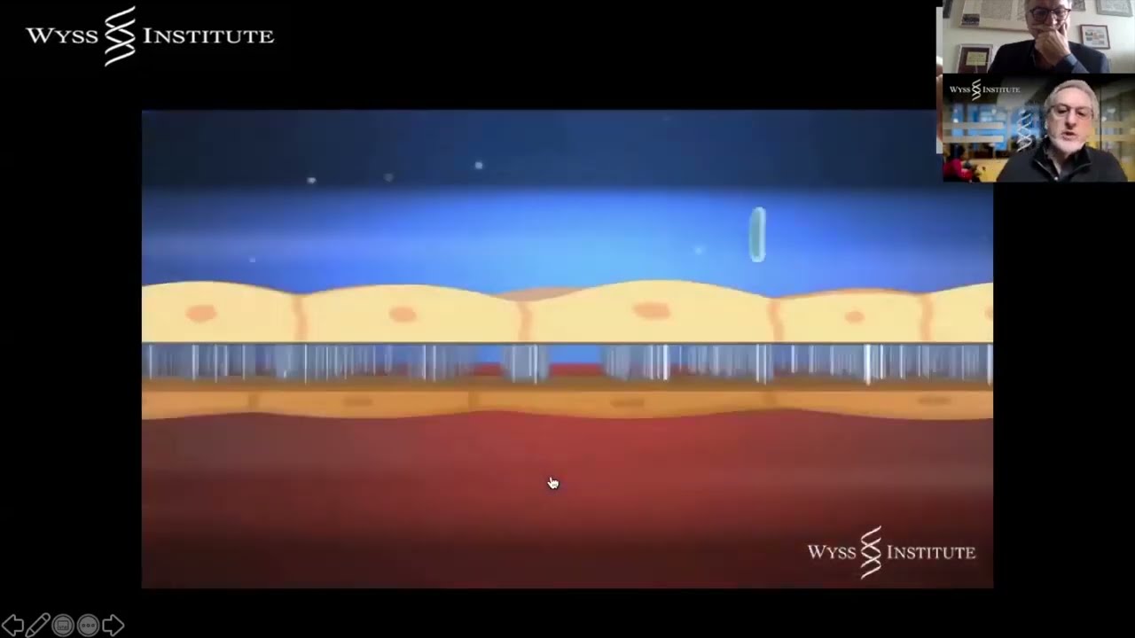
- Why this video: Dr. Donald Ingber explains how the Wyss Institute developed human intestine chips. He discusses the critical physiological need to co-culture complex, living microbiomes with human cells by establishing steady oxygen gradients across the membrane.

- Why this video: Before seeding anaerobes on-chip, they must be cultured off-chip. This lecture details standard laboratory methods for anaerobic growth, including anaerobic jars, gas packs, thioglycolate reduction media, and nitrogen flushing.
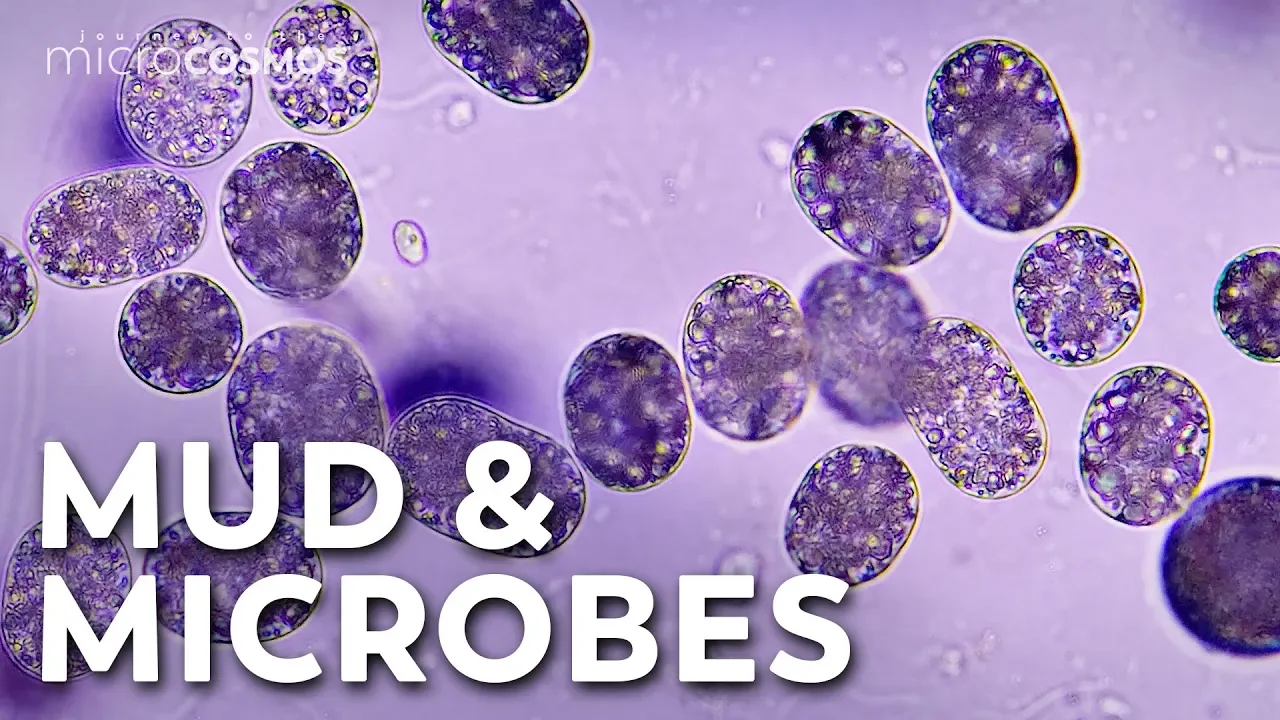
- Why this video: A microscopic visualization of the oxic-anoxic interface—the highly precise, sub-millimeter boundary layer where oxygen concentrations transition from high to zero, serving as a biological analogy for the microenvironments designed inside dual-channel gut chips.
Curriculum Gap Acknowledgment & Self-Study
⚠️ Curriculum Gap: The existing video pool demonstrates general anaerobic biology and macroscopic culturing techniques but does not feature step-by-step videos of setting up nitrogen gas sweep lines or oxygen-scavenging membranes on dual-channel PDMS chips in the lab.
To close this gap, search YouTube or academic literature for:
- "Anoxic aerobic interface microfluidic gut chip co-culture setup"
- "Oxygen gradient control in microfluidic organs on chips"
Knowledge Checkpoint
- Explain why obligate anaerobes (e.g., Bacteroides or Clostridium species) are highly sensitive to oxygen and cannot survive in standard cell incubators.
- Describe the design of a dual-channel microfluidic gut-on-a-chip, explaining how the PDMS membrane separates the aerobic (human) and anaerobic (bacterial) microenvironments.
- Understand how nitrogen gas perfusion or chemical oxygen scavengers are utilized to establish an active oxygen gradient on-chip.
- Define the biological consequences of co-culturing bacteria under static conditions versus dynamic flow conditions.
Course Map
Key People Index
- Dr. Donald Ingber (Wyss Institute/Harvard/Emulate): Pioneer of organ-on-a-chip technology. His lab developed the primary mechanical lung-on-a-chip and gut-on-a-chip designs, demonstrating that mechanical force and microfluidic flow are vital for human cellular differentiation and co-culturing the human microbiome.
- Dr. Dan Huh (University of Pennsylvania): Leading bioengineer who worked closely with Dr. Ingber on the lung-on-a-chip. His laboratory at UPenn continues to push the boundaries of microphysiological systems (MPS) for drug screening and disease modeling.
- Dr. Geraldine Hamilton (Emulate/Wyss): Former researcher at the Wyss Institute and leading industry voice who helped translate organ-on-a-chip technology from laboratory benches to commercialized testing platforms.
Final Self-Assessment
Complete this comprehensive, practical self-assessment to verify your competence in designing, fabricating, and running a gut-on-a-chip experiment.
- Design and Lithography: I can draw a 2D photomask with fluid channels of known height and width, and successfully pattern an SU-8 master wafer using a mask aligner and developer chemistry.
- Soft Lithography Fabrication: I can scale base-to-curing agent ratios, degas PDMS, cure it in a convection oven, cut clean slabs, and punch fluidic inlets/outlets without tearing the elastomer.
- Surface Modification and Bonding: I can explain how oxygen plasma changes PDMS and glass surfaces to create permanent covalent bonds.
- ECM Functionalization: I can select and coat microchannels with an appropriate extracellular matrix mixture (e.g., Collagen I and Fibronectin) to promote epithelial cell attachment.
- Sterility and Seeding: I can cleanly culture, harvest, and inject a bubble-free suspension of Caco-2 cells into a sterile microfluidic device inside a biosafety cabinet.
- Dynamic Flow Calculations: I can calculate and set fluid flow parameters on syringe pumps to supply physiological wall shear stress to the cells.
- Electrode Integration: I can successfully build, integrate, and align platinum or silver wire electrodes into a microfluidic device to connect to a voltohmmeter.
- TEER Quantification: I can record raw AC resistance values, subtract system background resistance, and apply mathematical normalization () to determine barrier confluence.
- Oxygen Gradient Establishment: I can explain the physical setup required to supply anaerobic media to a microbial channel while maintaining oxygen levels for human epithelial cells.
- Microbiome Co-Culture Execution: I can safely culture, isolate, and introduce anaerobic microbial strains alongside epithelial cells in an active, stable gut-on-a-chip device.