



















![突破血脑屏障 美国脑瘤治疗新希望 [20160527]](https://i.ytimg.com/vi/d70NI4NAIEc/sddefault.jpg)


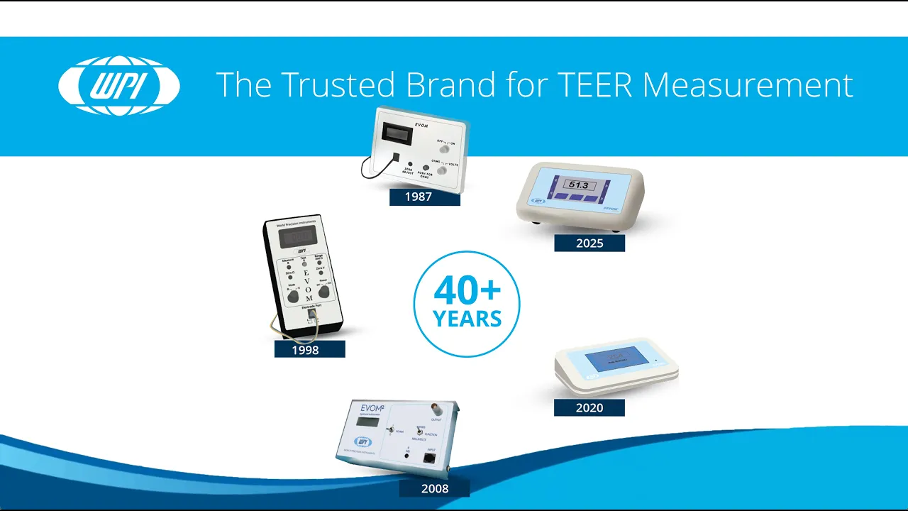
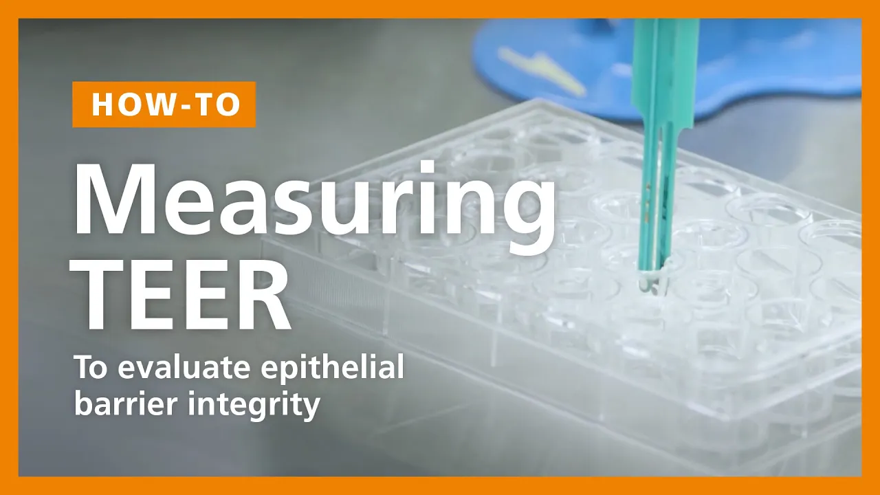
TEER (Trans-Epithelial/Trans-Endothelial Electrical Resistance) is a non-invasive method to measure the electrical resistance across cell monolayers, providing real-time monitoring of tissue barrier integrity in organ-on-chip platforms. Unlike traditional permeability assays that require endpoint measurements and can interfere with cells, TEER offers rapid (seconds to minutes) and continuous monitoring of barrier function under dynamic conditions. In organ-on-chip systems, semi-transparent planar gold electrodes are integrated into the chip design, enabling stable and accurate measurements without disrupting the cellular microenvironment. This technology enables researchers to observe how barriers respond to treatments, disease conditions, and physiological stimuli in real-time, making it particularly valuable for studying inflammatory bowel disease models and other barrier-related research.
Real-Time TEER Monitoring for Organ-on-Chip Barrier Integrity
Added:Hello and a warm welcome to our webinar today. Um really appreciate you joining us for today's topic of real time monitoring of barrier integrity with tear and or chip. Um also we'd like to thank everyone who submitted their questions in advance. We really used them to shape the content of this webinar and they will be the first ones that we're going to answer once is going to be finished with her presentation. Um now on to just a bit of housekeeping. So you probably have seen your control panel. There's a chat and a Q&A function. And we would really appreciate if you use the chat function for any queries that you have throughout the webinar and I will try to get back to them very quickly. Um and then use the Q&A function for the questions that you have to our speakers that we will um attend to after the presentation is finished. So now without further ado, I'd like to introduce our speakers for today. So first up we have um Sufi Pasa.
She is uh part of our R&D team. She's a scientist here and she holds a bachelor of engineering and biotechnology and a master of science in pharmaceutical biotechnology. Uh in April 22, Sophie began her master's thesis here on the emulation of an inflammatory bowel disease model and then she transitioned into a full-time position in August 23.
Um, also joining us today is our CEO CEO and co-founder Martin. He'll be joining for the Q&A session later on. Um, he's been working and researching with onrip cell culture applications since 2010.
So, a very long time. Um, and at University Hospital, he and his co-founder Kun Renet um, first experienced the need for fisological human organ models that really properly represent the situation in Vivo. and since 2021 he's also board member of the European network for organ on chip euro.
So now with this I'd like to hand over to Sufi. Hello everyone. Um yeah so first maybe a few words about our company. Um we provide organ on chip technology and also education around it training and we are also a service provider. Um our company combine a lot of scientific expertise and comprehensive biology in various fields with the ease of use system and we also provide lots of community support around it if you're interested.
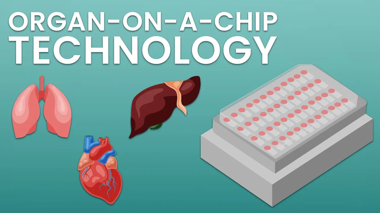
Um we are located in central Germany in Yenna and here you can see uh a nice picture of our team who's doing all the great work. Um in this brief webinar uh I will first introduce the concept of organship and then I mainly focus on today's topic the tier measurement. uh we will cover what tier is and how it is integrated into our organal chip system using the intestinal chip model as an example and at the end we are looking at useful applications of tier in organ on chip platforms. So for anyone who's not yet familiar with this technology, um what exactly is organ on ship? Here you see a general definition from the European organ on ship society. Um it uh describes a fitfor-purpose micrfluidic based device with different chambers and channels um allowing the setup of living organ substructures.
Um this also enables the creation of a controlled micro environment within the chip and once the tissue has developed it can be connected to a perusion system to mimic then uh blood flow and nutrient supply similar to the conditions in the human body. And with this comes also biomechanical stimulation which then affects receptor activation and downstream analysis or downstream signaling uh within the tissue and moves it uh towards a more physiological and invivo like state.
uh and with this we want to improve the translation of data between in vitro and invivo and contribute to close this gap. Yeah. So what are the current possible uh readouts with organ chip in this case especially with our D42 biochip. I can tell you um that the D42 platform is highly flexible and you can perform most downstream analysis you know already from standard cell culture.
So first it is suitable to sample super natan at any time point to check for biomarkers such as clinical parameters or cytoine grow feelings possible or you can check for compound turn over yeah or viability or cytotoxic cytotoxicity. You can also easily recover the tissue from the chip either as a single cell suspension or you can lice the tissue inside the chip for downstream analysis. But you can also take out the whole tissue um for immune flow sense staining or histoological staining. Um our platform is designed in the microscopic slide format so that it easily fits into existing lab workflows and you can do beautiful life cell imaging and uh life imaging is also suitable to investigate other parameters like reactive oxygen species formation or mitochondrial activity. And uh last but not least, you can check for uh enzyme activity uh protein and transporter functionality. There are plenty of kits on the market that are available and all protocols or the most of the protocols are all adaptable to our platform and you can also use them to do your readouts.
Um, but there's even more to get out of your experiment. Uh, we can also now monitor real-time tissue barriers. The fundamental aspect of organ on chip technology is the implementation of uh compartmentalized tissue barriers under dynamic conditions. And by recreating these barriers in a controlled environment, the organ on chip platforms allow us to better understand their roles and the interactions in the body. But what exactly are tissue barriers and why are they so important?
So tissue barriers are specialized cellular layers like epithelia or endothela layers that um control what can pass between two compartments in the body. In this way they act as a selective filter.
uh they allow essential substances like uh nutrients, oxygen and hormones uh to pass through. Um but they also block um harmful agents and in doing so they help maintain homeostasis and ensuring a stable and a balanced internal environment.
Um, another point is that they prevent toxins and pathogens from reaching and damaging uh the vital organs or some vital organs. What makes them a key part of the body's defense systems? So, um they're very important and the key structures of uh these barriers are tight junctions, adherence junctions and desmosomes.
Um there are several vascular and also epithelia barriers in the body that are of high importance. For instance, a very well-known barrier is the blood brain barrier. Um the blood brain barrier restricts a molecule transport into the brain. Um but we also have a gut epithelia barrier that helps us to separate the lumininal located microbes in the body to make us uh not sick but stay in the intestine and have a beneficial effect uh on our health. And these are just a few examples. There are many more but these barriers can be monitored in an organ on chip via tier.
And this is and this is what we implemented in our chip platform. So short tier is the transep and trans endothelial electrical resistance.
It is a widely used non-invasive method to assess how well epithelia or endothela cells form a functional barrier in vitro. And basically the electrical resistance which is measured uh across the cell layer uh gives us an idea of how intact or differentiated the monollayer is especially if the cells are forming strong tight junctions. And it's also uh a simple and fast bleed out. It's commonly used in static culture and it gives an first indication uh of barrier quality before moving on to more complex analyzis before tier became more widely used. um permeability essays uh were a standard method to evaluate barrier integrity uh in vitro and while there are still valuable tools um comparing both methods reveal key advantages of tier measurement. So let's take a closer look at how tier and permability essays differ in this case in a transfer. So um permeability essays rely on a label tracer molecule a molecule to see how much of this tracer molecule passes really through the cell layer over uh time. Um but te on the other hand uses an electrical current to measure the resistance uh across the monollayer which then reflects how tight uh the the uh cell junctions are.
And in terms of timing um permeability essays can take anywhere from 1 to 24 hours depending on the setup. uh whereas T measurements are much quicker often just a few minutes or seconds. And when it comes to compatibility, the permability essays could interfere with cells or drugs depending on the tracer used and tear is a less invasive method. uh in that sense it has a limited interference with cells and drugs but uh its accuracy can be quite sensitive to experimental conditions.
Things like electrode type as you can see here the chopstick electrodes uh the membrane area or media temperature that can all influence the outcome.
So both methods have their strengths and limitations and they are often used to together to get a fuller picture of barrier function. But compared to the uh to these invitro methods like the use of chopstick type electrodes and transfer cultures, the tier electrodes in our chip platform are incorporated at a fixed position without being movable and therefore it provides a more accurate and stable measurement with less sensitivity distribution. So that's a big advantage in comparison to the chopstick tier measurement.
And here you can see is a schematic of the chip where we equipped our biochip with a semi-transparent planer gold electrodes. And in this explosion view you can see that the electrodes are positioned at the inside of the chip of course attached to the top and uh the bottom bonding foil. Um, this enables you to get a readout throughout the tissue and check if the barrier is intact or not. And we also added an additional circuit board that is to attach to the chip. And this circuit board enables uh the contacting of the electrodes and the signal transmission.
And here you can see how it looks uh in real life. How our chip looks in real life from the front, the top view and the bottom view. Um that's not all. Also here we laid a focus on ease of use and designed a measurement device for contactless readout. The measurement device can operate up to three chips in parallel.
[Music] Um uh on each chip we have two models.
So we can measure six models with uh one device. And the measurement interval can be adjusted from very short intervals in range of seconds up to 1 hour. And here on the left side you see an exemplary setup how it can look like in the incubator. And here on the right side is a real life impression from an incubator setup.
Yeah. Um the signals from the tear chip are transmitted via Bluetooth to a computer with a data plotter software.
And this is of course designed as a open platform. So every opensource data plotter software can be used for this. And S tier is a temperature sensitive technique. We thus avoid uh biasing the experiment by keeping the incubator closed at all time. And before I present you one tier use case on our gutton chip, um I briefly want to introduce our intestinal model and explain it to you.
When you see inside a chip, it begins with the top bonding foil on a chip and it ends with the bottom bonding foil and everything between is inside the chip.
There's a porous membrane that compartmentalized the chamber into a top and a bottom channel. In the top channel, we set up a vascule and in the bottom channel, we set up the intestinal compartment.
And when we start um perfusing it, it also starts to outgrow beautifully uh in a vill like structure as we find it in in the human body. And in the intestine there are many parameters that are of interest. Uh but for instance, one is the barrier and that's why we chose the gutton chip for our first experiments with the uh tear chip.
So how is our body model exactly assembled? Um first um endothela cells the uh ux are seated into the upper chamber of the chip and after 48 hours um macrofasages are added and 24 hours later then the epithelia cells the kako 2 cells are seated into the lower epithelia chamber and once the entire model has been statically cultured for another 20 24hour sorry um both the vascular and the epithelia chambers are connected to the profusion which uh induces the formation of the vill's uh 3D structure and here in the image on the right you can see how it looks at the end inside the chip with the planner electrodes um during this model assembly uh I mentioned uh we also recorded tier values to see how the barrier in these chips uh changes every time we see the new cells. Uh upon seeding dubex the tear values slightly increased seating then the MDMs led to a small elevation in tier but um during seeding and formation of the confluent endothelia the tier values remained mainly low. Um a major increase in tier was then observed after seeding in the cargo 2 cells in the bottom channel. They formed a really tight barrier over 24 hours as you can see at the tier values. And uh when we connected the chips to the perfusion, the tier values at first increased but then remained stable over 24 hours per fusion but also at a high point.
And further and very important um we can show that the cell viability is not affected. So it's the same as it is in chips that do not have the T electrodes integrated. You can see it here by ATP measurements of the tissue that shows us um the biompatibility uh of the electrodes.
Um after our standard model assembly as I previously shown you the models were perused for an additional 5 days. Um then as shown here um the experimental barrier disruption was performed using EDTA followed by a recovery phase which was also monitored via tier measurements and the EDTA was uh applied to the intestinal bottom chamber for 30 minutes and afterwards the uh EDTA containing medium was replaced um with fresh medium without ED.
DTA which uh then induces tissue recovery within 24 hours. But first shortly uh what exactly is EDTA? How does it affect the cellular barrier? Um EDTA is a strong metal iron chator. It is known uh to bind magnesium and calcium and thereby uh disrupting the epithelial barrier.
So as you can also can see here uh when we treated the model we saw immediate uh effects upon EDTA administration um that the tier values dropped down close to zero as you can see on the left graph and uh when we remove the EDTA we can then also nicely see uh that the tissue barrier recovers over the time to the control tier values. So the tier measurement were able to detect the EDTA induced barrier induction and also the tissue recovery. Um we also compared it uh to a standard technique as mentioned for barrier measurement and permeability essay in this case the fits. In this physic, we also tested every condition, the control model, the EDT airr treated model and also after recovery time overnight. And uh we saw that this was not that sensitive uh to really detect significant changes or small changes in the tissue barrier as we saw uh really in the tier measurements.
So here you can see really the advantage of the T measurement in comparison uh to standard techniques like uh permeability essay. Um we can also beautifully capture the EDTA effect via brightfield imaging. Uh the light microscope images uh revealed also cell rounding and detachment uh from the membrane after 30 minutes of EDTA treatment. And after recovery you see again the uh 3D morphology with the villas and the cryptlike structures as you can see it also in the control model. And these images also show that the electrodes are semi-transparent. So you still have the possibility on a brightfield microscope to look at the tissue and to investigate it. As you can see here in the lower pictures, um the arrows show the borders of the T electrodes under the microscope. So it's really semi-transparent. Of course, to um complement this data, we stand for tissue barrier related markers such as eatherarine or set one in the intestinal cells. Um both junction proteins were disrupted after the treatment with EDTA for 30 minutes. Uh which can be clearly seen in the highlighted boxes uh with the white arrows. And the good distribution of the dappy signal shows us also that um the treatment is affecting the junctional complexes but not the nuclear integrity.
And after 24 hours of recovery, you seen similar marker expression as at the beginning as in the um untreated models, the controls. Um the expression of cell markers in the vascular cell layer showed similar results. Um we stained for CD31 and penilopran factor. Um and both markers were also disrupt disrupted um after EDTA treatment. Uh but they could also be restored after a 24-hour uh recovery phase with the help of a fresh medium.
So um to sum this up, we demonstrated here the integration of semi-transparent gold electrodes and the operation works via simple plugandplay workflow with the uh flexible intervals for measurement in our proof of concept study that we run for uh two weeks. we could see the changes that appear when we administer treatment in this case for instance with EDTA. Um the large electrode area favors also the quantification and uh the denormalization of tier values uh which is really nice to make it more comparable um to other systems or applications. For instance, also for trans health and the system shows um a high biological reproducibility. During several experiments we did, we saw only minor changes in tier levels that we recorded.
Everything was pretty close together. So our model offers several key advantages. First of all, our system is completely PDMS free, which helps to avoid the typical problems with molecule absorption that are often seen in PDMS based systems. It's um designed for simple fabrication making it easier to manufacture and scale and the scalable electro design ensures flexibility for different application and with the wireless contactfree readout via Bluetooth.
um measurements are noninvasive in keeping your sample intact and the system is also fully compatible with uh incubator environments and offers intuitive operation that makes it user friendly for everyday use. And additionally it is also uh very easy to sterilize and u maintain. Yeah. Um this slide summarizes the main application of tier measurements in organship platforms in general.
First T is widely used to assess the tightness of cell junctions, the permeability of tissue barriers and the overall structural integrity or the epithelia or endothelial layers of the epithelia or endothelial layers.
Um second uh it can provide a clear indication of how drugs and compounds affect the barrier including changes in transport mechanisms or how cells respond to phmological stimuli. And finally, care enables also real-time monitoring of barrier function under disease-like conditions um including here inflammatory responses, infection models um or tissue damage. And this makes tier a powerful non-invasive tool to study um dynamic physiological and pathological uh processes.
uh in a highly controlled uh environment. So um if you want to learn more more feel free to visit our website, follow us on LinkedIn or Blue Sky. Um, our general technology is available through the dynamic organ product line and we also offer both onsite and online trainings as well as detailed application notes to help you get started or deepen your understanding. And with that, I give to you back.
Yeah, thank you for for the slides. Um, definitely a lot of stuff that was also new to me. So, very interesting. Um, now we're moving over to our Q&A session.
So, I can already see a lot of questions coming in through the Q&A function. But as mentioned, we're going to first answer all the question that been submitted before we started this webinar. So, uh, Sophie prepared some slides for this, I believe.
So uh one question was is it possible to harvest the cells from the organ on chip device. Um as you saw in my presentation yes it is possible as you also can see here in the images even with the electrodes on the bonding foil. that the membrane can be removed using a scalpel or you can recover the tissue from the chip either as a single cell suspension or lies the tissue inside the chip and from there the cells can be processed as needed for example for immune flow sense staining or other things. Um a second question was um this method in OC still is unstable and varies. How to achieve stability in tier measurement? Um I would say we achieve good stability in our tier measurements in two main ways. Um first we use fixed planner electrodes with uh which stay in place and and um avoid variation that can happen if the electrodes are moved or not positioned exactly uh the same each time and this helps make the reading more reliable. And second, T is very sensitive to temperature changes.
That's why our system is designed to uh work directly inside the incubator and both the chip and the measurement device stay in the controlled environment and then thanks to the vireless contact free readout um we can measure from outside without opening the incubator or disturbing the cells. And as you can see in our results here, uh the experiment was done uh three times and the measurements are very consistent showing that uh our system provides stable and repeatable data. Um another question was um uh if there are examples for both human and animal cell usage. Uh I only can say so far we only tested it uh with human cells. So no animal cells used before. Then there was a question I use D42 BC00002 biochips. Is there a way of measuring the tier in these biochips? Um unfortunately I have to say it's not possible with the standard BC00002 biochip. It requires the T upgraded version with with the electrodes in it. But since it's still based on the PC00002, um your existing protocols and workflows um do not uh to be ch need to be changed. Um another question was how much is the tear chip? uh do you need any additional material for example a voltmeter uh and cables and um since we currently only offer the chips as a part of fee for service projects I can't tell you the pricing details yet but um we would let you know as soon as they become commercially available which will likely be hopefully at the end of the year and in the meantime uh we're also looking for beta testers So if you're interested um feel free to reach out to us. Um uh yeah and to answer the second question uh you don't need any voltmeter or any additional cables. All you need is the chip, the measurement device and any laptop running an open-source plotting software. It doesn't matter which software you use. So you can choose every every software for this.
Um what is the influence of holes in cell tissue layer on obtained signal and overall current distribution? Um I have to say because or gaps in the cell layer can strongly affect the tier measurements. Um the current flows through these gaps and then there's no resistance which leads uh to lower tier values. So you're not really longer measuring the true barrier function. That's why we always make sure the cell is fully confluent before we starting the measurements or treatment or um yeah manipulating it. Can we develop a simulation of the model to develop the chip response in in silico? Um we haven't worked on that directly yet but we've had exper experience with similar approaches.
Earlier this year we published some something on this topic um in co collaboration with fire. Maybe you can have a look at this. Uh the air code will take you uh to the paper. Yeah, that were all questions who were submitted up [Music] front. Hey, thank you Sophie. So we're going to start with a question that been submitted now. Um the first one that actually came in during your presentation was why did tear increase during the perusion?
Um yeah um it's only increasing because um it have we put on the perfusion and something change inside the chip but then you saw that it gets stable again and then there were no other changes.
They were really stable.
Thank you Martin. Did you want to add anything?
Uh yes um uh it is most likely also a reaction of the cells to the profusion um to the sheer forces so that the tissue and the cell layer is uh tightening and that's why we see an increase in in tier upon perusion.
Excellent. Um so the next question is how many cells can you recover from the trip for downstream molecular essays? If the cell number is not high enough it may be difficult to form qPCR or flowymetry.
Um this is a bit thank you for the for the question. Um this is a bit depending on the model used. Um in the intestinal model um we have plenty of cells because of the intestinal layer that is outgrowing. So here we're talking about uh 6 million cells or more in in total.
But for other cells like lung on chip or liver on chip um it's less. But still um between um 700K and 1 million cells because the um overall culture area in our standard biochip is um similar to uh 24 well. Uh yeah to the size of a 24 well.
Um and so um we can introduce quite a lot of material uh into the chip um and yeah therefore um ensuring um yeah compatibility with a lot of readouts not just for um uh cell loes um to to get proteins out of it but also for the biomarker secretion of cells.
Excellent. Um and the next one is for confocal imaging. Would you provide the protocols for the experimentation? Um of course um just uh please uh get in contact with us and uh we can um set up a separate meeting and and talk about uh your specific application and how you can realize this. So um we're very open to share our experiences here and to guide you through this.
Thank you Martin. Um next one is very nice work. Thank you. I'm new to this area of research. Uh can real time tear be measured in transfer systems or is it only possible in this type of systems?
No it's only it's also possible in transfer systems but we are not doing it here. So uh in our company we're only doing it in the organ on chip system but a lot of um research group do it also in the trans well.
Thank you Sophie. Um will the probe at the bottom side block the view of the microscope?
Um I guess my probe um also the the second electrode um it's referring to the second electrode of the on the bottom foil of the chip and no um also our pictures are made with a bright field microscope where um the light comes from beneath the chip. So it goes completely through the chip and nothing is interfering here.
Excellent. That's very good news. Um, next question is from Rita.
Hi, I'm new to this topic. How do you sterilize the tear electrode, 70% ethanol or other approaches?
Um, yeah, we ster sterilize with uh 70% ethanol.
Cool. Short and sweet. I like it. So, next one is from Chase. Um, is the tier measurement device tuned to a specific frequency based on the electrode material for measurement or can this be changed?
Um, at the moment it is tuned to a specific specific frequency. Um, but I think it can be changed if if needed, but yeah, at the moment it's only a specific uh frequency.
Cool. Um, so for now we've actually reached re reached our very last um, questions. So if you've got any more questions, feel free to submit them now.
Otherwise, I will likely conclude the webinar after this. So the last question is from Arita. Um, is there any clear trustable source where to look for normalized tier values for particular cell models? As seen in literature, the difference is too significant due to different hair setups. So do Martin did you want to take this one or you are unsure?
Um to be honest I'm a bit unsure about um a good literature source for normalized tier values. So we we've noticed this also that this um uh is varying throughout uh various publications. Um that's why we uh put um yeah special attention to um providing something where you can normalize um the tear values too um in yeah in reference to the um size of the electrodes. Um but yeah that's a good question but I don't have an answer for this. Sorry.
That's all right. um we can't always answer everything but actually if you take a look in the chat so Martin wrote something but also Tim who was um besides Sophie the other lead scientists who developed this platform he's posted a link in there as well so I believe this will be quite helpful as well um oh actually we've got another question coming in so um from Caris do you see alternating current in your tier setup if so what's the effect of different frequencies getting very technical now. I see we're getting deeper into the tier topic here. So, we have someone who's doing this technical stuff and um he was also uh the the the guy for the frequency um setups and and so on. So, I can't really answer this question. That was uh his job.
Okay, that's fine. But we can um try and forward this question to him and then just get back to you afterwards. So, now that I see there are no questions left um and we're well within time, I will conclude this webinar. I thank you um a lot for joining us today. We really appreciate this. There was really a lot of interested in this and I will send the recording after this so that you could share with your colleagues or just revisit some of the slides and should you have any more questions um you can always reach out to us and we will get back to you. Thank you and goodbye. Goodbye.
Up Next

RNA Interference: Nobel Lecture by Fire & Mello | Gene Silencing
@NobelPrize
6K views•2022-04-29

Graphic Medicine: Comics for Collaborative Healthcare Communication
@nationalpatientadvocate
189 views•2023-12-04

Neuroanatomy: Central and Peripheral Nervous System Divisions Explained
@AKLECTURES
136.2K views•2014-09-20

Stages of Labor and Vaginal Birth | Childbirth Animation
@nucleusmedicalmedia
52.1M views•2017-08-18
Related Study Plans & Knowledge Roadmaps
Structured learning paths in Medicine