Abnormal LFTs: Diagnostic Workup & Pathology
Learning Goal: Mastering the Workup of Abnormal Liver Function Tests: A Systematic Approach to Diagnosing Hepatocellular and Cholestatic Liver Diseases
Prerequisites
- Basic undergraduate biology or pre-medical level biochemistry.
- Familiarity with elementary gastrointestinal anatomy.
Estimated Study Time
- Total hours: 12 hours (including video lectures, supplementary reading, and self-assessment exercises).
Module 1: Liver Anatomy and Basic Physiology
Module Overview
This module establishes the structural and physiological baseline required to understand liver pathology. You will explore the macroscopic anatomy of the liver, its unique dual blood supply (portal vein and hepatic artery), and its primary physiological roles in protein synthesis, metabolic homeostasis, and detoxification.
Gap Note: High-resolution microscopic histology videos (demonstrating hepatic lobules, portal triads, and the zones of Rappaport) are limited in this pool. For comprehensive mastery, we highly recommend independently researching "Liver histology hepatic lobule portal triad animation" alongside these resources.
Recommended Videos

- Why this video: This video provides a crucial anatomical foundation, describing the liver's physical characteristics (weighing approximately 3 pounds), its division into four main lobes and seven clinical segments, and its multi-faceted operational role in metabolism and detoxification.

- Why this video: A concise, high-yield clinical overview detailing the liver's role as a master synthetic factory. It explains how hepatocytes manage glucose, fats, and protein metabolism while synthesizing crucial systemic markers like albumin and clotting factors.

- Why this video: Explains the physiological "gut-liver axis," focusing on how 75% of the liver's blood supply arrives directly from the portal vein. This highlights the liver's filtration role and how its physical connection to the intestinal tract influences metabolic and immune responses.
Knowledge Checkpoint
- Understand the dual vascular supply of the liver (hepatic artery vs. portal vein ratios).
- List at least five synthetic proteins produced exclusively or primarily by the liver.
- Explain how hepatocytes process carbohydrates, fats, and toxic waste compounds.
Module 2: Introduction to Liver Function Tests (LFTs)
Module Overview
This module covers individual blood markers that compose a standard "Liver Function Test" (LFT) panel. You will learn to differentiate between markers of hepatocellular injury (aminotransferases), markers of biliary/cholestatic function (alkaline phosphatase, GGT), and true indicators of hepatic synthetic function (albumin, PT/INR, and bilirubin).
Recommended Videos
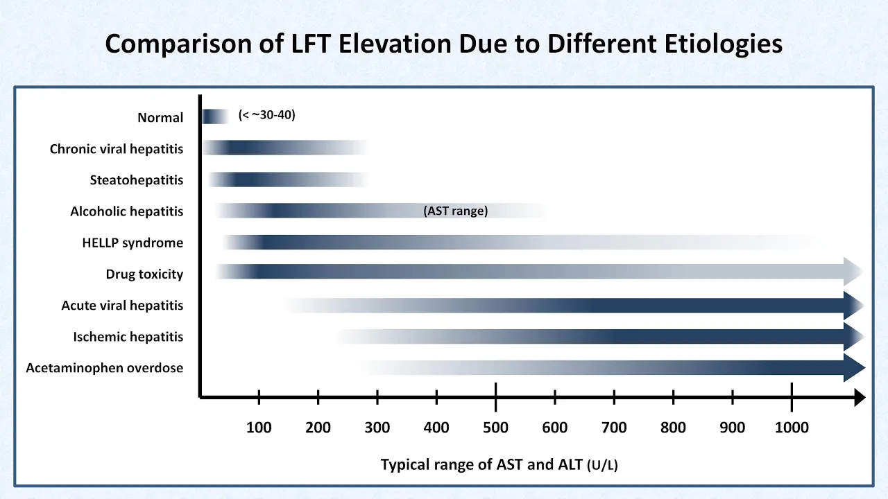
- Why this video: This is a gold-standard clinical lecture. It methodically steps through the biochemistry, normal physiology, and diagnostic utility of ALT, AST, ALP, GGT, albumin, and bilirubin, highlighting how to categorize these values before initiating a diagnostic workup.

- Why this video: Highly visual and clinical, this video clarifies enzyme tissue specificity. It explains why ALT is considered highly liver-specific, whereas AST is found in skeletal muscle, heart, and kidney tissues. It also outlines the diagnostic value of matching ALP elevations with GGT.

- Why this video: A masterclass in clinical pathology. This video is ideal for students who want a deep-dive, blackboard-style walk-through of LFT pathophysiology, exploring absolute enzyme elevation levels and the underlying cellular release mechanisms during tissue injury.
Knowledge Checkpoint
- Distinguish between ALT and AST in terms of cellular localization and organ specificity.
- Understand why Albumin and Prothrombin Time (PT/INR) are the only true biochemical measures of liver synthetic capacity.
- Explain why a concurrent elevation in GGT is used to confirm the hepatic origin of an elevated Alkaline Phosphatase (ALP).
Module 3: Hepatocellular vs. Cholestatic Patterns
Module Overview
The critical first clinical decision when analyzing abnormal LFTs is categorizing the injury pattern. This module teaches you how to classify LFT patterns as hepatocellular (predominant ALT/AST elevation) or cholestatic (predominant ALP/bilirubin elevation).
Gap Note: Although specific animations demonstrating the calculation of the R-ratio () are sparse in the pool, you will see its clinical utility applied in Faizan's clinical lecture below. To master this calculation, verify the formula:
- : Hepatocellular
- : Cholestatic
- : Mixed pattern
Recommended Videos
- Why this video: Directly addresses clinical pattern recognition. It introduces the application of the R-value to categorize liver injury into classic patterns, while outlining initial diagnostic steps for isolated bilirubin elevations (such as Gilbert's syndrome vs. hemolysis).

- Why this video: This video offers a step-by-step diagnostic workflow, dividing LFT abnormalities into three clinical columns: Hepatocellular, Cholestatic, and Synthetic/Mixed. It explains how to interpret these combinations efficiently at the bedside.
Knowledge Checkpoint
- Calculate the R-ratio given absolute patient enzyme values and laboratory upper limits of normal (ULN).
- Classify a patient's clinical pattern as hepatocellular, cholestatic, or mixed based on raw lab values.
- Describe the physiological mechanism behind isolated hyperbilirubinemia in Gilbert's syndrome.
Module 4: The Workup of Hepatocellular Injury
Module Overview
Once a hepatocellular injury pattern is identified (ALT/AST elevated out of proportion to ALP), a systematic diagnostic workup must be initiated. This module covers the differential diagnosis of transaminitis, focusing on Metabolic Dysfunction-Associated Steatotic Liver Disease (MASLD/MASH), Alcoholic Liver Disease, and Toxic/Drug-Induced Liver Injury (DILI).
Gap Note: This module has highly comprehensive coverage of fatty liver progression, but is missing dedicated lectures on viral hepatitis serologies (A-E) and step-by-step drug-induced liver injury (DILI) workups. We highly suggest supplementary searches for "Viral hepatitis serology interpretation made easy" to secure your diagnostic foundations.
Recommended Videos

- Why this video: Provides a highly practical clinical checklist of the seven most common causes of isolated ALT elevations in outpatient settings, detailing the impact of insulin resistance, chronic medication use, and metabolic strain.
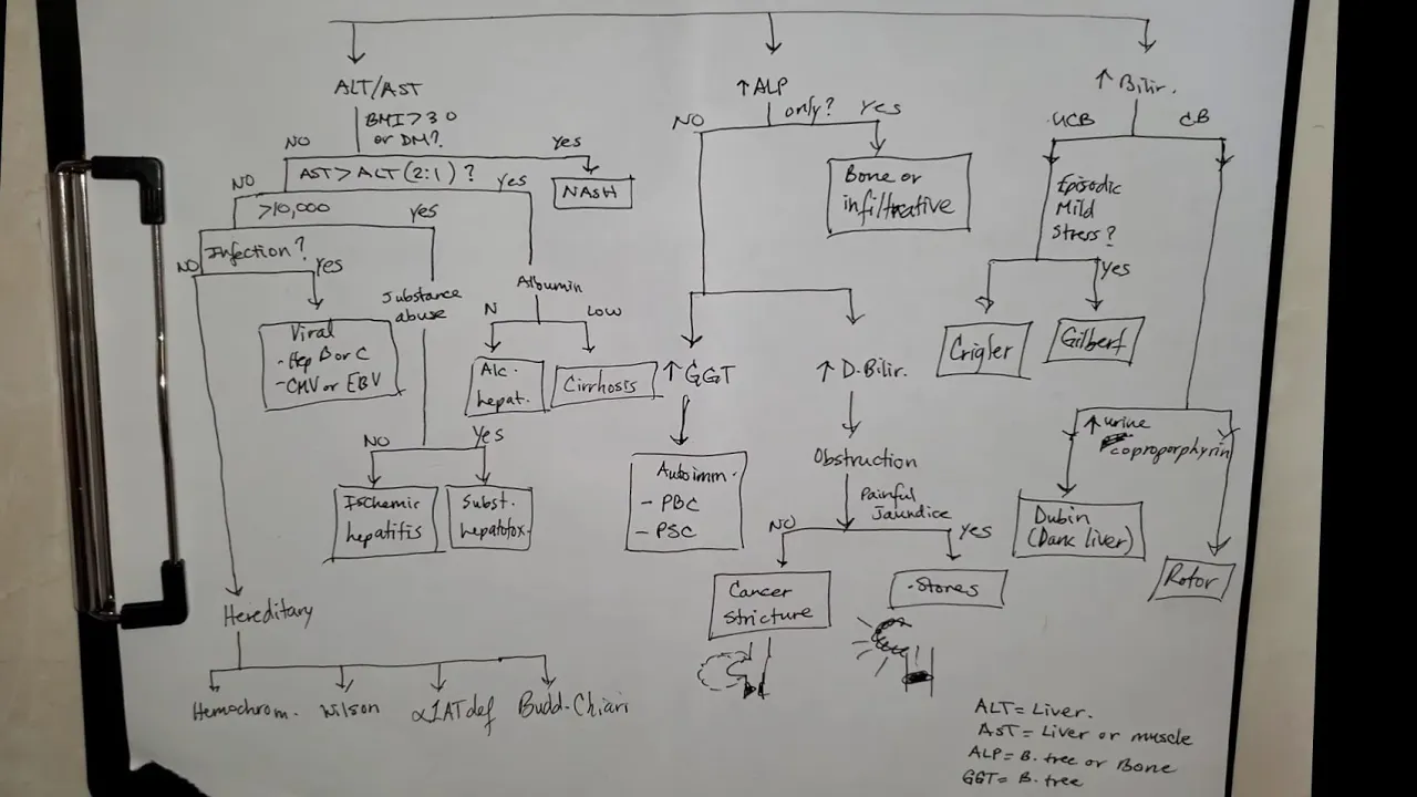
- Why this video: Walkthrough of a systematic clinical algorithm. It shows you exactly how to approach transaminitis by first screening for NAFLD/MASLD risk factors, then ruling out toxins, viral panels, autoimmune profiles, and structural abnormalities.
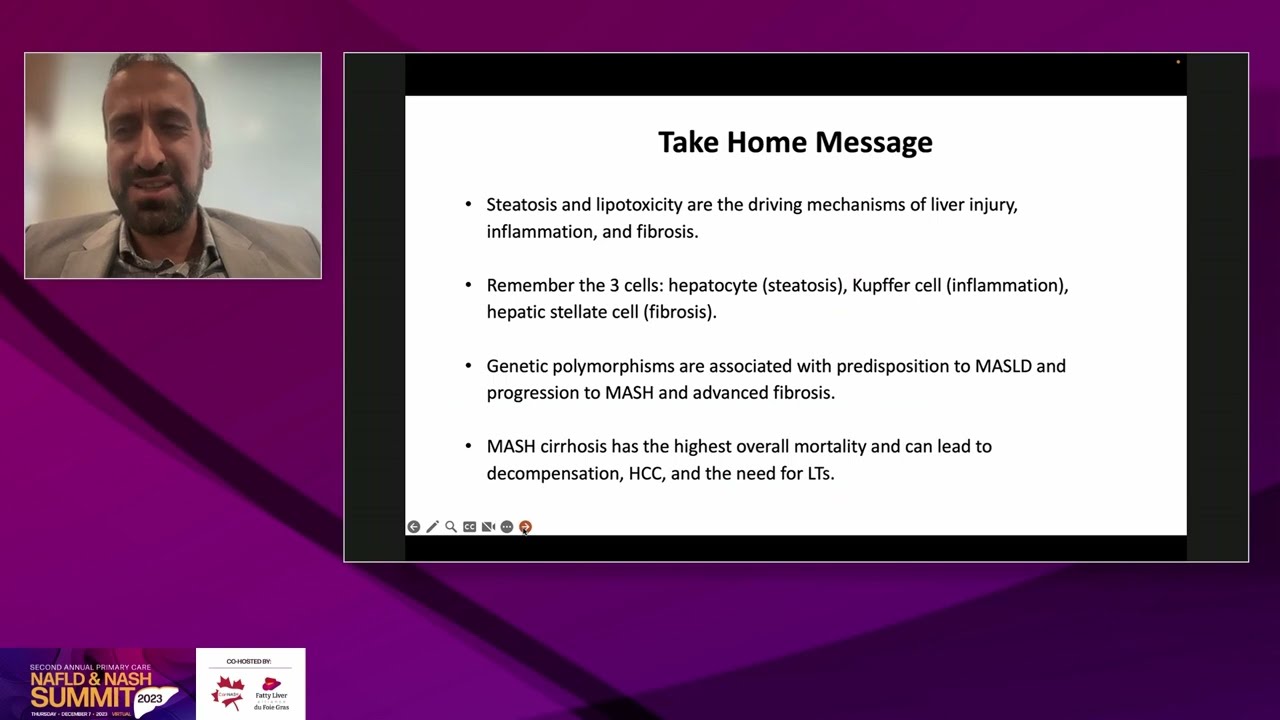
- Why this video: An expert clinical lecture illustrating the pathophysiology of MASLD (formerly NAFLD) and MASH (NASH). It covers how visceral adiposity and systemic insulin resistance drive free fatty acid accumulation, lipotoxicity, and progressive hepatocyte inflammation.
Knowledge Checkpoint
- Understand why an AST:ALT ratio > 2:1 is highly characteristic of alcoholic liver disease (due to mitochondrial damage and pyridoxal-5'-phosphate deficiency).
- Map the progression of metabolic liver disease from simple steatosis to steatohepatitis (MASH), fibrosis, and cirrhosis.
- Identify common medications (such as acetaminophen, NSAIDs, and statins) capable of inducing severe transaminitis.
Module 5: The Workup of Cholestatic Injury
Module Overview
When Alkaline Phosphatase (ALP) and Bilirubin elevations dominate the clinical picture, the underlying pathology involves a disruption of normal bile flow. This module covers the systematic workup of cholestasis, differentiating intrahepatic causes (e.g., Primary Biliary Cholangitis [PBC] and Primary Sclerosing Cholangitis [PSC]) from extrahepatic structural obstructions (e.g., choledocholithiasis, biliary strictures, or pancreatic malignancies).
Gap Note: While the physiological processes behind biliary jaundice are well-illustrated below, specific diagnostic comparisons for PBC and PSC are limited in this selection. For exam preparation, supplement this module by searching for: "Primary Biliary Cholangitis PBC vs Primary Sclerosing Cholangitis PSC Osmosis" (focusing on AMA antibodies for PBC vs. the magnetic resonance cholangiopancreatography [MRCP] beaded-duct findings for PSC).
Recommended Videos

- Why this video: Breaks down the pathophysiology of obstructive (post-hepatic) jaundice. It explains how biliary tract obstructions block the excretion of conjugated bilirubin, leading to pale stools, dark urine, and systemic pruritus.
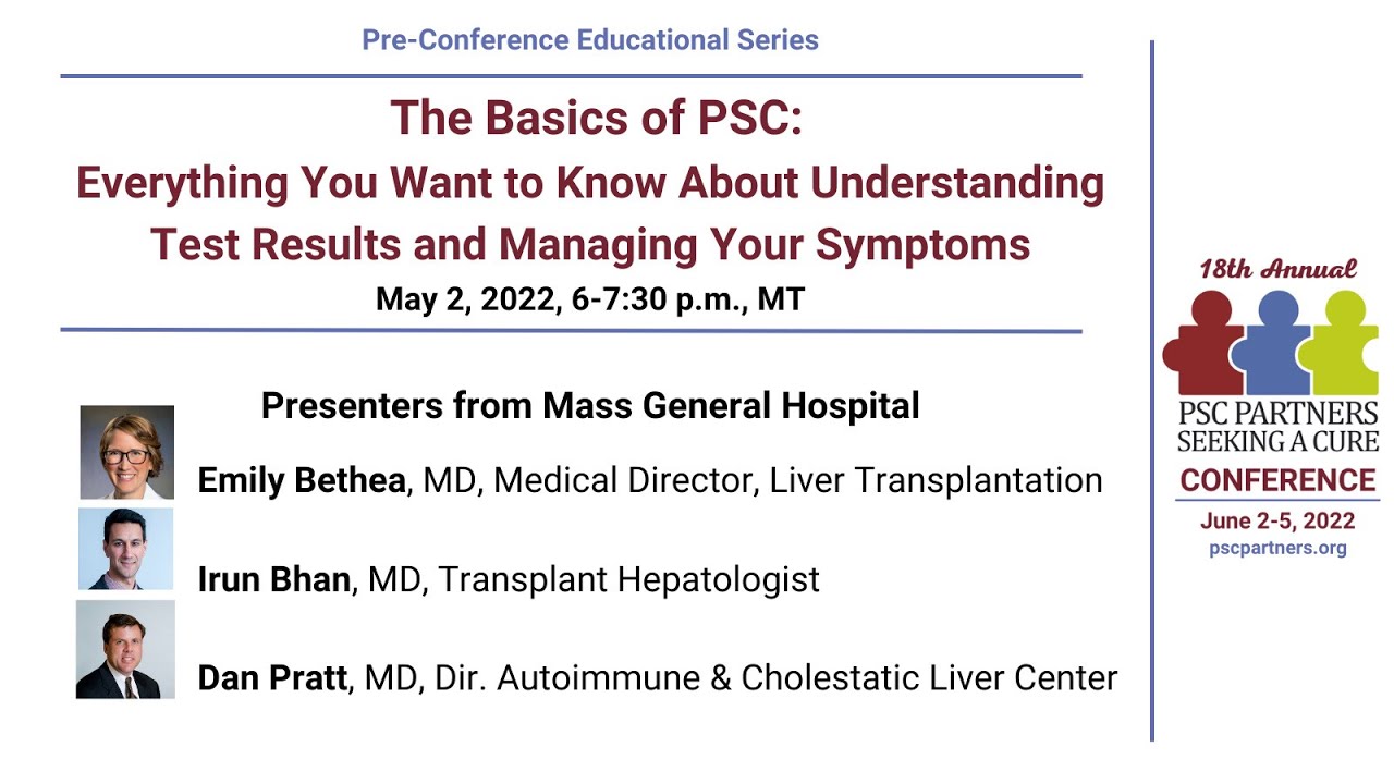
- Why this video: This short webinar clip provides vital context on how biliary disease destroys hepatic parenchymal function over time, highlighting how chronic cholestatic diseases like PSC ultimately lead to synthetic dysfunction.
Knowledge Checkpoint
- Detail the anatomical difference between intrahepatic and extrahepatic biliary obstruction.
- Explain why obstructive jaundice leads to direct (conjugated) hyperbilirubinemia, pale stools, and tea-colored urine.
- Understand the role of right upper quadrant (RUQ) abdominal ultrasound as the first-line imaging modality in cholestatic injury.
Module 6: Clinical Integration and Case Studies
Module Overview
This final module integrates biochemical, anatomical, and diagnostic concepts. You will study complete gastroenterology lectures that utilize real-world clinical presentations, complex or mixed LFT panels, and physiological variants (such as physiological ALP elevations during skeletal growth phases) to refine your diagnostic instincts.
Gap Note: Dedicated interactive student case vignettes are sparse in the video pool. To complement this module, we recommend independently searching for: "Abnormal LFTs clinical case interpretation practice" to test your diagnostic speed on sample patient charts.
Recommended Videos

- Why this video: An outstanding, highly academic clinical review that ties all previous modules together. It guides you through the interpretation of complex, mixed patterns and highlights crucial benign physiologic variants (such as bone-growth-driven ALP spikes in healthy adolescents).

- Why this video: A specialized gastroenterology lecture that reviews the practical bedside workup of abnormal LFTs. It emphasizes how to combine patient history (e.g., travel, toxins, alcohol, metabolic risk) with targeted ultrasound ordering.
Knowledge Checkpoint
- Formulate a diagnostic algorithm for an asymptomatic patient with isolated mild transaminitis.
- Correctly recognize when an elevated ALP is physiological (e.g., third-trimester pregnancy, adolescent bone growth) rather than pathological.
- Select appropriate follow-up tests (e.g., viral serologies, iron studies, ceruloplasmin, or autoantibodies) based on a patient's LFT pattern.
Course Map
Key People Index
- Dr. Naim Alkhouri
VP of Academic Affairs and Chief of Transplant Hepatology at Arizona Liver Health. Highly regarded researcher in metabolic liver diseases, featured in Module 4 discussing MASH progression pathways. - Dr. Gary Wu
Professor of Gastroenterology at the Perelman School of Medicine, University of Pennsylvania. Pioneer in gut-liver axis research, featured in Module 1 exploring the systemic impact of portal circulation. - Dr. Susruta Sen
Clinical Biochemist and Laboratory Medicine Specialist. Featured in Module 2 explaining LFT laboratory ranges and underlying metabolic pathways.
Final Self-Assessment
Test your clinical reasoning skills with these diagnostic criteria checklist items:
- Biochemical Differentiation: Can you explain why a patient with severe cirrhosis can have completely normal AST/ALT levels (due to "burnt-out" parenchymal loss)?
- Calculations: Given () and (), what is the exact R-ratio, and what diagnostic pathway does it warrant?
- Ratio Interpretation: What is the clinical significance of an AST:ALT ratio versus a ratio ?
- Cholestatic Imaging: Why is abdominal ultrasound the immediate next step in a cholestatic LFT pattern, and what visual cues are you looking for?
- Autoimmune Serology: Which specific autoantibody test would you order for a middle-aged female with pruritus, fatigue, and an elevated ALP?
- Bilirubin Conjugation: Can you map out why hemolytic anemia leads to unconjugated hyperbilirubinemia, while gallstones cause conjugated hyperbilirubinemia?
- Drug Toxicity: What is the standard hepatotoxic threshold for acetaminophen ingestion, and what clinical lab markers indicate acute toxicity?
- Synthetic Evaluation: Why does acute liver injury often present with normal albumin levels but abnormal PT/INR values? (Hint: Consider half-life differences).
- Physiologic Deviations: How would you clinically reassure an active adolescent presenting with isolated ALP elevation and completely normal GGT and transaminases?