Mastering the Localization-Based Neurological Examination and Acute Stroke Syndromes
Learning Goal: Establish a world-class clinical foundation in functional neuroanatomy, execute a precise localization-based neurological examination, differentiate the pathophysiological cascades of acute stroke, and rapidly apply the NIH Stroke Scale (NIHSS) to localize and manage acute vascular brain injuries.
- Prerequisites: Basic knowledge of human biology, neuroanatomy, or clinical terminology.
- Estimated Total Study Time: 18 Hours
Module 1: Foundations of Neuroanatomy and Vascular Supply
Module Overview
To diagnose and localize neurological disorders, clinicians must first possess a spatial map of the central nervous system (CNS) and its vascular architecture. This module covers the structural divisions of the cerebral cortex, key functional lobes (frontal, parietal, temporal, and occipital), the brainstem, and the complex vascular network known as the Circle of Willis.
⚠️ Video Pool Coverage Gap: There are no direct introductory neuroanatomy videos in the current video pool. To master this foundational module, please independently search for the recommended queries below on YouTube.
Recommended Independent Search Queries
- “Neuroanatomy basic overview animation”
- “Circle of Willis anatomy 3D explanation”
- “Functional neuroanatomy cortical lobes lecture”
Essential Theoretical Core
- The Cerebral Cortex: The brain is divided into four major lobes. The Frontal Lobe houses the primary motor cortex and Broca's area (expressive language). The Parietal Lobe processes primary somatosensory input and spatial awareness. The Temporal Lobe controls hearing, memory (hippocampus), and receptive language (Wernicke's area). The Occipital Lobe contains the primary visual cortex.
- The Circle of Willis: This arterial ring at the base of the brain provides collateral blood flow. It is fed by the bilateral Internal Carotid Arteries (ICA) anteriorly and the Vertebral-Basilar System posteriorly. Key branches include the Anterior Cerebral Artery (ACA), Middle Cerebral Artery (MCA), and Posterior Cerebral Artery (PCA), coupled with the anterior and posterior communicating arteries.
Knowledge Checkpoint
- Diagram the flow of blood from the vertebral and internal carotid arteries through the Circle of Willis.
- Identify which cortical lobes are primarily supplied by the MCA, ACA, and PCA.
- Describe the primary clinical function localized to the precentral gyrus versus the postcentral gyrus.
Module 2: The Neurological Examination: Step-by-Step
Module Overview
The neurological examination is a systematic clinical tool used to map subjective symptoms to objective anatomical structures. It comprises assessments of mental status, cranial nerves, motor function, sensation, reflexes, and cerebellar coordination.
While a complete screening neurological exam is not fully represented in the video pool, we have extracted highly specific clinical segments illustrating key visual, motor coordination, and diagnostic screening techniques.
Recommended Videos

- Why this video: This video provides an excellent, high-yield look at visual field examination. It demonstrates visual confrontation testing to identify field deficits (like hemianopia), which are essential when performing cranial nerve assessments (CN II) in both routine exams and suspected strokes.

- Why this video: This resource demonstrates the exact physical maneuvers required to test cerebellar coordination and identify limb ataxia. It shows the proper clinical execution of the finger-to-nose and heel-to-shin exams, contrasting normal coordination with dysmetria.

- Why this video: This video walks through the bedside application of basic cranial nerve, sensory, and motor exams to screen for signs of a cerebrovascular accident (CVA), illustrating how generalized exam maneuvers adapt to emergency clinical presentations.
⚠️ Video Pool Coverage Gap: The video pool lacks a comprehensive video covering deep tendon reflexes, detailed sensory mapping, or a complete non-stroke cranial nerve walkthrough. To fill this gap, search YouTube for: “Cranial nerve examination clinical skills OSCE guide” and “Motor and sensory neurological exam demonstration”.
Knowledge Checkpoint
- Explain how to perform confrontation testing for visual fields in all four quadrants.
- Differentiate between cerebellar dysmetria (ataxia) and weakness during a motor exam.
- Identify which cranial nerves are responsible for extraocular movements (EOMs) and how to evaluate them.
Module 3: Principles of Neurological Localization
Module Overview
Neurological localization is the clinical art of determining where a lesion lies within the nervous system based on a patient's examination findings. The chief starting point is distinguishing between lesions of the central nervous system (Upper Motor Neuron) and the peripheral nervous system (Lower Motor Neuron).
⚠️ Video Pool Coverage Gap: The current video pool does not contain direct video tutorials on localization principles or UMN vs. LMN lesions. You are encouraged to supplement your study with the search queries below.
Recommended Independent Search Queries
- “Upper vs lower motor neuron lesion physical exam findings”
- “Neurological localization rules explained”
- “How to localize lesions in clinical neurology”
Essential Theoretical Core
- Upper Motor Neurons (UMN): UMNs originate in the motor cortex or brainstem and carry signals down to the spinal cord. Lesions in the UMN pathway (e.g., stroke, spinal cord injury) lead to a loss of inhibitory control. Clinical signs include hyperreflexia, spasticity (increased tone), the presence of pathological reflexes (like the Babinski sign), and pronator drift.
- Lower Motor Neurons (LMN): LMNs originate in the anterior horn of the spinal cord (or cranial nerve nuclei) and travel directly to target muscles. Lesions (e.g., peripheral neuropathy, radiculopathy) result in a loss of the motor pathway. Clinical signs include hyporeflexia (or loss of reflexes), flaccidity (decreased tone), muscle atrophy, and fasciculations (muscle twitching).
| Sign | Upper Motor Neuron (UMN) Lesion | Lower Motor Neuron (LMN) Lesion |
|---|---|---|
| Muscle Tone | Spastic (increased) | Flaccid (decreased) |
| Reflexes | Hyperreflexia (clonus) | Hyporeflexia / Areflexia |
| Atrophy | Minimal (disuse only) | Severe and rapid |
| Pathological Signs | (+) Babinski, Pronator Drift | Fasciculations |
Knowledge Checkpoint
- Contrast the physiological mechanism of hyperreflexia in UMN lesions with the hyporeflexia seen in LMN lesions.
- Describe the clinical presentation of "crossed signs" and explain why they point directly to a brainstem lesion.
- Define pronator drift and explain how to test for it at the bedside.
Module 4: Pathophysiology of Acute Stroke
Module Overview
An acute stroke is a medical emergency caused by a disruption of blood supply to the brain. This module explores the structural and cellular differences between ischemic and hemorrhagic events, the metabolic cascades triggered by oxygen deprivation, and the physiological significance of the ischemic penumbra.
⚠️ Video Pool Coverage Gap: No direct animations or conceptual lectures explaining the cellular pathophysiology of stroke exist in the video pool. Use the search queries below to supplement your study.
Recommended Independent Search Queries
- “Ischemic vs hemorrhagic stroke pathophysiology animation”
- “Stroke penumbra and collateral circulation explainer”
Essential Theoretical Core
- Ischemic vs. Hemorrhagic Stroke: Approximately 85% of strokes are ischemic (caused by a thrombotic or embolic occlusion of an artery). The remaining 15% are hemorrhagic (caused by rupture of a blood vessel leading to intracerebral or subarachnoid bleeding).
- The Ischemic Penumbra: During an acute ischemic stroke, the region of brain tissue directly supplied by the blocked vessel dies rapidly—this is the ischemic core. Surrounding the core is a zone of hypoperfused, dysfunctional, but structurally intact tissue called the penumbra. This tissue is kept alive temporarily by collateral circulation (leptomeningeal vessels) and is the primary target of acute reperfusion therapies (IV thrombolysis and mechanical thrombectomy).
[Arterial Occlusion] ──> [Ischemic Core (Infarcted/Dead Tissue)] └──> [Ischemic Penumbra (Hypoperfused/Salvageable)] │ (Maintained by collateral vessels; │ target for emergency reperfusion)
Knowledge Checkpoint
- Differentiate between a thrombotic ischemic stroke and an embolic ischemic stroke.
- Explain the clinical and therapeutic significance of the "ischemic penumbra" in the setting of acute stroke treatment windows.
- Describe the cellular mechanisms (e.g., excitotoxicity, calcium influx) that lead to rapid neuronal death in the ischemic core.
Module 5: Acute Stroke Syndromes & Clinical Localization
Module Overview
Stroke syndromes present with highly predictable patterns of deficit based on the vascular territory affected. This module teaches you to associate specific neurological exams with vascular anatomy (such as the MCA, ACA, and PCA) and key brainstem pathways.
Recommended Videos

- Why this video: This video introduces the clinical presentation, diagnosis, and emergency assessment of acute stroke. It links clinical presentation directly to vascular territories, showing how to approach a patient presenting with hyperacute deficits.
⚠️ Video Pool Coverage Gap: The video pool has limited coverage of specific vascular stroke syndromes (e.g., detailed breakdowns of MCA vs. ACA vs. PCA, and specific brainstem syndromes). Please use these search queries on YouTube to learn more: “Stroke syndromes MCA ACA PCA basilar artery localization” and “Brainstem stroke syndromes rule of 4s localization”.
High-Yield Stroke Syndrome Reference
- Middle Cerebral Artery (MCA) Syndrome: The most common stroke syndrome. Characterized by contralateral hemiparesis and hemisensory loss (predominantly affecting the face and arm more than the leg). If the lesion is in the dominant hemisphere (typically left), it leads to aphasia (Broca's expressive or Wernicke's receptive). If in the non-dominant hemisphere (typically right), it leads to hemispatial neglect.
- Anterior Cerebral Artery (ACA) Syndrome: Characterized by contralateral hemiparesis and hemisensory loss affecting the leg more than the face and arm. Patients may also exhibit abulia (apathy, slow motor/verbal responses) and urinary incontinence due to frontal lobe involvement.
- Posterior Cerebral Artery (PCA) Syndrome: Characterized primarily by visual deficits, specifically contralateral homonymous hemianopia (often with macular sparing). Dominant-hemisphere lesions can cause alexia without agraphia (the patient can write but cannot read what they wrote).
- Brainstem Stroke Syndromes: Often present with the "Rule of 4s" or crossed signs.
- Wallenberg Syndrome (Lateral Medullary Syndrome): Caused by occlusion of the Posterior Inferior Cerebellar Artery (PICA). Symptoms include ipsilateral loss of facial pain/temperature sensation, contralateral loss of body pain/temperature sensation, ipsilateral Horner syndrome (ptosis, miosis, anhidrosis), dysphagia, and ataxia.
Knowledge Checkpoint
- A patient presents with severe weakness in their right leg, mild weakness in their right arm, and apathy. Which blood vessel is most likely occluded?
- Explain why an MCA stroke in the dominant hemisphere causes language deficits (aphasia), while a non-dominant MCA stroke causes neglect.
- List the classic clinical symptoms that make up Wallenberg (lateral medullary) syndrome.
Module 6: Emergency Stroke Evaluation & The NIH Stroke Scale (NIHSS)
Module Overview
In acute stroke care, "time is brain." The National Institutes of Health Stroke Scale (NIHSS) is the gold-standard, highly structured physical examination tool used to rapidly quantify neurological deficits in acute stroke. This module provides comprehensive training on each item of the scale, correct scoring rules, and practical patient simulations.
Recommended Videos

- Why this video: Led by clinical expert Lisa Klein, this video from Johns Hopkins Medicine provides a detailed and professional review of the 11 components of the NIH Stroke Scale. It is an excellent tutorial on how to score each deficit accurately at the bedside.

- Why this video: This video is a thorough, official training manual for administering the NIHSS. It walks through each item step-by-step, explaining the precise clinical criteria and pitfalls of scoring, making it perfect for certification preparation.
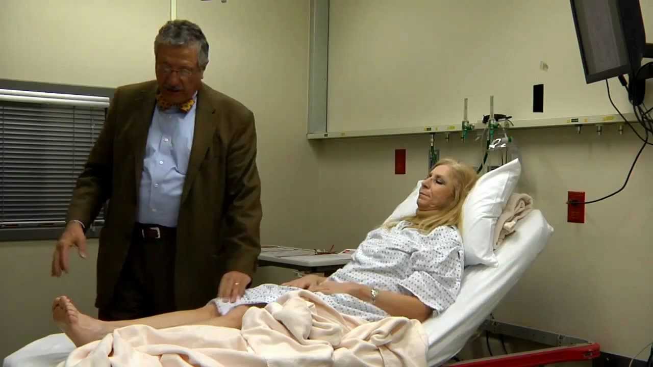
- Why this video: This simulation video provides a highly visual demonstration of the NIHSS exam being performed in real time. It offers a clear, concise visual reference for applying the assessment in a clinical environment.
- Why this video: Led by Dr. Sami Harik, this interactive video simulates real patient scenarios. It helps learners test their clinical decision-making, apply scoring rules to challenging patient responses, and learn to make consistent, objective evaluations.
Summary of the NIHSS scoring categories (Total Score: 0 to 42)
- 1a, 1b, 1c: Level of Consciousness (LOC) (Alertness, questions, commands)
- 2: Best Gaze (Horizontal eye movements)
- 3: Visual Fields (Confrontation)
- 4: Facial Palsy (Symmetry on smile/grimace)
- 5 & 6: Motor Arm & Leg (Drift test: 10 seconds for arms, 5 seconds for legs)
- 7: Limb Ataxia (Finger-to-nose and heel-to-shin)
- 8: Sensory (Pinprick response)
- 9: Best Language (Describe picture, name items, read sentences)
- 10: Dysarthria (Articulation of words)
- 11: Extinction and Inattention (Double simultaneous sensory stimulation)
Knowledge Checkpoint
- What are the exact time requirements for testing motor drift in the arms versus the legs during the NIHSS?
- Explain the golden rule of NIHSS scoring: why you must score the patient's first response and avoid coaching, even if you think they can do better.
- How do you score Item 11 (Extinction/Inattention) using double simultaneous sensory stimulation?
Course Map
Below is the recommended pathway for navigating the modules, illustrating how foundational anatomy leads directly to clinical examination, localization, and acute emergency management.
Key People Index
- Dr. Sami Harik, MD (UAMS Health): A leading neurologist known for his clinical instruction in neurodegenerative and vascular diseases. His simulation-based videos help demystify the nuances of bedside neurological exams.
- Lisa Klein, MSN, RN, AGCNS-BC (Johns Hopkins Medicine): A clinical nurse specialist in stroke care who provides clear, standardized instruction on the NIHSS to help healthcare providers maintain accuracy and consistency.
Final Self-Assessment
Test your mastery of localization-based examinations and acute stroke syndromes by checking off the learning milestones below:
- 1. Identify the key anatomical vessels of the Circle of Willis and explain how they provide collateral blood supply.
- 2. Perform a complete visual field confrontation exam in four quadrants and accurately identify a homonymous hemianopia.
- 3. Differentiate between an Upper Motor Neuron (UMN) and Lower Motor Neuron (LMN) lesion using clinical signs like reflexes, tone, and Babinski testing.
- 4. Explain the metabolic cascade of the ischemic penumbra and why rapid recanalization (thrombolysis/thrombectomy) is critical.
- 5. Identify a Middle Cerebral Artery (MCA) stroke syndrome, including differentiating between dominant (aphasic) and non-dominant (neglect) hemispheric presentations.
- 6. List the key clinical findings in lateral medullary (Wallenberg) syndrome and trace them to their anatomical structures in the brainstem.
- 7. Demonstrate how to perform and score all 11 categories of the NIH Stroke Scale (NIHSS) on a standardized patient.
- 8. Explain the correct scoring response on the NIHSS when a patient has a pre-existing amputation or severe joint contracture during motor testing.
- 9. Successfully localize a stroke to the Anterior Cerebral Artery (ACA) territory based on leg-dominant hemiparesis and behavioral changes.
- 10. Apply the "first response" rule of NIHSS scoring to a complex patient scenario without coaching or assisting the patient.