


![[Neuroanatomia] 11 - Vascularização do Sistema Nervoso](https://i.ytimg.com/vi/-TPL37ijrzQ/hqdefault.jpg)




















Acute ischemic stroke management requires rapid identification of stroke type through clinical assessment (NIHSS score >4 indicates major stroke requiring intervention) and imaging protocols (CT for initial hemorrhage exclusion and ASPECTS scoring, MRI for diffusion-perfusion mismatch to identify salvageable tissue beyond 4.5 hours). Treatment options include IV thrombolysis (alteplase or tenecteplase) within 4.5 hours for eligible patients, and mechanical thrombectomy for large vessel occlusions, with the key principle being that tissue preservation matters more than time elapsed since onset.
Acute Ischemic Stroke: Diagnosis & Management | KGMOA Webinar
Added:Neuroanatomy and cerebral vascular supply (e.g., Circle of Willis, major cerebral arteries, and their clinical territories).
Pathophysiology of ischemic versus hemorrhagic brain injury, including the concepts of the ischemic penumbra and core.
Fundamentals of the clinical neurological examination and the principles of localizing neurological deficits.
Basic interpretation of emergency brain imaging, particularly non-contrast head CT scans, to rule out intracranial hemorrhage.
The physiological mechanisms of blood coagulation and the endogenous fibrinolytic pathway.
Secondary stroke prevention strategies, including antiplatelet therapy, anticoagulation for atrial fibrillation, and carotid revascularization guidelines.
Management of acute post-stroke complications, such as cerebral edema, hemorrhagic transformation, and reperfusion injury.
Advanced neuroimaging modalities, including CT/MR perfusion imaging, to assess tissue viability beyond standard time windows.
Comprehensive etiological workup of stroke (e.g., TOAST classification, cardiac monitoring for occult arrhythmias, and patent foramen ovale evaluation).
Principles of stroke rehabilitation, multidisciplinary care, and the physiological basis of neuroplasticity during recovery.
241 views6likes1:32:58Original Release: 2025-08-27
[Music] And uh um I I first of all thank uh KJMO uh for inviting me for giving this talk um in in this wonderful platform. Um so as Dr. said acute stroke is a really a menace to the society. Right now the incidents and the epidemiology of stroke is rising maybe more because of the early identification of the stroke or maybe because of the lifestyle diseases have gone up and resulting in uh reducing the age of or of onset of the stroke. Now the uh people are getting strokes at much earlier age as compared to the uh earlier generation around people are getting strokes at 40s or 50s.
So which contributes much to the not only to the mortality and to the morbidity and the suffering of the family. So uh shall I I'll start uh I'll start by sharing my slides.
Uh Dr. Can I share my slides? You can show up.
>> Okay. Okay.
So, so is it visible uh now?
>> Visible. Visible.
>> Okay. Okay. So, um the topic uh given to me today is acute eskeemic stroke large vessel and small vessel disease. I'll try to compreh uh comprehensively go through the whole spectrum of stroke ranging from the small vessel to large vessel stroke its diagnosis um imaging protocols and um uh some of the evidence to stud uh state u management uh in the acute setting in both medical management and endovvascular management.
So this is our uh team. Uh so stroke uh by by who definition the stroke is uh said to be a clinical syndrome consisting of rapidly evolving clinical signs for of focal or global uh disturbance in cerebal function last more than 1 hour. Uh so anything which lasts less than 1 hour is now classified into TIA. AHA also gives a similar definition and u and it includes uh brain spinal cord retinal uh cell death attributable to eskeemia uh with evidence with pathological evidence and radiological evidence and eskeemic stroke is defined as an episode of neurological dysfunction caused by focal cerebral retinal or spinal infection. All three are categorized as uh in the category of acute stroke. Um so the uh initial assessment of an acute stroke involves history taking detailed physical examination and investigations. So what are the protocols uh usually followed?
Most important thing is onset of stroke.
So you should know when the deficit when when the uh sudden onset deficit has started that is that has paramount importance in determining the treatment decisions imaging decisions all the further decisions regarding management of the stroke is dependent upon the onset of the stroke. Some as you know most of the strokes also present late night uh in the middle of the night or many any time it may be the onset may not be clear because there will be no direct uh eyewitness to the stroke. So stroke of unknown onset also poses a serious challenge in the diagnosis and management.
And uh in addition we need to know the past history of similar episodes of stroke and u we need to know the past surgical history any recent surgical history because that has implication in the management in the like thrombolysis.
uh thrombolysis is contraindicated in many of the surgeries uh major surgeries if the patient has undergone in the last 2 weeks or 3 weeks. uh and past medical histories of paramedic importance like coronary artery disease uh presence of atrial fibrillation whether the patient is on any anti-arithmia drugs whether the patient is on oral anticoagulation or antipllets all these will have implications in the uh management and the major symptoms of carot carotin TIA or minor stroke will be transient ipsilateral monocular blindness contrateral body weakness you have to ask for contrateral body sensory loss paristhesia sensory it especially if there is an dominant hemisphere is involved hemopia or visual defects especially in posterior circulation strokes PC posterior cerebral artery strokes involving the occipital area parietal oxal area oxal association visual association areas and whe and disarthria symptoms of vertebra ga includes bilateral weakness or clumsiness maybe a shifting weakness or a crossed weakness that means ipsilateral phase is involved. Contrateral body involved with sensory loss with or without sensory loss and paristhesia. Uh then you have to think about posterior circulation involved. Hemi anesthesia maybe aamic stroke involving phase and the he bilateral contra bilateral hemopia or contrlateral homoous hemiopia maybe a cortical involvement posterior cerebral articulator region. Then trans uh symptoms like vertigo, dipopia, dysphasia, disarthria in isolation, it doesn't have a meaning. But if two or more of these symptoms are simultaneously involved, then it might be a pointer towards a posterior circulation or probasular stroke.
Symptoms which are not acceptable as evidence of TIA includes syncopy, dizziness, confusion, urinary and fecal incontinence alone. Suppose a person comes with only history of incontinence.
There is no it cannot be deemed as a GIA unless proved unless there is associated other deficits in the form of visual deficits or sensory deficits or motor weakness cross weakness um or disarria dysphasia unless if it is associated with these symptoms uh isolated urinary incontinence confusion isolated confusion isolated episodes of syncopi or vague dess and vague generalized weakness cannot be classified as da many of the times patient comes such with these symptoms in the casuality isolated occurrence of vertigo, diplopia, dysphasia has no meaning. But if two or more occur in combination then you have to think about these posterior circulation uh a typical presentation of strokes like posterior circulation ta stroke. Now uh public can be educated regarding the uh assess assessment of early assessment of stroke using fast face arm speech test like uh teach them educate them regarding facial deviation arm weakness and speech difficulty and then ask them to come to the nearest health uh nearest healthcare area healthcare center. So uh TIA the TIA in isolation is not of any uh grave consequence to the patient but if you neglect a TIA he may come up with a stroke in the next few days or months.
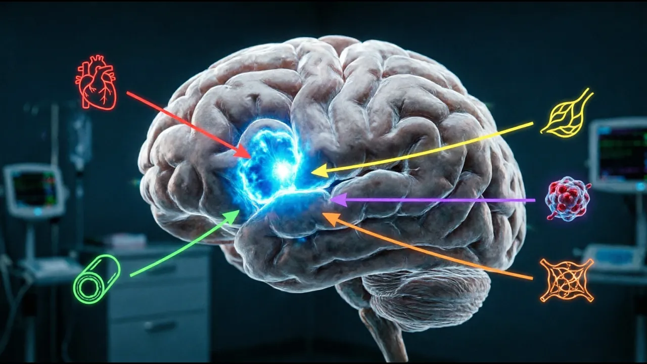
Uh so for risk stratification of TIA is very important. TAA is any focal deficit uh lasting less than 1 hour. So this risk stratification is based on age more than 60 years. Blood pressures, high blood pressure last more than 140 90. Uh clinical features like unilateral weakness, speech disturbance, duration of symptoms lasting more than 60 minutes or around 60 minutes. Uh all these classify as a high risk TIA behav TIA episode which might result in a uh stroke in near future. So for risk stratifications, a scoring system is there. ABCD2 score any score around four or five has a risk of stroke. As you can see here, there is a risk of stroke for four or five ABCD score is around 10% in the next 90 days and score of six or seven has a risk of 80 18% in the next 90 days. So as soon as you see a TIA then ask for a baseline imaging like a CT to rule out any hemorrhage or any other contra indications for antiplatelets then immediately initiate antiplatelet at least at the earliest and within and then you need to have a secondary prevention why the patient has developed a t there must be a reason either it can be an artery to artery emolism because of a proximal critical stenosis of a large vessel or it can be a cardiamolic so we have to evaluate for the secondary causes then uh then only the adequacy of the therapy can can be ensured to that patient because TA alone doesn't mean anything but if you do a vascular imaging further you can know the cause of the TAA and take adequate steps to prevent a major stroke catastrophic stroke ideology of acute stroke um we have to look for otheries of acute stroke in the case of an acute stroke toast toast classification is a pra pragmatic classification which has been developed for classifying the ethiology of acute stroke majority of the acute stroke falls in the atherosclerotic cerebrovas disease, large artery disease 20%, around 20% falls in this category. Cardiamolism constitute 20%. Small vessel disease constitutes 25%, cryptogenica and idopathy other causes constitute around 30%.
E is eskeemic stroke. Here this is an example of a large artery stroke. You can see a proximal critical stenosis in the ICA. This is a CT angagram reconstructed image showing a proximal critical stenosis of the ICA bifurcation. This is the bifurcation.
This is the ECA and this is the ICA. You can see a proximal critical stenosis uh on the right ICA here there's a focal critical narrowing of the middle cerebral artery. You can see the terminal ICA of the this is the right internal coroted artery. This is the left internal carroted artery and this is the basil coming in in the posterior basil artery here which is not well visible but uh these are the internal carroted arteries. So the right internal carot artery divides into a middle cerebral artery here and an anterior cerebral artery here in the midline which supplies the midline and here this internal left internal carot artery divides into a left middle cerebral artery here and left anterior cerebral artery here. So here you can see the terminal ICA where it go becomes the middle cereal artery. There's a focal critical construction of narrowing of the middle cerebral artery here. So uh any occlusion more than 50% or 60 60% is classified as significant with you need to do a plaque morphological analysis whether that plaque is an unstable plaque or a stable plaque just like a coronary plaque. Uh so how do you know the whether it is unstable or stable?
You do an MR plaque imaging to see whether the plaque is stable or unstable. Now there are methods to assess the stability of the block and if it is an unstable plaque the chances of a next DIA or chances of it getting rupture o oluding occluding is very high plaque rupture and occlusion rates are very high if in an unstable block. So you have to characterize the morphology of a plaque in the case of a large vessel critical stenosis.
So the the cardiamolic stroke the major causes of cardiamolism every most most of the people are well aware of the cardabolic causes like left atrial thrombus atrial fibrillation uh transient BT 6 syndrome recent myioardial inffection any boprothetic valves rheumatic heart disease uh congestive heart failure with low ejection fraction less than 30% dilated cardiammyopathy all these and bacterial endocarditis all these are the well-known causes of cardiamotic stroke uh Other causes small artery stroke, penetrating artery stroke or perforator stroke. What we call as perforator stroke or arteropathic stroke. Uh this is caused by a branch occlusion of a small perforating artery. A tiny artery and not the major vessels are involved.
The tiny perforators are involved. Here you can see the perforator getting involved in the basil. A basil perforator artery stroke resulting in a ventral pondine inf. This bonds this is a ventral pondine infog. Here you can see a small perforating artery stroke resulting in a thealamic infog. This is aamus. Here you can see a laconar infot any any infot less than two c 2 cm is class class classified as a laconar perforating artery stroke uh or a branch artery stroke the major ideology being lipohilyosis the path physiology is lipohilyosis because of uncontrolled diabetes metabolic derangement uh uncontrolled diabetes hypertension dysipidemia or hyperomosmia age all these factors contribute to lipohinosis of small arteries which result in these type of small vessel strokes Other causes of strokes al including uh uh includes cryptogenic stroke that is embolic stroke stroke of unknown source.
We don't know the source where it comes from. Some embolic suddenly throw up into the brain from somewhere maybe mostly in the from the arch of iota and large artery large proximal vessels. um many of the many a times the large vessel screening including angio CT andogram and the routine MR an angiogram may miss these type of strokes because arch of iot may not be covered in most of the angioraphies CT andogram or MR angio so you in in some of the cryptogenic stroke where you don't find any other cause you have to screen for these arches to find the emolic source and there are other causes of idopathic strokes and rare cause of strokes like hypercoagulable states antifphospholipid antibbody syndrome protein uh Prothroin 2210 mutation hyperroysmia polyythemia high hemoglobin more than 16 or 17 it warrants treatment for it because patient is liable for a uh eskeemic stroke. So uh we we we as part of the treatment routine treatment protocol we allow them to do blood letting so so that and to reduce the hemoc concentration and hemattocrit so that uh as a secondary prevention strategy to prevent a stroke and then use of hormonal pills contraceptives venus sinus thrombosis all these and vasculitis CNS vasculitis non non-inflammatory vascularities like fabric disease RCVS all these are the rare causes of stroke uh which need to be evaluated when the source of stroke or the cause of stroke is unknown and the major risk factors of stroke as we all know is lifestyle factors like uh high BMI uh heavy smoking blood pressures high uncontrolled blood pressures dysipidemia obstructive sleep apnoa cardiovascular risk factors family and family history of stroke any uh if you have a family strong family history of eskeemic heart disease or stroke You have to evaluate for all the rare causes of strokes like um metabolic causes, hyperomosis nemia factor deficiencies everything has to be screened. Um now the goal of diagnosis of stroke is to identify the stroke type within 60 minutes within as early as possible not within 60 minutes. There is no criteria or threshold as 60 minutes as early as possible so that the treatment can be initiated at the earliest because time is green. um you lose uh 2 million neurons in a span of minutes if you don't identify early and the greater some people so how do you know whether your brain will survive this long or your brain doesn't survive some people's brain survive for some are fortunate that even after 10 hours some people used to say okay he went after 6 hours 8 hours uh revascularization strategy worked like thrombolysis worked and patient became completely okay some people even after reaching two to 3 hours. They are unfortunate people. They lose the they they develop massive infection in spite of getting revascularized and thrombolized. Why the difference? So this difference is mostly because the collateral status is different for different patients, different people. My my collateral status will be different from yours and that will be different from a third persons. So each person has it his own collaterals and these collaterals are the ones which are helping the brain to sustain this eskeemic insult for a long duration.
Some may sustain it for hours. Some some there are people who have come to me with acute stroke who has last sustained eskemia for 18 hours 16 hours. We revascularized it after at around 20 hours or 18 hours became completely normal. But some unfortunate ones even 2 to 3 hours revascularization within 2 to 3 hours their brain is gone. In fact, volume has completely become huge and the the patient has developed a permanent mobility and some of them developed permanent deficits.
So acute so early identification is the key. So for that acute stroke teams should be ready and stroke rating scale everyone should be in that acute stroke team who is dealing with the emergency acute strokes should be well versed with the stroke rating scale. It is called NIHSS National Institute of Health Stoke scale and the baseline investigations of course chest X-ray blood sugar ECG everything is warranted blood sugars more than 400 is a contra indication for thrombolysis. So you need to have a blood sugar at the time of presentation and blood pressures any high blood pressures around shooting around 210 220 is a contra indication for thrombolysis.
So you need to be aware well aware of the uh blood pressures also uh and if the patient is in within window period we need to optimize the blood pressure before giving license. So baseline management is very important at the in the emergency department and um low low n so what is NIHSS as I national institute of health stroke scale many of many are well versed with this the scale so but for I'm briefly uh narrating uh the major factors included in the NIHSS uh stroke scale is consciousness level of consciousness how do you assess based on questionnaire if if patient person answers both the questions correctly, he is given a two marks and if uh and whether we'll assess whether the patient is obeying commands if he performs two tasks correctly then he is given two marks two points that means uh lift upper limb or protrude the tongue close the eyes you can give commands like this and see how the patient obeys if the patient obeys both the commands properly promptly then you can give a score of two then gaze deviation if there's a force deviation the score is Uh if there's no deviation the score is zero. Visual hemi anopia if there is bilateral hemopia score is three.
Complete hemianopia on one side score is two. Uh facial pulsy major complete facial paralysis is given a score of three. Motor arm weakness. If there is mild drifted the arm uh then the score is one. If there is no effort against gravity and the limb falls then the score is three. If there is no movement the score is four.
uh like similarly leg power is also taken into account power of the leg then limb at taxia if the taxi is there present in two limbs then the score is two if it is present only in one limb score is one sensory deficit if there is a mild to moderate sensory loss in one side then score is one there is severe total sensory loss the score is two then see if there is patient is having aphasia mild aphasia that means patient is able to tell some words broken words then score is one if There is severe aphasia. He's not able to reproduce anything and there's he's going in for a global aphasia that's mute and global aphasia score is three. Severe disardria score is two and sensory inattention.
You can simultaneously touch both the sides. If the patient is not a facicic you can ask simultaneously you can touch both the sides and ask whether he's perceiving both the feeling on both sides or you can show both fingers together and see if he's seeing both the fingers simultaneously. This is called simultia. So if there is sensory in attention or visual in attention uh then the score is um two. So total score is around 42. So and any score around more than four is considered as a major deficit major uh stroke. uh and any any score of eight or nine is usually large vessel occlusions, large artery occlusion like IC occlusion or a middle cervical artery occlusion or anterior cervical artery occlusion will have NHS of around 7 8 or 9. So anything more than seven is significant. You have to uh suspect a large artery occlusion and you have to do a vascular imaging to know the site of occlusion. Which artery is occluded? You need to know a large Now as I said blood pressures uh monitoring blood pressure is paramount importance the blood pressure should be brought less than 180 100 uh 100 before thrombolysis it's a it's a level one indication before thrombolysis it's mandatory to bring down the blood pressures to 180 100 but don't attempt to minimize the blood pressures to much lower levels because if it is a hemodynamic stroke the patient will have will have worsening of the perfusion deficit the brain perfusion and the flow into the brain brain will reduce further and he'll develop a secondary stroke. So um uh we have to be very cautious not to reduce blood pressures too much. So 180 less than anything less than 180 100 is okay and like blood glucose around four more than 400 is a contra indication for thrombolyis.
Uh so next next next in the line of management is imaging. How to diagnose the stroke? Now we have identified that it is a stroke clinically. Now you have to go about with imaging. CT is the baseline imaging major workforce in most as we as we have CT in all the major uh all the peripheral centers and all the center uh even I I'm sure it'll be available in many of the government hospitals also um and so CT is a very valuable tool uh to assess uh in patients with acute strokes because once the CT once you assess the CT and you deduce that there is no ma major infection but the patient has clinically there is deficit definite def deficit of the to there's a definite deficit in the form of either aphasia or visual difficulty hemianopia or par parasis of the limbs upper limb weakness or lower limb weakness then you can definitely thrombolize the patient immediately if provided there is no bleed in the CT so CT uh is a basic C uh baseline imaging basic mandatory requirement for a for all for the management of all acute strokes.
So the major purpose of CT is to exclude intracraanial hemorrhage so that you can thrombolize the patient immediately to establish an aspect score. So I'll tell you what aspect score is. So the infact volume inside the in the CT assessment of the inf volume in the CT uh we use a score called aspect.
So this is the aspect score. It is developed by the Alberta strokes uh uh stroke school Canada uh Calgary. So uh it's very simple. You can see this is the CT brain. You take CT brain in two cuts. One is at the basal ganglia level.
This as you can see this is the thealamus. This is the internal capsule.
You have cordate here. Uh is my arrow visible?
>> Yes. Yes.
>> Okay. So uh so this is the cordate. This is the internal capsule. This is thealamus. So this is the ganglionic cut. So in the ganglia cut you can identify the cordate. This is the internal capsule. You can see the lentififor nucleus here and this is the insular area and this is the cortical area M1 M2 M3 at the level of basal ganglia. Now you go take another level above the basal ganglia and the superior cut M4 M5 M6 in the cortex. So assessing these two if there is infaction or hypodensity this is a normal CT. You can see you can see clearly see the gray white differentiation is made here. The white gray matter is seen as lighter light and white matter is seen as a little bit dark.
That's how it looks in CT. So the here the gray white differentiation is clearly maintained everywhere. You can see the salsa you can see the guy here and you can clearly differentiate between the gray and white matter. So if it is obliterated here on this side uh suppose in the M1 area it is obliterated that means one score is less. So totally there are 10 areas is taken into account 1 2 3 4 5 6 7 8 9 10 above the super gangleionic level three levels and gangleionic level there are six uh areas. So adding together you will get a score of 10 out of 10 you can uh for each area which is gone because of infection because of hypodensity you can subtract that area from 10. So if m1 is gone 10 minus one if m1 and m2 both are gone 10 minus 2 so the aspect score will be 8. It's very simple calculation only thing you have to identify the infracted tissue from the CT. This is a very important uh tool because uh this is going to uh you are going to thrombolize based on this aspect score. Any any any aspect score more than seven that means 8 9 10 is safe to thrombolize but ideally 9 and 10 is the safest to thrombolize. So you can do it at any center uh any center in the periphery any physician can do it uh provided that he knows how to read aspects just learn aspects and uh be thorough and confident in assessing aspects in the CT brain know know what are the contra indications for thrombolysis assess the patient's national NIHSS scale and then you can lie the patient immediately then and there that will be beneficial 100% beneficial for that patient uh but you have to give the risks also there is a 6% bleeding risk for IV thrombolysis. So the aspect score is very important uh for understanding and for delivering the stroke care in all the centers. You need you just need to have a baseline CT CT machine to give a CT brain. Um and you should know how to calculate the aspects how to see the gray white differentiation to see whether the gray white differentiation is obliterated uh or not. Uh and subtract that area from 10 and uh you will get the aspect score. Any aspect score more than eight is safe to the thromb.
So this is a posterior circulation aspects which is not routinely used.
It's for research purpose mostly. This is the midbrain area. This is occipital area. Theamic area, pond area, cerebellar area. These are the posterior circulation areas we have to look for in the CT.
Usually posterior circulation areas are not well visible. In fact are not well visible in the CT. For that you need an MRI. For posterior circulation strokes you prefer to do an MRI rather than CT.
So if you are suspecting a posterior circulation stroke go directly for MRI.
Don't waste time on CT.
So this is MR uh if there is if there is a facility for MR you can directly go for acute stroke imaging MR protocol.
There are centers following MR protocols also. We follow uh for early window strokes we follow MR protocol and for late winter strokes we follow uh CT proof protocol. Uh so the it varies from center to center. So how do you assess inf fox in the m this is the diffusion weighted image. This is a flare image.
You can see diffusion hyper inensity here. Can you see a bright area here?
This is the diffusion restricted area.
Yeah. So here also you can see a bright area. Diffusion restricted area. This is the flare image. Here there is a bright a bright signal intensity in diffusion.
That means it is diffusion restricted area. But flare is preserved. There is nothing in flare. flare is clean that means this infot is less than 4 hours and it is not evolved not completely mature that in fact is not established.
So still that brain area can be salvaged by proper revascularization. This candidate is an ideal candidate for ivropolysis. But here if you see diffusion there is a diffusion restricted area and here you can see the flare changes have already started appearing. You can see the flare white areas here in the same areas as that of diffusion. Some of the areas has become white in the flare also. That means that area is already going. It is undergoing permanent damage. Uh flare edema has set in. That is cytotoxic edema has started to set in. That area is gone. You cannot revascularize or salvage that area. So thrombolizing these patients will be risky because this damaged area is the area going to bleed also. So there is a chance of reperusion bleed. If you do either mechanical throctomy or IV thrombolysis or whatever rep revascularization strategy you do, there is a chance of bleed. So uh this is this area is gone. So what does what does it mean? Diffusion is if diffusion shows a hyperensity and flare doesn't show a hyperensity that means this is called diffusion flare mismatch. So if there is a mismatch that means that area is salvageable. You can revascularize the patient. You can thromalize the patient.
Usually it indicates that the stroke is within 4.5 hours. Suppose the patient gets up from from sleep and comes comes to your suddenly comes comes with the deficit into the casualty. You don't know when the onset of the stroke.
Somewhere in the sleep he developed a stroke. So you do you take an MR and you can see the diffusion flare mismatch then you can infer that that stroke is within 4.5 hours. That means that patient has developed a stroke somewhere in the sleep within 4 and a half hours of coming to your casualty.
Now uh so the main as we said the major recommendations is either you can use a CT based protocol or a multimodality CT or MRI protocol but vascular imaging is a must in acute stroke. Now you cannot stop with CT alone. So if you don't have a facility for vascular imaging what vascular imaging includes angography like CT andogram and MR angograph. If you don't have a facility for angogram then that patient has to be uh referred to a center immediately with the vascular imaging facility like an ang and anagram facility either a CT andogram or a manogram should be there.
Um but for after thrombolysis you can do a thrombolyis if if the after after seeing the CT as once the CT aspects is more than eight there are certification programs for conduct assessing aspects many of the physicians can take part of certification program and work with the stroke units for some time or like one or two months or three months do observership and you can get a aspects uh certification and you can um assess and you can initi initiate the thrombolysis fac in peripheral facilities also but uh it doesn't stop with thrombolysis what after thrombolysis what you don't know whether the clot has lied with your whether your thrombolysis has worked or not for that you need a vascular imaging because if it is a large vessel occlusion only 1/5if of the 1/5if of the cases only the thrombolysis is going to work 80 remaining 80% patients are going to have a evolving infaction or a frank stroke or a large vessel stroke, major stroke. So you are losing time then and unless you have a clear idea whether that patient is going to worsen or not for that you need a vascular imaging CT and or MRI. So after for thrombolysis can be instituted with CT but then you have to go for a vascular imaging CT multiodality CTO or MRO. Before 1991 the only treatment was aspirin aspirin aspirin and aspirin physiootherapy the so there are only two outcomes death and bedbound state and or eventually leading to death. Why it is so morbid the strokes are so morbid because as I said the neurons lost per second is 32,000 and per hour you are losing 120 million neurons and that accelerates your aging by uh three 3 years per hour you are losing 3 years 3.6 six years per hour in the case of a large vessel superendtorial stroke. That's how morbid the strokes are. So all this so the treatment for acu uh the search for a treatment for acute stroke started in '90s. All the trials were uh starting from ECAS, Nins 2, EAS 2, epithet, IST3 uh all the trials initial trials were all negative trials but then IST3 became positive 3 became positive. Uh so slowly the IV thrombolysis people are people started understanding that IV thrombolysis started working in strokes.
Now we have wake up stroke extend strokes. So we are trying to increase the uh benefit uh of stroke to even patients who are present very late who present very late in the window. Uh patients who develop stroke in the sleep presenting at in the stroke in the sleep also may get a benefit provided the tissue is not damaged completely. If the tissue is damaged then there is no point in revascular rising. So how do you know whether the tissue is damaged? Your imaging will tell you whether the tissue is damaged. So Altiplace initially all the trials used to give the IV thrombolysis. Ultipplace is the agent you have two agents mainly Alultiplace and tenative place. Alultiplace was the initial agent which was available in the in the initial part when it got approved. Uh it was approved for 3 hours initially. Then later trials showed benefit up to 4.5 hours. Now it has shown benefit even beyond 4.5 hours. Now it can be used up to 9 hours provided you do a CT perfusion and show I we'll come to that later that the tissue should be salvageable the tissue should be preserved in a gone tissue in an already infected tissue you cannot give multiplies it will result in bleed or a catastrophe so after 4.5 hours you definitely need a perfusion imaging so we'll come to perfusion imaging uh later so this is the number needed to treat uh with IV thromb IV thrombolysis every four to five people treated with uh within window of 90 minutes. If a patient presents within 90 minutes into a window into uh into an emergency every one patient out of four will benefit with IV thrombolysis. If a patient presents after 90 minutes, one and a half hours to three hours in the casuality you have to treat nine patients to de show IV thrombolysis. If you treat nine patients, one patient will benefit out of the nine with IV thrombolysis. Rest may not benefit. This is in the uh this is in the case of a large vessel occlusion especially in the large large vessel occlusion because usually large vessel occlusions are not responsive to ivy thrombolysis and in late window up to 4.5 hours you have to treat 14 patients so that one patient can get the benefit of ivy thrombolysis so what beyond 4.5 hours or in stroke of unknown time of onset what is the strategy MR MRI has to be done in all strokes beyond 4.5 hours.
What does MR show? As we said earlier, diffusion is showing a restriction hyper inensity flare. There is nothing. That means this area is preserved, salvaged, salvageable area. So that means the stroke is within 4.5 hours from the onset. There is a diffusion flare mismatch. So the onset is less than 4.5 hours from the onset. So this patient is going to benefit from IV thrombolysis.
Here you see diffusion is showing a hyperensity. Flare also is showing a hyperensity. This patient is not going to benefit from any form of revascularization IV thrombolysis. But if it is a large vessel stroke still there will be other areas here which may be salvageable. You don't know. So you have to do a vascular imaging in this case and see like CTU or MRU and see whether there there is a salvageable area around this area. If perfusion imaging and will show a penumbra that is called penumbra penumbra is the salvageable area. This is called core.
Core is already dead tissue. It is gone tissue of the brain that that brain tissue is already gone. So this is core around the core you can see the penumbra that is a salvageable tissue. So how much area is salvageable and how much is the core you can know only by perfusion imaging and so after 4.5 hours of from onset you have to do a perfusion imaging to see how much area can be saved and how much area is already gone.
So this is the perfusion image which helps in identifying of the this is the core area. You can see the CBB uh this dark area is the area which has already got core area. Whereas all this yellow area behind this is the right right hemisphere of the brain. This is the left hemisphere of the brain. So CT you can this is the corresponding patient CT. You can see the core area corresponds to the CT hypodensity. You can see hypo can you see hypodensity here uh in the CT right frontal area the high parietary right high frontal area.
This is the area this hypo density. You can see a black area here. This is the hypodens area. Corresponding area in the perfusion is appearing as core. But behind the core there is a large area which is dark yellow. This is the normal per normally perfused hemisphere which should be green. But here color coding can be different in different perfusion softwares. This this software this is this yellow this area is the area which is devoid of blood flow. That means it is in a relative eskeemic state and it can go into infection and dam permanent damage in near future. That means this artery this whole artery is supplying this whole area is occluded. Uh so by by opening up this artery we we'll know that this much area can be saved. This area cannot be saved. This is gone area.
It's a core. So this is a moderate core with a large penumbra. So this is called penumbra. Penumbra is a salvageable area. Savable area behind the core. This area is gone area. That is a core. So this is a perfusion imaging based on which after 4.5 hours we come to a decision whether to open the artery whe whether to give IV thrombolysis whether to do mechanical throctomy and open the artery all these decisions are based on this perfusion imaging so thrombolysis in a wake up stroke you don't know the time of onset after 4.5 hours you do a diffusion MRI and see the diffusion flare mismatch and if there is any flare edema if there is already a core which is formed then you go for a CT perfusion Now what are the indications of thrombolysis? Any stroke which is a which has a NIHS scale of more than four. Earlier we talked about NHS stroke scale uh anything more than four is said to be a major stroke. Sorry.
So any uh any any anything with with an NHS score of more than four is is an indication for thrombolysis. That means in simple terms any major deficit to the patient he is uh he will benefit with IV thrombolysis. For example in a dominant hemisphere speech if there is a speech defect like aphasia he has to be liced.
Aphacasia score alone may not be qualifying for a thrombolysis because the NHS will be very low. But that is why the scores score for aphasia is a a bigger score is given for aphasia. So if the patient is a facasic he already has a score of three or four and then if in addition patient has a mild facial deviation then the score is two. So 4 + 2 uh 3 + 2 is around five. So NH is more than five and he is he is a candidate for IV thrombotis he's going to benefit because it's a major deficit a is a major deficit without which his whole uh the future is uh doomed. So any any stroke with NS more than four is a candidate for thrombolysis. Age should be more than 18 years. Even now pediatric age group this is not an absolute criteria. Now even uh the we are doing thrombolysis at borderline ages also in the young adolesence also and blood pressurees less than blood pressure should be less than 180 100 as we said we have to achieve the target of less than 180. If it is more than 220 or 210, you have to optimize the BP before thrombolizing. You have to give the medicines like bis like levital oil or uh amloopen or or or you have to start NTG drip and reduce the blood pressures to optimize the blood pressures to 180 less than 180 100 and then you have to license and in CT aspects as we discussed CT aspects more than seven is a candidate for thrombolysis onset within 4.5 hours within 4.5 hours if the onset is very clear you ask the history clearly to the patient arriving witness they'll tell that at 7:00 he was found to have ask the time ask when he was seen last normal that that will give you a clue to the onset time don't ask when he got when he when the deficit developed or when because the bystanders and the patient family always tells the the onset of deficit when they have noticed but you go beyond and ask when was uh he seen last seen to be normal at the time of that that should be taken as the onset time from that time if it is 4.5 hours onset is less than 4. If you don't if they don't know the time then it becomes stroke of unknown time onset then you have to go for MRI. If the CT is showing a hypodensity go for MRI and look for diffusion flare mismatch. If the onset is very clear with the patient family then uh as less than 4.5 hours then take a CT and look at the aspects.
If it is more than seven you can thrombize eight. Usually for being safe better thrombolize. If it is more than eight uh less than eight you refer to a nearest stroke center so that vascular imaging can be done to rule out large vessel occlusion.
Uh now major contra indications for ivy thrombolysis any acute subaractid hemorrhage intra cerebral hemorrhage GI tumors any GI bleeding within 3 weeks in the past 3 weeks if the patient had a GI bleed contraindicated for thrombolysis uh iotic dissection is a contra indication as per a guidelines 2019. uh any all others are relative contraindications like head trauma less than 3 months is a relative contra indication nowadays u recent in any past history of stroke in the last 3 months is a relative contra indication there is no absolute contra indication for thrombolysis even if the patient has developed a stroke one month ago or two months ago it was a minor stroke there was no large deficits or large infacts which are seen in the CT then you cannot deny thrombolysis to that patient telling that in the last two months you had a stroke you are not liable eligible for thrombolysis. You cannot say that that patient can be liced provided the in fact volume is less and he has completely recovered from that deficit.
So uh the absolute contra indications for ivy robotis is reducing nowadays.
Hemorrhage definitely is a contra indication. Any intraranial surgeries or surgeries done in the last uh one month is a definite contra indication. GI bleed within 2 to 3 weeks is a definite contradication. Major surgeries in 1 month is a definite is a contra indication.
So after IV thrombolysis what next things to look for post thrombolysis blood pressure control in look for BP as we said we have to have a tight BP monitoring for the next 24 hours once you lice the patient because he is liable for reperfusion bleed. What is the rate of reperfusion bleed after IV thrombotis? 6 to 8%age. So you have to counel the family that the patient may get a hemorrhage there is a risk of hemorrhage of 6 to 8% after IV thrombolysis and the hemorrhage risk increases if if his blood pressures are uncontrolled and not monitored. So you have to have a strict blood pressure monitoring uh mechanism for when you are starting an IV thrombolysis facility. So blood pressure should be monitored every 15 minutes in 2 hours every 30 minutes for 6 hours and every hour for 24 hours.
Aim of the blood pressures is to lower it to less than 170 and IC is a devastating complication which can develop in post uh in thrombolysis after thrombolysis in 6 to8% patients uh especially patient taking patient who has qualopathy but but if you uh it is not mandatory that you have to check PTNR in all the cases of ibthroposis but if there is a history of if there is a deranged liver function or patient on anticoagulation better be safe. Check check for coagulopathy before thromalizing the patient. PTR should be made available rapidly before thromalizing in in such patients not in all patients.
Overall risk can vary up to 6 to 8%. Uh symptomatic hemorrhages is around 3% 2.5 in large trials it has been shown to be 2.5%. To 3%.
uh what are the risk factors for developing large uh postthrombolysis hemorrhage? One is development of a large infected area. So a large vessel occlusion thrombolysis which is not completely opened up the artery is not completely opened up and the area and the large area of the brain is affected after thrombolysis then that area is going to be going to bleed. There is a possibility high risk of bleeding in uh infected large infected area.
Uncontrolled blood pressure is another risk factor. Qualopathy is a risk factor. So this is the complication you can expect after thrombolysis. Parangal hemorrhage. This is a par hemorrhagic in infark. This is a parangal hemorrho. You can call it ph2. This is a hemorrhagic infog. Hi and h1 and h2. Hi1 and hi2.
Hemorrhagic in type one. Type two. Uh this is you can see within the infected area there are picical hemorrhages. There are small conglo. Here you can see hematoma occupying the infected area. Here there is a large hematoma parang hematoma. So once you once there is a bolopathic bleed or postis hemorrhage look for mass effect midline shift and see if if there is a deterioration GCS immediately patient goes for decompression hemocraintomy your neurosurgery codings should be available immediately.
So, so in large artery occlusion you have thrombolyze small artery occlusion once you thrombolize patient gets better then blood pressures are optimized and patient remains in the ICU the management is in the acute period is over but in large artery occlusion only as you said as we discussed earlier only 1/5if of the patients are going to have revascularization that means artery will be opened only in 20%. Rest 80% artery is remaining closed. Artery is not open.
In that case you have to go for a mechanical thromboctomy. So up to what time you can go for mechanical throctomy? You can go up to six. Earlier trials showed that up to 6 hours it is beneficial.
So this is a 2015 was the year of throbectomy. That is the year when all the throbctomy trials became positive and the a new treatment has era has started for acute stroke in the in the name of mechanical throctomy for large vessel occlusions. So all these trials including MR clean this MR clean data MR clean showed was a positive trial revascal later showed that up to 8 hours you can do mechanical throctomy. Then uh subsequently uh other other trials like extend trial and uh uh yeah extend extent trial became positive. All these all these became positive for mechanical. So a new era came up for uh for acute stroke. This is 2015.
So in that era also the role of thrombolysis remained the same. It is called bridging thrombolysis. So you thromalize the patient within window take a baseline CT as as we said aspects aspect is high more than eight that means inferted area is very small uh you can liize that patient and then shift the patient for throctomy that is called bridging thrombolysis drip and ship first drip give the drip injection for thrombolysis and then ship the patient to a throbectomy center the nearest throbectomy center for large vessel occlusions How do you know whether it is larglution? As we said, NIHSS is more than seven or eight.
You have to suspect largest seclusion. A large NIHSS score that means the patient has a major deficit, aia, becoming drowsy, developing gaze deviation. All these are markers to show that it patient is having a large vessel occlusion. Immediately shift to a give uh take a baseline CT lies the patient at the earliest. If it takes more time to shift to you lie immediately and then shift the patient. If the CT is not showing any hemorrhage and if the aspects is more than eight. Now uh what are the throctomy techniques? Some of the thrombctomy techniques I will just show you. Suction throctomy.
So this is how you do a throctomy.
This is a schema for a throctomy. I'll just briefly describe. This is a clot.
This is a internal coroted artery. You can see antior cerebal artery. Middle cable artery. There's a thrombus inside the middle cervical artery. You are going in with a catheter. This is a guide catheter which is placed in the internal coroted artery and through the guide catheter there is an intermediate catheter which is taken in. Through that a small microcatheter is taken and passed into the clot. Here you apply a stain retriever or you can do a suction. This intermediate catheter is then railroaded and taken up into the thrombus and engage the thrombus and give us the other end of the tube will be this tube will be connected to a suction apparatus pump a vacuum pump and that will suck the clot in. This is called suction throctomy.
So how do we consider suction throbectomy in a pre-procedural plan? We do an imaging and you can see the clot here. You saw this clot. This is a middle cerebral artery thrombus.
MCA is occluded. Right MC is secluded.
This is the right hemisphere. This is the left side. So you can see the right MCA is occluded by a large thrombus.
It's a short clot. So what we did? So this is the angiogram. We took up the patient for angiogram DSA for throbectomy. You can see the internal coroted artery. You can see the anterior cerebral artery giving the middle of the brain in the center part of the hemisphere. In the ant this is the antior ceral artery territory. Here you can see leptomin collaterals going and supplying the MCA. These collaterals are going to sustain some of the tissues for some more time. That means you will get time. The more the collaterals, the more time you will get for revascularization.
If the collaterals are very less that that brain is going to die off fast and you he's a rapid progressor and you have to go in fast and you can see the MCA.
This is the middle cerable artery which is occluded. This is anterior cerebal artery. This is the middle cervical artery which is occluded. Here another internal coroted artery. This is a right MCA occluded. This is an ICAD. Actually this occlusion is an iicad. It's called intraranial athoscllerotic disease because there's a plaque here and occluding. It's not a thrombus which is occluding but there's a plaque here which has occluded the uh vessel. So the IATS are very difficult to treat even suction mechanics may not work in these cases. The because the artery vessel wall is damaged vessel wall itself is having it's a ruptured plug. So these management is more complex in these cases. Intracural angoplasty has its own risks not like coronary angoplasties. So when to consider suction throctomy in tandem occlusions uh this is another case of tandem occlusions like um you the whole ICA is occluded this is the neck of the patient this is the brain you can see the teeth uh you took up the catheter here into the ICA and you are doing a suction in the whole ICA to revascularize so this is uh suction throbectomy in a proximal occlusion so occlusion can be proximal or distal Here the IC is secluded.
A few cases I'm showing. There's a 61 year old lady who presented in the hospital. She she inhos stroke in inside the hospital she developed a stroke. She collapsed in front of the ICU where her daughter was admitted. She had leftia grade 2x5 power upper limb lower limb with gaze deviation. So 2x five power means the NIH is already is having score three in upper limb and lower limb. So already six gaze deviation. Another three uh 6 + 3 n right side disoriented patient confused. So again another score of around two.
So 9 + 2 11. So here you can see the n's score is 11. Now you got an idea of how to calculate an it takes only 10 seconds, 15 seconds, 20 seconds or 30 seconds. When you examine the patient you can calculate an IS very easily. So acute stroke pathway is activated and this is uh CT baseline CT is taken. CT shows what this is called dense MCSI.
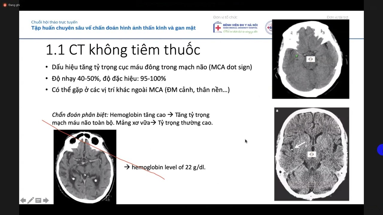
You can see hyperensity inside the middle cable artery. that means it's a thrombus inside the middle cable artery.
Uh the so the thrombus can be identified even in a plain CT. You don't need angiogram to identify thrombus. Even in a plain CT sometimes you can identify the thrombus. It's called 10 MCSI. So that means this right hemisphere is going to have the injury. Uh still it is not infected. The CT brain is still preserved. Aspects is good. You can see the whole right hemisphere here is preserved. Gray white is preserved. No you cannot there are no big hypodensity seen. No hypotensity seen in the cortical areas. So the cortex is largely preserved. So this is a ideal candidate for IV thrombolysis followed by thrombctomy if needed. Suppose IV thrombolysis sometimes IV thrombolysis with IV thrombolysis the patient revascularizes well and good. You don't have to do thrombctomy.
So we did a CT angio. You can see the occluded artery here. Here you can see the internal coroted artery.
Here internal coral artery is going up dividing into ACA and MCA. Here the middle cervical artery on the left side you can see on the right side the middle cervical artery is cut off here you can see the cutff blocked blocked middle cerval artery. So we predicted this in the CT.
So CT itself was showing the right MCA had a that right MCA is occluded angiogram confirmed CT andogram confirmed that the occlusion is there.
Then we went ahead directly for a an angiogram DSA cerebral angiogram intracraanial cerebral angiogram. You can see uh this is the vertebral artery injection. The posterior we assess the posterior circulation. You can see the posterior cerebral artery is collateral giving collaterals to the middle cerebral territory. This is the collateral coming into the middle cery.
That means the middle cerebral artery is surviving on borrowed borrowed collaterals from the posterior cerebral artery. So this collaterals may not last long. So you have to immediately revas.
Some of them are robust collaterals.
This is a mild moderate collateral. So it may not last long. So patient definitely has to be taken up for throbectomy. You can see the occluded artery in the ICA.
[Music] We went in. We took a guide catheter plac in the internal can see the internal coral artery. You can see a tube inside the internal car. This is the guide catheter and through that you took another intermediate catheter into the middle cerebral artery. This is the intermediate catheter. We sucked out the clot.
This is the this is the thrombus which came out and this is the artery which is open. You can see the middle circle artery is completely open. Now the you see the perfusion anterior circle was already open earlier. Here you can see anterior circle was already flowing and it was some of the collaterals were contributing to the middle circle here but MCA was cut off here. Here you can see the same MCA is opened up. Now here this was the site of occlusion. The thrombus came out and the artery started flowing. Patient clinically improved post empty. Another 65 year old male coronary artery disease type 2 diabetes acute left hemi parasis disarthria. NHSS 9. So largest occlusion onset IV license 2 hours 14 mig. Okay. For regarding IV thrombolysis there are two agents which are available for ivy thrombolysis which is approved by the DG the director general of health service India. one uh uh uh DCGA sorry the DCGA India has approved two molecules one is IV altiplace which was initially approved now IV tenective place India was the first country to where connective place was approved uh for use um and tenative place is advantage of tenective place is that we can give a ball full dose uh altiplace you need an infusion regimen so altipl is alultiplace the dose is.9 mig per kilogram tenct place the dose is 025 mig per kilogram but we can give a lower dose connective is also 2 mg per kilogram which is approved by DCTI.
So uh here we give based on the weight body weight 2 mig 025 mg per kilogram based on body weight we gave 40 mg consented for DSA and this was the CT CT aspects is good you can see all the cortical area the gray white area is maintained you don't see any hypodensities gross hypodensities anywhere here maybe a mild hypodensity in the posterior part maybe in the posterior internal capsule here there a small hyper density and the posterior parietal Rest all areas are preserved.
So maybe one one less aspects maybe one less. So one area gone means you subtract one point out of 10. Maybe nine or eight maybe nine. So in this case it was nine. So it was a favorable aspects for a thrombolyis.
This was M1 MCA. You can see the CT angio. This is CT andog showing MCA cut off. Middle cervical artery is cut off here. The left medal artery is completely flowing. Right metal artery is cut off here. This is the internal coral artery. This is the middle circular art which is cut off.
So we assess the collaterals CTO. We took the CTO for collateral. You can see collaterals filling. There are white all these are vascular markings arterial collaterals. So there we understood that there's there are good collaterals. So that this side of the brain right side of the brain is surviving on collaterals that we understood through CTIO. Then we have to go in and head and uh then this is the thrombus. You can see the thrombus in the middle cervical artery.
We went in with the catheter. This is the middle we are in the middle cervical artery. We are in this point now. Here you can see the catheter tip here.
Intermediate catheter here. This is a clot. So we sucked out this clot. We connected it to the now the flow is full. You can see the fully artery is revascularized. The thrombus is completely gone. The flow is reinstituted. M1 thrombus resolved.
6 months followup patient is full.
smoking three uh she had some deficits at the time of leaving because as because there were some hypotens there were some infox which is already established but in 6 months she started walking now bas artery occlusion whatever we have discussed is for the anterior circulation like internal coroted artery middle cerebral artery now for basil artery things are different it's a post the posterior circulation things are different there is no window period for basil artery there is no window period for posterior circulation um even up to 24 hours 36 hours or 48 hours we have done throbectomy with because the purpose is to save life because post basal artery means the patient will be GCS3 and mortality rate will be very high 60 to 70% will die so for the bas artery occlusion we don't look at the window period even at 24 hours we can take up the patient for angiogram and thrombctomy if needed but IV thrombol is we don't give beyond 4.5 to 6 hours. So here also you look at the infodium whether the area is gone or not MRI and CT will give an idea. 69 year old lady with atrial fibrillation presented with progressive giddiness at taxexia. So these are the posterior circulation symptoms. Vertigo, diplopia, atexia, giddiness, hemianopia, homonymous hemianopia that means inability to see on one side of suppose a patient says I cannot see the right side. There was a bluring of vision, blackening on the right side.
Left side I can see but right side I'm not able to see. That is homonyous hemi anopia and patient has a walking difficulties cerebrar type of speech scanning stato like speak speech is like a drunkard with kittiness dipopia tossis there will be bilateral tossis also look for tossis and hemi anesthesia maybe talamus. If thealamus is involved patient will be drowsy also. So all these are pointing towards a posterior circulation stroke.
Uh bas occlusion. So here the NHS was 12.
You can see plain CT even in the plain CT we can identify the thrombus in the basil artery. This is the dense basil sign. The basil artery is thrombos. You can and MR shows a diffusion restriction in the pond area. You can see he left hemipons is having a diffusion area diffusion restricted area and the cerebellum also is having a diffusion uh restriction and here you can see the p posterior communicating artery pon collaterals. So from internal coroted artery there are collaterals going into the basil artery and supplying the posterior cerebral artery territory because basil arteries occluded the collaterals come from the pecom into the bas posterior cerebral artery territory.
So such patients may have may can be salvaged uh if there is a peacock the their the brain brain uh the oxipital area will not be gone and you will get more time up to 24 hours or 36 hours here this patient fortunately had good pe presented at a late window. This this this patient came at around around 18 hours or 24 hours or something one day later. Earlier she was in she was in some other hospital got got transferred to a hospital. Then this is the basil artery which is occluded artery. You can see the occlusion of the artery system.
Uh here through PCOM basil artery is retrograde filling. The ICA you can see this is ICA. Internal cored artery is fine flowing. Right MCA is seen left MCA through aircom left MCA is also filling.
The whole anterior circulation is preserved. Only thing is posterior basil artery is gone.
So we did a suction throctomy and the artery opened up. This is the bas artery. Earlier it was occluded here.
Here you can see after this curve we took the you can see the catheter here catheter and micro catheter and the intermediate catheter. You can see the artery was occluded here at at this point. It was occluded. Now it is revascularized and flow started appearing in the still bas circulation.
So the basal arteries opened up 5 months followup MRS 1. Now she started walking. After 5 months patient had an atrial fibrillation. So patient is on anti-coagulation oral anti-coagulation.
Uh so the for secondary prevention the cause of stroke is has to be completely evaluated. If it is a cardio cardiac source like low ejection fraction atrial fibrillation VT the patient should be put on oral anticoagulation later later on after two weeks. If it is a major stroke we'll start or anticagulation usually after 2 to 3 weeks not early because there is a risk of hemorrhagic transformation. If it is a very large stroke, we wait for one maybe one month.
But secondary prevention has to be initiated in a later date like oral antiquagulation or a dual antiplate and that dual antiplate has to be continued for 3 to 6 months um and then can be converted to a single antiplate.
But if the patient has arithmia and cardiac high risk cardiac risk factors and recurrent stroke then better to continue on dual antiplate and if it is cardiac source better to continue on anticoagulation.
So is there evidence of throbectomy after 6 hours? Up to 6 hours is okay.
After 6 hours is the throbectomy okay.
So there are two trials which help which answered this. One is dawn trial and diffuse trial. So after 6 hours also you can do throctomy up to 24 hours but you cannot the patient selection should be based on MRI MR perfusion and CT perfusion.
As we discussed earlier this is the perfusion scan. This is the penumbra area. This is the gone area. uh this area is gone. This is already a core.
You can see a CBF flow reduction here.
But this yellow area, the whole yellow area, this is salvageable area. Here it is blue on this this side. This is the affected side. Right side is affected side. Left side is the unaffected side.
Here you can see the salvageable area.
So u another example of extended thrombctomy that means late throctomy.
We have done late throbectomy up to 24 hours. Uh this this patient arrived after 10 hours but u somehow got delayed some somewhere in another department and uh we started doing mechanical at around 14 hours 13 to 14 hours. So we lost a lot of time. This was a tribal officer.
Um she developed while she was at work she developed hemoplegia. See she was having rheumatic heart disease severe MS on or anticagulation left hemoplegia and seizures. Um so NIHS was 14 this was the MR for you can see even after 10 hours or 14 hours the whole MR uh brain is preserved only this much area is gone you can see the MR diffusion the whole brain is preserved what is it why is it preserved because of the collaterals the whole collaterals were sustaining that much brain so we had hope after seeing this image we had very we had a good hope that we can revascularize the patient and we save the patient. So we did an an MR angio.
You can see the MCA is flowing on the left side. The right side MCA is cut off. You can see the cut off on the right MCA. This is the right hemisphere.
So this right MCA is cut off here.
So perfusion we did a perfusion scan here. You can see this area is gone.
This black area is gone. On the this is the right hemisphere, left hemisphere.
In the right hemisphere this area is gone. But this red area you see this whole red area this is the salvageable area. This is the area which is under high risk of getting infection. So uh with hours delay or maybe uh next day or the next uh the by next day this whole area will become infected and he she will develop complete hemoplegia and she'll develop going for decompression heavy cranectomy. That area is gone that uh she will become permanently morant.
So we have to save this area because some area is gone but there is a large salvageable area. So we proceeded as we did an angiogram. We found the occlusion.
We took the catheter in we are in the middle cervical artery. Now in this part in the occluded this is the occluded part here. Internal car artery is dividing into med cer. We are this is the occluded part. Here the catheter is inside. You can see the catheter inside the skull. The internal inter intermediate catheter. You can see the stent. This is the stent which we use for retrieving the clots. So we use the stent retriever. Now the artery is fully open. You can see the artery completely opened up. It was occluded here. There was no flow distally here. It was completely occluded here. There is no flow here.
Now the artery started opened up. Artery completely opened up after sucking out the clot. So this area is gone. We predicted that this area will go earlier itself in the by imaging. And this is this much area will be salvaged.
Exactly. That's what happened. This much area got salvaged. This area is gone.
This is the posttop day two. This area is the hypotens area which is gone. And this much area we we could salvage after 14 hours or 16 hours of onset of stroke coming from galur.
So this is a CTN at discharge. You can see the medal artery beautifully flowing and perusing the whole right hemisphere at discharge.
This is the patient. She recovered completely.
So take home message it is not the time which matters it is the tissue which matters. So we need to know whether the tissue is salvageable or not. So imaging matters but uh we need to thrombolysis can be done at the earliest and time because time is brain you have to thrombolize at the earliest if possible if possible within 3 hours. If within 2 hours or 1 hour that is the best for the patient that is called golden hour. that time you will not get uh you cannot uh uh get that result with a thrombolysis done at a later time maybe around 6 hours 8 hours definitely the patient is going to have a residual deficit now is it usually we do bridging thrombolysis as I said that means um we do thrombolysis and then shift the patient for throbectomy this is evidence showing that bridging is helpful all evidence showing bridging is helpful So I hope you got an idea of a brief idea of uh how a schemic stroke is assessed imaging protocol and what is to be done at the earliest uh for a small vessel and for a large occlusion um and the agents which are used are connective place and ultip place for ivy thrombolysis within 4.5 hours 4.5 hours without perfusion you can do only with uh uh with just MRI based on MRI you can do or CT you can based on CT you can do thrombolysis within 3 hours based on MRI you can do up to 4.5 hours or up to uh and based on perfusion scan you can go up to 9 hours even now thrombolysis can be done up to 9 hours based on perfusion scan you have to demonstrate that the large area can be saved and a small area is already core core is formed in a small area when large salvageable area is there then you can thrombolize that patient up to 9 hours. So this is the data available and now there are a lot of trials happening in newer molecules like retip place uh don alpha which is noting to the market.
Thank you everyone for the patient listening.
[Music] >> Thank you Dr. Anand. It is an excellent talk.
>> Thank you. Thank you.
>> Uh am I open?
>> Any questions? I'm ready. I'm ready to take if there are any questions.
>> Uh can am I audible?
>> Yes. Yes, you can. You are audible.
>> Okay. Uh the first question is when a stroke patient come to a peripheral tal hospital, what medication to be given before referral to a stroke center?
>> Yes. Uh good question. If the patient is already on antiplatlets, you need not load the antiplatlets again. But if on dual antiplatlets, suppose the patient is on ecosin or a clo and patient is not on clidog, then you can load the second medication like clidogram 600 migram. You can give uh load the patient and then send to refer to a higher higher center. If the patient is n antipllet nave but for that you need a imaging imaging is a must because you don't know whether you are dealing with a bleed so CT is a must so without take without seeing the CT you cannot even load the patient also >> most of the hospitals we don't have CT so we can only uh determine clinically we suspect a stroke >> yes if it is if there's a strong suspicion of eskeemic stroke that's what if if there is a anticident history of atrial fibrillation cardiac risk factor is eskeemic heart disease patient uh and and there is no typical presentation of that of bleed like there is no uh sudden thunder headache there's no vomiting no raised ICP sudden raised ICP symptoms no anopora no but but large vessel strokes also can mimic some of the bleeds that's where you falter some of the large vessel strokes will look like a bleed in that case you can you need not give anti load antiplate Unless you you can just refer to a higher center where there where there is imaging available and you can load from there also it doesn't matter there is no yeah >> can we give statins >> yes h highdy statins can be given 40 to 80 migram 40 to 80 milligram can be given but never reduce the blood pressures too much because you don't know if it is a large vessel occlusion the patient may be surviving on collaterals and the collaterals are dependent on high blood pressure so if you reduce the blood pressure the collaterals will suffer and that area is going to have a faster infraction. So the progression of the stroke will be fast up to 180 100 BP is okay. 180 110 BP is okay. Uh don't reduce beyond less than 180 in the acute phase.
>> Can we give normal saline to improve the perfusion? Yes, definitely.
Uh even we try noradenile also in some some cases uh when there is a delay and if the blood pressures are too low especially for hemodynamic strokes we do it. Suppose there is a proximal corroted artery critical stenosis and the perfusion is impaired we in such cases to improve the perfusion we give a small dose of noranal so that perfusion improves and we mostly we use normal saline only to improve the perfusion.
Another question is any upper NHS score beyond which thrombolysis is contraindicated.
>> Uh NIHSS score per se is not taken as a guidance for contra for IV thrombolysis.
It is but but CT aspects is taken as a guidance for contra IV thrombolysis. NHS is high NHS just means that it is a large vessel stroke but it can be a bleed also. So that's that's where the fallacy is. Uh you have to rule out the clinical uh features of a bleed. If there is a severe thunderclap, there was a preceding headache followed by sudden collapse and ano sudden rapid development of an isocoria and raised ICP better not to give antipllets also.
But uh NHS high high NHS is not a contra indication. You have to give dual antipllet. You have to load with echosprin and chloropod both before sending to a higher reference center and hydrostatic.
>> What are the complications that can develop after thrombolysis? What is your opinion about thrombolysis at a peripheral institutions where there is no ICU or surgical backup?
>> Uh tricky situation but uh it uh if you have a uh if you can take a CT is mandatory for all tropolises. If if you are starting a IV thrombolysis in a peripheral setup, CT is mandatory. A basic CT machine is sufficient. Not a hi-fi CT machine is not needed. A basic secondhand CT machine is also sufficient.
But that should be there. Uh that assessment in that CT is to rule out hemorrhage once the C and and to know the aspect score. But within 2 hours that is called the golden hour of stroke. one hour one hour or two hour that time cannot be reproduced later. If it one one at one hour and two hour you need not see the aspects also at 1 hour if a patient comes to one within one hour you take the CT you find no infection no hemorrhage nothing you just li the patient that that is going to save that patient u but CT is a must for IV thrombolysis I I believe >> do we have to counel about possibility of decompression cranctomy in thrombolysis to all patient >> that's what I said 6 to 8% is the bleeding risk which in all large major large trials have come which has been uh the uh the bleed rates have been around 6 to 8%. But symptomatic hemorrhage rates as low as 3% is also reported. But better to be uh better to counsel the patient on the higher side like 8% 6% or 8% bleeding risk after thrombolysis in at least 70% of those quagalopathic breeds might need a decompression chemical activity 60 to 70%. If it if it is an elderly person he may survive a decompression because there will be lot of atrophy brain atrophy and brain can accommodate more edema. So he may we have seen many people many people come out without decompression elderly people but young brains usually need decompression because brain will be full and edema can develop rapidly and patient can decelerate uh immediately.
Nowadays it's quite common to see chronic small vessel eskeeia on CT and MRI. Any specific management consideration for this small vessel disease uh per se the management of small vessel disease is uh mainly the main state of treatment will be antiplatlets and risk factor modification. Major thing is risk factor modification. Ask the patient definitely the patient is going to have some sometimes the small vessel disease patient may have only mild cognitive impairment patient will say that I am of late I'm forgetting things off late I'm developing on and off headache I'm forgetting things I'm uh recent memory loss or sometimes there will be transient focal deficit in the form of a facial weakness which just disappeared CT will be showing a diffuse hypodens perventricular hypodensities or some laconar in fox here and there you many times if you will get get a The report says chronic lacon or inox in the bentricular area support white matter area. So all these small vessel disease is due to uh vascular athosclerosis.
Uh sometimes it may be due to lipohilyosis. So vascular the major main state of treatment will be risk factor modification. Control the diabetes control HB1C blood pressure control uh dysipidemia control and if look for other risk factors. If there is a family history, look for hyperomocia, other factor deficiencies and vasculitis or other risk factors also.
>> Thank you. When to start antiplatlet after a post thrombolysis bleed?
>> Usually after 24 hours we take a repeat CT, repeat imaging. Uh and after that repeat CT imaging only we'll start antiplatelets. First 24 hours we don't do any uh we don't give any antipllets after arthropolis but uh loading prior loading uh antipl if an anti if antiplates are loaded prior to the that means the patient from the periphery got received antiplate loading and then reached you your facility then that's not a contra indication for ivy thrombolysis per se even even in a loaded patient we give ivrombolysis but we'll usually go for a low dose ivy thrombolysis in those cases there that because DGCI has also approved 02 mig per kilogram. Usually we give 025 milligram but we go for a lower dose like 02 or less than that.
>> When to start antiplatlet in patients after hemorrhagic transformation >> uh depends on the volume of the hemorrhagic transformation. uh usually moderate hemorrh volume of hemorrhagic transformation as I showed you earlier parangal hematoma in the case of parangal hematoma you delay for maybe around 3 weeks or four weeks you take a CT and once he is resolved then only you will start but for hemorrhagic infox you can start after mild to moderate hemorrhagic infox that means hemorrhagic infact type one mild hemorrhagic in you can start after two weeks and hemorrhagic infact type two maybe after two to three weeks hematoma definitely one after resolution. Okay. And you can start with single antiplatelet and then go for double antiplate not straight away dual antiplatelet. But if the pat the most of the complex situation arises when the patient has a cardiac underlying cardiac pathology like atrial fibrillation cardiac arhythmia low ejection fraction which warrants a anticoagulation then the management becomes tricky because already patient is having healic inf then you have to give anticoagulation otherwise patient will develop a second thrombotism that's called secondary stroke again a secondary stroke will develop another thrombolism from the heart uh that will cause another stroke so the management becomes tricky in hemorrhagic infacts inf patients with the cardiamolism uh the anticoagulation may not be full-fledged anti-coagulation cannot be given for next two two weeks at least.
>> Which all important clinical test we have to do in stroke patients?
>> Um baseline you mean baseline?
>> Which all important clinical test we have to do? It's a query the chat box >> depends on the test depends on uh whether uh if you want to for management purpose you need to do a uh complete blood hemogram uh ptr ag uh >> for the clinical test NHS stroke scale >> clinical test NHS stroke scale okay uh the clinical uh test just on admission what is the NHS is a scale of the patient that that that determines whether you want to treat the patient or not if NH is less than four you don't thrombolize more than four you take the risk of thrombolysis because there is a risk of thrombololis 6 to8% risk of thrombolysis you don't you can take that risk if it if the NH is more than four less than four low NHS strokes you don't usually take you should usually don't thrombolize because the deficit is very less so you just antib and secondary prevention strategy will be sufficient In those cases maybe hearin will be also helpful.
Other clinical the clinical tests include NIHSS and uh um uh then uh ABC definitely ABC for posterior circulation you have to look for ABC because it's a basel stroke the patient is drowsy talamic stroke patient will be drowsy he would have aspirated so ABC would should be made ABC should be documented and uh if there is a desaturation you have to intubate the patient and um The other clinical parameters you have to look for is the localization. Suppose a patient has a hemoplegia and proceeding uh with a gaze deviation. The sight of gaze deviation tells you the sight of stroke. Suppose if it is a left MC in fact the patient will have a gaze deviation towards the left.
Uh if it is an eskeemic stroke in hemorrhage contal forced forced eye deviation can also happen. Supp usually in in a left MCA infant the patient looks towards the side of the infant. So a forced eye deviation that will tell you an idea which side the patient is having stroke with a contal hemipllegia >> and then anoor look for anoria whether the patient is developing rapidly progressive edema and raised ICP symptoms. Then uh definitely NHS includes limb weakness uh facial deviation um uh lower limb weakness conscious state ask you can ask questions to the patient like simple axial commands you can ask lift the limb lift the leg how are you from what is your name you can ask simple commands and look for the response if two at least two response for a prompt uh for a for a normal uh speech For a normal conscious conscious level designation at least two response should be proper and commands also patient should be able to obey two at least two commands to to get a score of two at uh zero.
>> Okay. Another question is when to start heperin in a patient with AF and stroke.
Do we need to start antipllets in hemorrhagic stroke after acute phase?
When to start heparent in a patient with atrial fibrillation and stroke? Do we start antiplatlets in hemorrhagic stroke after acute phase?
>> Yeah, anti-coagulation in acute stroke as I said earlier if it is a large volume stroke involving more than 2/3 of the MC territory or hemisphere then we wait we wait for at least one to two weeks so that the edema results because a hemorrhagic transformation risk is very high and hematoma formation is very high. So we wait for one to two weeks.
um at least but if it is hematoma we wait for for we wait for the for maybe for for around 3 weeks or four weeks it all depends on how how how much there is a sec secondary stroke risk suppose the LV is showing an LV clot echo is showing an LV clot in that case even if there is a hemorrhage we try to start anticagation early maybe we'll wait for one week or 10 days take a repeat CT and then see that hemorrhage is resolved and then we start small dose at least small dose antic vagation because there is always a second uh there is a large LV thrombus waiting there for to go up and cause a second stroke.
Another question is what is the cost of mechanical throctomy?
uh mechan mech the mechanical throattomy see it depends on the institution each institution has a has its own uh you mean if the if the whole cost is uh uh taken up by the patient and if the institution doesn't support uh a mechanical throctomy then the cost can go up very high like around uh it can vary up to four to five lakhs but if the institution supports if there are mechanisms by which Or if the public uh if there is a stroke fund which has been constituted by the institution or by a group or by an NGO or by somebody then the cost of the stroke probectomies can be reduced to a drastically to maybe 1 1.5 lakhs we can complete a procedure less than one lakhs also one lakh also we can do a procedure around each because the cost of the materials has to be bon if the materials are also provided free of cost you can do stroke stroke throctomy free of if the materials are available.
So majority of the hospitals it will be around private hospitals major if if the if the patient is going to bear the whole cost it goes up to 3.5 to five lakhs 3.5 to five lakhs but if there is a mechanism we have a mechanism for for as a stroke fund and we have mechanisms where poor patients and all the affordable patients we have a we can reduce uh there are people who can bear the cost and also and NOS's also who who can bear the cost and reduce the cost to less than 1.5 lakhs Okay, thank you. Another question is loading do in acute thrombotic stroke as a acute coronary syndrome is it needed?
>> Loading dose. Yes, in acute stroke you have to load the patient. If the patient is antiplate nave in eskeemic stroke, if you are suspecting an eskeemic stroke, you can load the patient 300 and 600 global. Yes, you can load. Another question loading dose to of antiplate in antiplated n patients.
So 300 aspirin and 600 mg clidog.
>> Yeah, it depends on the body weight. If the patient is very fragile and less than 45 kg, then you can reduce the dose also because there is a bleeding risk.
Suppose the patient needs an IV thrombolysis in a in the next center.
You you never know it is a largest location or if it is a major stroke and patient reached within window and is and her collateral status is favorable for IV thrombolysis. Brain is sustaining till that point of time core is very small. Pumba salvageable area is big. We can liize the patient in that scenario.
It will be helpful that uh if you have loaded completely for that 45 kg patient that is going to cause a serious threat and problem to the next physician who is going to liice the patient. So in that case you can uh give suboptimal doses also I believe like we do half half loading also sufficient provided the patient is going to get the IV thrombotis and promptly in the next >> what's the duration of treatment in TA and the role of antiplatments in TA?
Yes, as uh I discussed initially in the slides, the TIA any by definition is the focal deficit lasting less than 1 hour.
TA as per uh as such doesn't need any treatment immediately uh warrants treatment immediately but it has to be evaluated immediately for before the next it needs treatment in the sense that you have to start antiplatent you have to start the risk factor modification everything immediately but further evaluation has to be done for TA you cannot take TIA uh just as a minor stroke and leave it alone because that will be a precursor and a warning stroke for the next big catastrophe which is coming in the next few months or few weeks. So the risk stratification of TIA as I discussed earlier is based on certain factors like age more than 60 years, ABCD2 score age more than 60 years, blood pressures, high blood pressures more than 140, 90 and so and uh the focal deficits uh persistent focal deficits like duration of the deficits lasting for around six.
The longer the deficits, if the deficits are lasting longer, maybe deficit lasting one or two, three, four minutes, the ABCD score is less. You can give a score of one. If the if the duration of the focal deficit is lasting 20 minutes, 30 minutes for 40 minutes, then the score is two. You get a higher score. So any score around more than four is definitely warrants immediate attention to the cause of the GA.
Sometimes there will be a large proximal IC stenosis which is there uh which is modifiable risk factor causing a critical stenosis which has led to the TA of the patient. So we need to revascularize that. Suppose there will be a cardiamolic stroke the patient will be in AF a small amus has gone in and got lied and patient recovered but before the next emolis large embolis goes and patient blocks a major vessel we need to identify that atrial fibrillation and treat give oral anticoagulation. So TIA as as such doesn't is not important but TA the cause of the TIA has to be um verified immediate at the earliest and should be rectified but you need definitely to start antiplate therapy at the onset when you see a ta patient with vascular risk factors and you have to risk stratify the patient based on ABC2 score.
Can can we give IV manitol to any stroke patient with a high BP without CT before referral?
>> Uh man per se is not indicated in any acute strokes. Uh but if you are seriously suspecting a bleed a hematoma well and good that you give manitol and then refer. But if you are suspecting an eskeemic stroke there is no point in giving a manitol and referring because anti- edema is not the one which is needed because edema is setting in. It depends on when you are seeing the patient. Suppose you are seeing the patient after 6 hours or 8 hours then definitely man will be helpful because it is a time when edma started starts building up. one dose manitol is doing not going to do half but it is not indicated maybe after seeing the CT the second person may stop the manitol also because it's it's not warranted if there is no edema manitol is has to be used only when there is a edema when there is a crisis u otherwise there is no point in prophylactic use of manito but if you are seriously suspecting a hematoma you or I bleed you can definitely give manitol and for the patient that will be prudent because that will buy more time for the surgeon to decompress the patient also Uh only one more question. Uh the previously asked the difference in loading do between CAD and eskeemic stroke CVA.
>> Difference in loading do antiplated between CAD and CVA.
>> Uh you are more well versed with dealing with all the CADs. So you kindly enlighten me on how much you are giving for CAD.
We are giving 300 mg of uh aspirin and >> 300 300 huh >> 300 300 in n patients.
>> Okay, we can in uh CVA patients you can go give 300 600 that is a proposed loading dose for CVA patients.
>> Okay 300 and 600. Yeah 300 600 you can give full full dose loading. I I told you it depends on the weight also. If you are dealing with a fragile patient 45 kg better give half loading because patient is going to get a IV thrombololis also from the next center.
It should not cause bleeding. If there is no facility of IV thrombolysis anywhere nearby and the patient is in the well in the window period and it is going to take at least 3 4 hours 5 hours by the time they reach hospital you can give full loading otherwise give half loading.
Okay. Questions.
Hello Dr. Vibin.
Thank you Dr. An for enlightening us about the stroke. So we have to identify the stroke early. We
Up Next
6:51
NIH Stroke Scale Exam Demonstration | Neurology
@JumpSimulation
256.5K views•2018-01-12

48:47
Graphic Medicine: Comics for Collaborative Healthcare Communication
@nationalpatientadvocate
189 views•2023-12-04

10:28
Neuroanatomy: Central and Peripheral Nervous System Divisions Explained
@AKLECTURES
136.2K views•2014-09-20

3:05
Stages of Labor and Vaginal Birth | Childbirth Animation
@nucleusmedicalmedia
52.1M views•2017-08-18
Related Study Plans & Knowledge Roadmaps
Structured learning paths in Medicine
medicine
Abnormal LFTs: Diagnostic Workup & Pathology
01Liver Anatomy and Basic Physiology
02Introduction to Liver Function Tests (LFTs)
03Hepatocellular vs. Cholestatic Patterns
+ 3 more learning modules
12h6 modules
Explore Roadmap
medicine
Analyzing Blood Smears: RBC, WBC & Platelets
01Introduction to Blood Components & Smear Preparation
02Normal Morphology of Blood Cells
03Erythrocyte Pathologies & Morphological Variants
+ 2 more learning modules
12h5 modules
Explore Roadmap
medicine
Mechanical Ventilation: Waveforms & Alarms
01Anatomy and Physiology of Breathing
02Introduction to Mechanical Ventilation & Core Modes
03Ventilator Settings and Waveform Interpretation
+ 2 more learning modules
12h5 modules
Explore Roadmap
medicine
Neuro Exam: Localization & Stroke Syndromes
01Foundations of Neuroanatomy and Vascular Supply
02The Neurological Examination: Step-by-Step
03Principles of Neurological Localization
+ 3 more learning modules
18h6 modules
Explore Roadmap