Normal vaginal childbirth consists of three stages: Stage 1 involves cervical effacement and dilation over 12-19 hours, with symptoms including regular contractions, back pain, and possible rupture of membranes; Stage 2 begins at full cervical dilation (10cm) and lasts minutes to 3 hours, where the mother pushes while the baby's head rotates and molds during descent through the birth canal, followed by shoulder delivery; Stage 3 lasts 5-30 minutes as mild contractions help expel the placenta, allowing immediate bonding through skin-to-skin contact and breastfeeding.
Stages of Labor and Vaginal Birth | Childbirth Animation
Added:Deep Dive
Basic anatomy of the female reproductive system, specifically the structure of the uterus, cervix, and birth canal.

L2: Female Reproductive System | Human Reproduction | 12th Class Biology ft. Vipin Sharma #brilix
103.9K
The uterus has three layers: perimetrium (outer), myometrium (middle muscular), and endometrium (inner, sheds during menstruation). The cervix connects uterus to vagina, with cervical canal serving as birth passage. The uterus is pear-shaped, wider at top and narrower at bottom, with fallopian tubes attached at the top.
Click to watch →

Biology Human Reproduction part 5 (Female Reproductive System: uterus, cervix, vagina) class 12 XII
105.6K
The female reproductive system consists of the uterus (pear-shaped muscular organ where fertilized eggs develop into babies over 9 months), cervix (narrow passage connecting uterus to vagina), and vagina (tube-like structure serving as the birth canal and site of sperm discharge); the uterus has three tissue layers—the perimetrium (outer protective layer), myometrium (middle muscular layer that contracts during childbirth), and endometrium (inner lining that thickens to support embryo implantation or sheds during menstruation if no pregnancy occurs).
Click to watch →
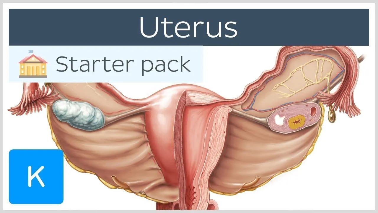
Uterus - Anatomy, Definition and Function - Human Anatomy | Kenhub
538.2K
The female reproductive system consists of interconnected organs working together for reproduction. The uterus, located in the pelvic cavity, is a thick-walled muscular organ shaped like an inverted pear, measuring approximately eight centimeters. It sits between the bladder and rectum, connected inferiorly to the vagina. The uterus divides into four parts: fundus (top portion above uterine tubes), body/corpus (main central section where blastocyst implants), isthmus (narrow constricted region between body and cervix), and cervix (connecting to vagina with internal and external os). Associated structures include the broad ligament (double peritoneal layer containing uterine and ovarian arteries), two uterine tubes (fallopian tubes) carrying eggs from ovaries, and two ovaries producing eggs. The vagina serves as a conduit for menstrual blood, accommodates the penis during intercourse, and serves as the birth canal. Blood supply comes from the uterine artery and ovarian artery branch, with venous drainage via uterine veins. Sympathetic innervation derives from the inferior hypogastric plexus, while parasympathetic supply comes from pelvic splanchnic nerves. Lymphatic drainage varies by region: body and cervix drain to iliac nodes, while fundus drains to para-aortic nodes.
Click to watch →
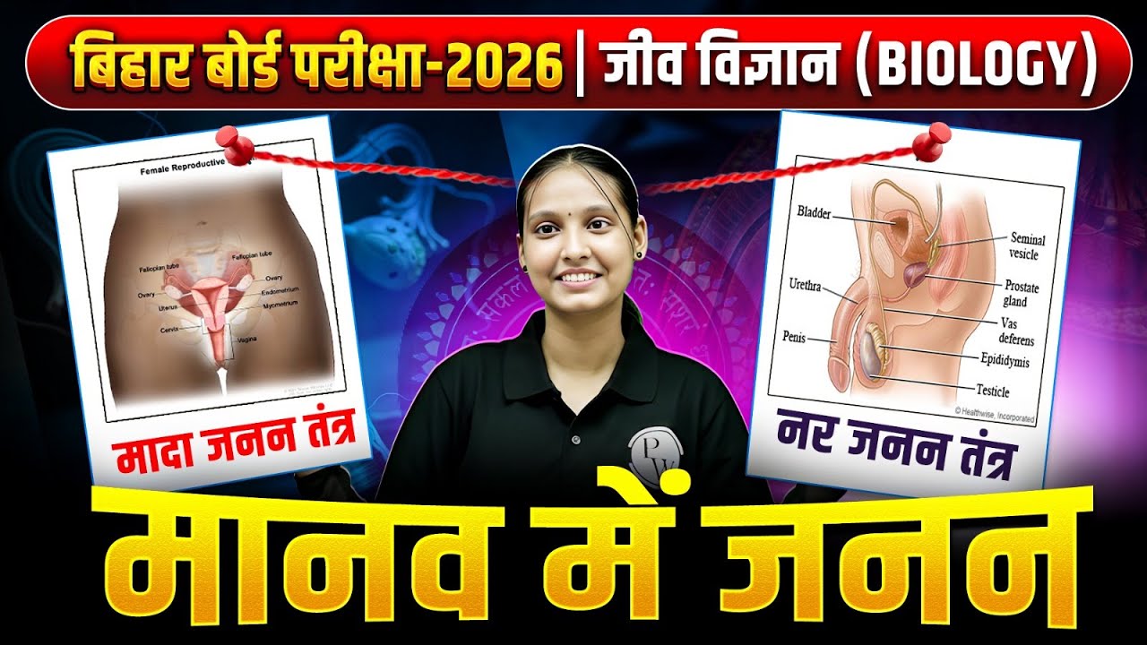
Class 10 Science मानव में जनन | 10th Biology Complete Revision Bihar Board | By Nidhi Mam
79.7K
The uterus (7-8 cm long, 5 cm wide) has three layers: perimetrium (outer), myometrium (middle muscular), and endometrium (inner). The endometrium thickens monthly in preparation for potential implantation. If fertilization doesn't occur, the endometrium breaks down and is shed as menstrual blood. The cervix (2-3 cm long) connects uterus to vagina and can stretch to 10 cm during childbirth. The vagina (8-10 cm long) serves as the birth canal and has slightly acidic pH (3.8-4.5) to prevent infection. The hymen is a thin membrane partially covering the vaginal opening. The external female genitalia (vulva) includes mons pubis, labia majora, labia minora, clitoris, urethral opening, and vaginal opening. The clitoris is homologous to the male glans penis.
Click to watch →

Midwifery And Gynaecological Nursing | AIIMS NORCET 10 | Aarogyam Infinity MCQ Series | Part 2
54.6K
This comprehensive section covers key anatomical concepts in female reproductive system. The cervix:corpus ratio changes through life stages: 1:2 before puberty, 2:1 at puberty, 3:1 during reproductive age, and equal after menopause. The uterine isthmus is 0.5 cm long, bounded by anatomical and histological internal os. The uterus is normally anteflexed and anteverted with 120-degree angle between body and cervix, and 90-degree angle between cervix and vagina. Uterine size is 7.5 × 5 × 2.5 cm (3 × 2 × 1 inch). During pregnancy, uterus length increases five times to 35 cm. The vagina is lined by stratified squamous epithelium, has 45-degree angle with horizontal plane, and pH of 3.8-4.5 maintained by lactobacilli.
Click to watch →
Deep Dive
The physiological role and structure of the placenta, umbilical cord, and amniotic sac during pregnancy.

5 รก
516
The placenta develops from the embryonic chorion and maternal endometrium, containing blood pools that facilitate gas and nutrient exchange between mother and fetus; the umbilical cord contains one large vein and two arteries surrounded by Wharton's jelly, which prevents kinking and maintains blood flow; amniotic fluid, produced from maternal plasma, fetal skin, and fetal urine, maintains fetal temperature, aids lung development, stimulates gastrointestinal development, allows fetal movement, and protects against external impacts, with production peaking at weeks 34-36 of pregnancy.
Click to watch →

Etüt-5 Çocuk Sağlığı Okulöncesi Öğretmenliği
1.9K
During pregnancy, three temporary organs form to support fetal development: (1) Placenta - a temporary organ forming 3-5 months into pregnancy, also called 'çocuğun eşi' (child's partner) because it attaches to the uterine wall behind the fetus; (2) Umbilical cord - connects the fetus to the placenta, enabling nutrient and waste exchange; (3) Amniotic sac - a fluid-filled membrane surrounding the fetus, providing physical protection against external impacts and facilitating childbirth through lubrication. The placenta performs three essential functions: metabolic (antibody transfer for immune support), transport (delivering oxygen and nutrients while removing waste), and hormonal (producing growth hormone and estrogen for fetal development and maternal preparation).
Click to watch →

Class 9 Biology | Annual Exam 2026 - ബയോളജിയിലെ മുഴുവൻ ചിത്രങ്ങളും ഒറ്റ വീഡിയോയിൽ. | Xylem Class 9
79.4K
The placenta is a temporary organ that connects the mother to the fetus, facilitating nutrient and gas exchange. The umbilical cord is the cord-like structure connecting the fetus to the placenta, containing blood vessels that transport oxygen and nutrients to the fetus and return deoxygenated blood. The amniotic sac is a fluid-filled membrane surrounding the fetus, containing amniotic fluid that protects the developing embryo and provides a cushioning environment.
Click to watch →
![[모성간호]3. 태아](https://i.ytimg.com/vi/9wNzxmil1n8/maxresdefault.jpg)
[모성간호]3. 태아
40.3K
This comprehensive section covers the anatomical structures supporting fetal development. The amniotic sac (chorioamnion) consists of three layers: chorion (outermost, maternal-derived), amnion (innermost, fetal-derived), and yolk sac (middle). The placenta forms from chorion and amnion fusion, completing by 3 months, weighing approximately 500g (one-sixth of fetal weight). The placenta performs critical functions: nutrient and oxygen exchange, waste removal, hormone production (hCG, estrogen, progesterone), respiratory support, and immune protection. It acts as a filter, allowing beneficial substances while blocking harmful ones. The umbilical cord connects the fetus to the placenta, approximately 50 cm long, containing three blood vessels: one umbilical vein and two umbilical arteries, surrounded by protective Wharton's jelly. The umbilical vein carries oxygenated, nutrient-rich blood from the placenta to the fetus, while the two umbilical arteries carry deoxygenated, waste-laden blood from the fetus to the placenta. Amniotic fluid is normally clear and colorless, becoming slightly cloudy or amber-colored as pregnancy progresses due to fetal debris. By term, volume reaches approximately 1000cc. Functions include: protecting the fetus from external pressure and trauma, maintaining constant temperature, allowing free fetal movement for physical development, preventing adhesion between fetus and uterine wall, lubricating the birth canal during labor, and cleaning the birth canal to reduce infection risk.
Click to watch →

2026 Okul Öncesi Öabt- Çocuk Sağlığı ve İlkyardım Konu Anlatımı (Part 3)
1.1K
The placenta is a temporary organ that develops in the uterine wall during pregnancy. It facilitates oxygen and nutrient transfer from mother to fetus, removes waste products, produces hormones (estrogen/progesterone), and prevents harmful substances from reaching the fetus. The umbilical cord (55 cm long) connects the placenta to the fetus, containing two arteries and one vein for blood exchange. The amniotic sac contains fluid that protects the fetus from trauma, prevents heat loss, allows movement, and supports symmetrical growth.
Click to watch →
Deep Dive
Endocrine regulation of pregnancy, particularly the roles of hormones like oxytocin, progesterone, and estrogen in initiating labor.

Parturition ( Labor ) In Female Pregnancy || Reproductive Physiology
81.8K
Parturition is governed by hormonal and mechanical factors. Three key hormones influence labor: (1) Estrogen and progesterone - released from corpus luteum and placenta, with estrogen increasing and progesterone becoming constant near term, disrupting the balance and initiating contractions; (2) Oxytocin - released from posterior pituitary, with both release and receptor numbers increasing to stimulate contractions; (3) Fetal hormones (cortisol and prostaglandins) - work with oxytocin to increase uterine contractions. The shift in estrogen-progesterone ratio near term is critical for initiating labor.
Click to watch →

SINH LÝ CHUYỂN DẠ
113
Labor initiation involves complex endocrine changes. Prostaglandins (PGE2, PGF2α) increase progressively during late pregnancy, reaching maximum levels before labor onset, promoting cervical ripening and uterine contraction. Estrogen increases uterine sensitivity to oxytocin and promotes myometrial development. Progesterone withdrawal (relative to estrogen) is a key trigger, with the estrogen:progesterone ratio reaching an optimal threshold to initiate labor. Oxytocin receptors increase progressively during pregnancy, becoming abundant by term. Oxytocin is released in pulsatile patterns, with concentrations peaking during active labor.
Click to watch →

Pregnancy, Parturition & Lactation | Iyliya Khan | ERP 112
107
Estrogen increases uterine blood flow via VEGF (angiogenesis) and nitric oxide (vasodilation). In the myometrium, estrogen increases gap junctions (for coordinated contractions) and progesterone receptor expression (making the uterus responsive to progesterone's relaxing effect). Progesterone maintains pregnancy by keeping the uterus relaxed, transforming the endometrium into a receptive lining, and suppressing myometrial contractility. Progesterone also inhibits lactation during pregnancy by blocking prolactin's effects on breast tissue. Labor initiation begins with fetal maturation: the fetal HPA axis activates, increasing cortisol, which stimulates placental CRH production (positive feedback loop). Cortisol is crucial for fetal organ maturation, especially the lungs. Cortisol increases fetal DHEA sulfate, which the placenta converts to estrogen. This increases the estrogen:progesterone ratio, critical for labor. Estrogen increases oxytocin receptors and prostaglandin production, while progesterone decreases or becomes less effective. Near term, the myometrium undergoes progressive changes: contractility increases due to estrogen, increased oxytocin receptors, and prostaglandins. Excitability increases as the resting membrane potential becomes less negative. Conductivity improves through estrogen-stimulated gap junction formation. The cervix undergoes enzymatic breakdown of collagen fibers due to relaxing factors, prostaglandins, and estrogen, softening and dilating it for fetal passage. Mechanical factors (fetal size and movements) stretch the uterus, activating mechanoreceptors that trigger oxytocin release. Oxytocin receptors increase 200-300 fold during pregnancy, reaching maximum at term, driven by estrogen. This makes the uterus highly sensitive to oxytocin, explaining why synthetic oxytocin is effective for labor induction. Circulating oxytocin levels do not increase until full cervical dilation, indicating labor initiation is due to increased uterine sensitivity rather than increased oxytocin production.
Click to watch →

OBG I Introduction Of Labor l Stage Of Labor l Nursing Officer Online Class l Utkarsh Nursing Live
3.1K
Labor initiation involves a complex endocrine cascade. The baby's hypothalamus releases CRH, stimulating the anterior pituitary to release ACTH, which activates the adrenal glands. The adrenal medulla produces catecholamines (epinephrine and norepinephrine), while the adrenal cortex produces mineralocorticoids, glucocorticoids, and androgens. Glucocorticoids increase glucose and decrease progesterone. Estrogen increases significantly, enhancing oxytocin receptor sensitivity. Prostaglandins synthesized from arachidonic acid facilitate cervical ripening and uterine contractions. Hormones require receptor binding to function.
Click to watch →
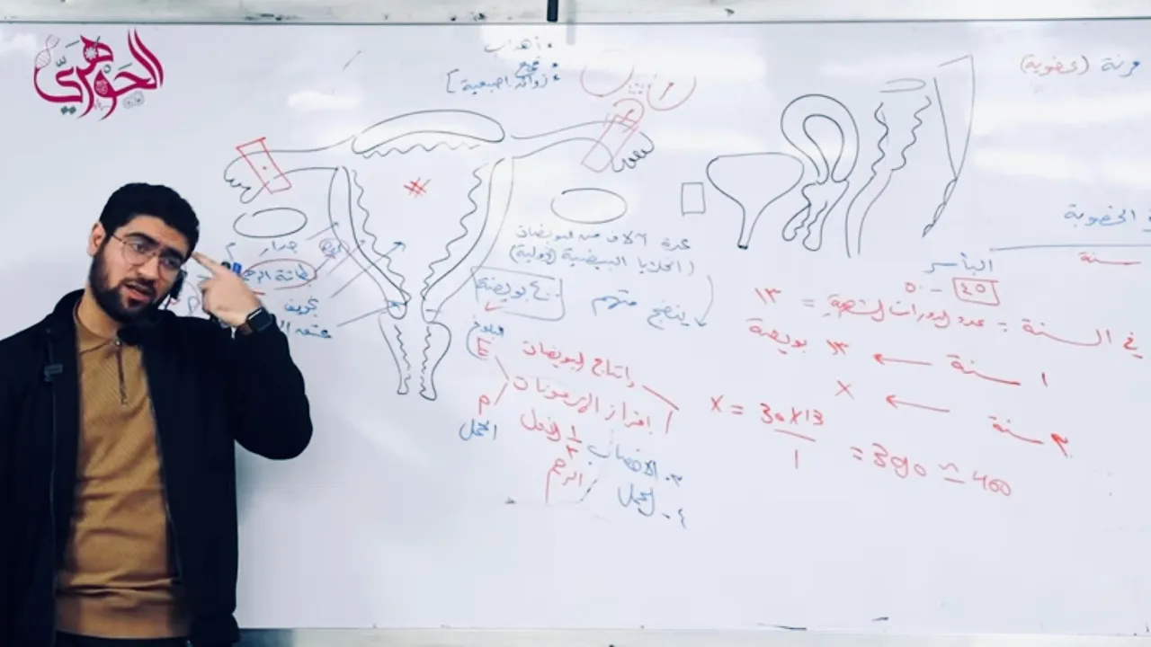
أحياء تالتة ثانوي ٢٠٢٣| الجهاز التناسلي الأنثوي ♥️✅
147.3K
Progesterone suppresses oxytocin production by the posterior pituitary gland. This prevents uterine contractions during pregnancy. When progesterone levels drop at the end of pregnancy, oxytocin can stimulate uterine contractions, initiating labor.
Click to watch →
Deep Dive
Fundamental medical terminology related to pregnancy, such as gestation, contractions, and fetal presentation.

Les termes et expressions liés à la grossesseالمصطلحات والعبارات المتعلقة بالحمل
149
This section covers fundamental medical terminology related to pregnancy and childbirth. Key terms include: pregnancy test (test de grossesse), gynecologist (gynécologue), obstetrician (obstétricien), uterus (utérus), embryo (embryon), fetus (fœtus), placenta (placenta), and the three trimesters (first: weeks 1-12, second: weeks 14-26, third: weeks 27-40). Additional terms cover miscarriage (avortement), contractions (contractions), false contractions (contractions fausses), epidural anesthesia (anesthésie épidurale), natural childbirth (accouchement naturel), premature birth (accouchement précoce), and Cesarean section (césarienne).
Click to watch →
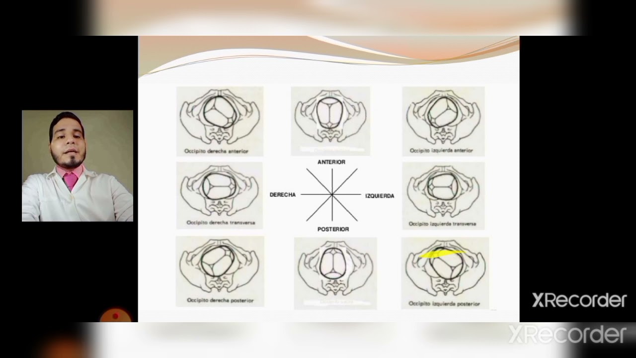
Trabajo de parto: PRESENTACIÓN CEFÁLICA DE VÉRTICE.
272
This comprehensive section covers the foundational concepts of labor and fetal presentation. Labor is defined as the physiological phenomena for delivering a viable fetus. Key concepts include fetal presentation (part of ovum contacting superior stricture), situation (relationship between fetal, uterine, and maternal axes), and position (relationship between guiding point and maternal pelvis). The three periods of labor are dilation (latent and active phases), expulsion, and placental expulsion. Fetal diameters include transverse (biparietal 9.5 cm, bitemporal 8.5 cm) and longitudinal (suboccipitofrontal 9.5 cm, suboccipitobregmatic 10.5 cm). Presentation varieties include vertex (most automatic), brow, face, and frontal. Position classification uses the occiput as reference with varieties like right anterior, left posterior. Diagnosis involves Leopold's maneuvers and auscultation. The most frequent position is occiput left anterior (60% of cases).
Click to watch →

Pregnancy Terminology with Dan & Phil! | MOTHERHOOD
2.1M
Pregnancy involves several key biological processes and terminology. Gestation is the nine-month period of development inside the womb between conception and birth, where the fertilized egg transforms into a baby. The placenta develops in the uterus as an organ that provides oxygen and nutrients to the growing fetus while removing waste products. Fundal height measures the distance from the pubic bone to the top of the uterus, correlating with fetal growth and increasing approximately two inches monthly. Understanding these foundational concepts helps demystify pregnancy for those unfamiliar with reproductive biology.
Click to watch →

محاضرة تمريض نسا terminology of obstetrics and gynecologyفرقة ثانيه
7.2K
This section covers fundamental pregnancy terminology: Fertilization is the union of sperm and egg. Gestational age is calculated from the first day of the last menstrual period, with full-term pregnancy being 40 weeks. Gravida refers to total pregnancy count, while Para indicates number of births. Primipara is a woman who has given birth once, and Multipara has given birth two or more times. Stillbirth is delivery of a dead fetus after 28 weeks, while abortion occurs before 28 weeks. The antepartum period spans from conception to labor onset.
Click to watch →

Module 8, Terminology of Woman's Health, Obstetrics and Gynaecology
370
Pregnancy terminology includes gestation (time from last menstrual period to birth, normally 37-42 weeks), trimesters (first: weeks 1-13, second: weeks 14-27, third: weeks 28-42), fundal height (FH) measurement from pubic bone to uterus top, gravida (pregnancy count), and para (birth count). Ultrasounds assess fetal development and health. Ectopic pregnancy occurs when fertilized egg embeds in fallopian tube instead of uterus. Rubella status checks immunity to German Measles. Proteinuria checks for preeclampsia (high blood pressure condition). Fetal heart rate (FHR) monitors baby health.
Click to watch →
Prerequisite Knowledge
- Concept 01Basic anatomy of the female reproductive system, specifically the structure of the uterus, cervix, and birth canal.
- Concept 02The physiological role and structure of the placenta, umbilical cord, and amniotic sac during pregnancy.
- Concept 03Endocrine regulation of pregnancy, particularly the roles of hormones like oxytocin, progesterone, and estrogen in initiating labor.
- Concept 04Fundamental medical terminology related to pregnancy, such as gestation, contractions, and fetal presentation.
Subsequent Learning
- Step 01Common obstetric interventions, including labor induction, epidural anesthesia, assisted deliveries (forceps/vacuum), and Cesarean sections.
- Step 02Potential complications of labor and delivery, such as shoulder dystocia, breech presentation, fetal distress, and postpartum hemorrhage.
- Step 03Immediate postpartum care and assessment of the newborn, including the APGAR scoring system and the process of uterine involution.
- Step 04Pharmacological and non-pharmacological pain management strategies used during the active stages of labor.
First Stage
Delivery
First Stage
0:05Playing Section
- 1
Early labor involves cervical effacement and dilation over 12 to 19 hours.
- 2
Signs include regular contractions, back pain, mucus plug, and water breaking.
- 3
Contact medical staff when symptoms arise to plan hospital arrival.
The Physiological Model and Critique of the Friedman Curve
While standard obstetric education relies on rigid, chronologically-timed 'stages of labor'—traditionally dictated by the Friedman Curve—critics argue this medicalized framework pathologizes normal variations in childbirth. Modern midwifery and progressive obstetrics advocate for the 'Physiological Model of Care.' This perspective views labor as a highly individualized, non-linear process influenced by hormonal, emotional, and environmental factors rather than a strict mechanical timeline of cervical dilation. Critics contend that adhering strictly to standardized stages often leads to unnecessary interventions, such as synthetic oxytocin administration or unplanned cesarean sections, when a birthing person's natural rhythm simply falls outside the statistical average.
Deep Dive
Common obstetric interventions, including labor induction, epidural anesthesia, assisted deliveries (forceps/vacuum), and Cesarean sections.

PODCAST PUNTO ENARM CAP 4/30. PARTO PRETERMINO/ PARTO/ RPM/ HEMORRAGIAS PUERPERALES
8.1K
Labor induction indications include lack of progression (no dilation after 2 hours), gestational hypertension, post-term pregnancy, and infections. Contraindications include pelvic/transverse presentation, uterine scar, cord prolapse, and active STIs. Bishop Score >6 indicates favorable cervix for oxytocin induction; ≤6 requires ripening (misoprostol, membrane sweeping). Instrumental delivery with forceps requires third plane of Hodge: Simpson for occiput posterior, Piper for cephalic presentations. Cesarean section types: classical (vertical, only for placenta previa/cervical cancer), transverse/low segment (preferred for most cases). Absolute indications include repeat cesarean after classical (uterus must be cesarean again), repeat after two transverse cesareans, pelvic presentation, cephalopelvic disproportion, hydrocephalus, third-trimester hemorrhage, active STIs, HIV >1000 copies/mL, and twin pregnancy.
Click to watch →

Obstetrícia - Esquenta ENARE 2022
4K
This segment covers labor management, cesarean section, and labor induction. Benefits of normal vaginal delivery include reduced risk of hemorrhage, infection, thromboembolism, postpartum depression, placenta accreta in subsequent pregnancies, respiratory distress syndrome, NICU admission, breastfeeding difficulties, and adult life complications. Labor admission criteria require active phase of labor (cervical dilation ≥5 cm with regular contractions 3-4 per 10 minutes) and multiprofessional team assessment. The four clinical phases of labor are: first phase (dilation), second phase (expulsion), third phase (placental), and fourth phase (puerperal). WHO recommendations for first stage include fetal heart monitoring every 15-30 minutes, vaginal examination every 4 hours, offering pain relief options, allowing oral intake, and encouraging mobility. Contraindicated: routine episiotomy, routine oxytocic use, routine tocolytic use, routine intravenous fluids, routine pelvic measurements, routine fetal scalp electrode, routine amniotomy, and routine internal monitoring. WHO recommendations for second stage include duration limits (<2 hours with epidural, <3 hours without), fetal heart monitoring every 5 minutes, perineal massage and warm compresses, spontaneous pushing, and upright positions. Contraindicated: routine episiotomy and Kristeller's maneuver. Third stage recommendations include universal oxytocin 10 IU IM after delivery of anterior shoulder, controlled cord traction, delayed cord clamping (at least 1 minute), uterine massage only if needed, systematic placental examination, and skin-to-skin contact. Perineal lacerations are classified by depth: first-degree (mucosa and muscle), second-degree (external anal sphincter), third-degree (rectal mucosa), and fourth-degree (rectal mucosa). The fourth stage requires maternal vital signs monitored every 15 minutes for the first hour after placental expulsion. Pain management includes offering both pharmacological and non-pharmacological methods, with regional anesthesia (spinal or epidural) as the gold standard that can be administered at any stage of labor. Shoulder dystocia management follows the mnemonic ALERT: call for help, McRoberts maneuver (hip hyperflexion), suprapubic pressure (Rubin's maneuver), episiotomy, and internal maneuvers (Woods screw, Rubin's II, Zavanelli). Umbilical cord prolapse requires emergency cesarean section. Cesarean section absolute indications include placenta previa, vasa previa, cord prolapse, uterine rupture, acute fetal distress, active genital herpes infection, previous classical cesarean section or uterine surgery, and maternal death with live fetus. Relative indications include multiple gestation, maternal medical conditions, macrosomia, malpresentation, prolonged second stage, cervical cancer, multiple previous cesarean sections, and urinary/fecal incontinence. Complications include increased risk of postpartum hemorrhage, uterine atony, infection, urinary tract injuries, hematoma, seroma, uterine dehiscence, placenta accreta in subsequent pregnancies, uterine rupture, ectopic pregnancy, incisional hernia, and chronic pelvic pain. Labor induction uses Bishop score to evaluate cervical readiness (≥6 favorable, <6 unfavorable). Methods for cervical ripening include mechanical (balloon catheter, membrane stripping) or pharmacological (prostaglandins, oxytocin, relaxin, nitric oxide). Prolonged pregnancy management: gestation ≥42 weeks is prolonged; from 40 weeks, begin fetal vitality assessment; from 41 weeks, consider induction; from 42 weeks, all fetuses must be delivered.
Click to watch →

2 Parto distocico y enfermedades intercurrentes
5.2K
Labor induction methods include Hamilton maneuver (membrane stripping), misoprostol (prostaglandin E1 analog) for cervical ripening, and amniotomy in active labor. Oxytocin infusion follows cervical ripening. Indications include post-term pregnancy (>42 weeks), preeclampsia without severe features at term, and fetal death. Contraindications include acute fetal hypoxia, placenta previa, prior myomectomy, prior cesarean section (especially classical), active genital herpes, and advanced cervical cancer. Instrumental delivery (vacuum/forceps) is indicated to shorten second stage for maternal (exhaustion, cardiac disease, preeclampsia) or fetal (distress) indications. Conditions include complete dilation, ruptured membranes, cephalopelvic compatibility, flexed head, and empty bladder. Cesarean section is indicated for emergency (fetal distress, abruption, cord prolapse), elective (podalic, transverse lie, cephalopelvic incompatibility, prior myomectomy, prior cesarean), and HIV with high viral load (>1000 copies/mL). Antibiotic prophylaxis with cefazolin 1 hour before procedure prevents infection.
Click to watch →

Obstetrics and gynaecology nursing questions MCQS
181.8K
This section explains labor and delivery mechanisms: effacement is cervical thinning during labor; oxytocin initiates contractions; identical twins result from a single fertilized egg splitting; hysterectomy is surgical uterus removal; assisted delivery uses forceps or vacuum extractors; cesarean section involves surgical incisions for childbirth; and miscarriage (spontaneous abortion) is pregnancy loss before 20 weeks. These processes are fundamental to obstetric care.
Click to watch →

दवाई से दर्द क्यों और कैसे चालू करते हैं ARTIFICIAL LABOR PAINS (HINDI)
799.8K
Labor induction carries certain risks compared to spontaneous labor. These include increased fetal heart rate variability, higher likelihood of instrumental delivery (forceps or vacuum), and increased probability of requiring cesarean section.
Click to watch →
Deep Dive
Potential complications of labor and delivery, such as shoulder dystocia, breech presentation, fetal distress, and postpartum hemorrhage.

Women's Health Obstetrics EOR Review
49.5K
Breech presentation is when baby is not head first (3-5% of term pregnancies). Types: Complete breech (hips and knees flexed, butt presents first); Incomplete/footling breech (one or both hips not flexed, foot presents first); Frank breech (hips flexed, knees extended, butt presents); Shoulder presentation (transverse lie, shoulder presents). Diagnosis by physical exam confirmed with ultrasound. External cephalic version may be attempted at or near term to manually manipulate fetal position. C-section considered if unsuccessful. Labor dystocia is abnormal labor progression causing fetal distress, maternal infection, uterine rupture, or postpartum hemorrhage. Categories: Power problems (uterine contractions), Passenger problems (fetal size/position), Passage problems (birth canal). Shoulder dystocia occurs when shoulders lodge at pubic symphysis after head delivery; presents with turtle sign (retraction of delivered head). Management: McRoberts maneuver (hyperflex maternal hips/knees + suprapubic pressure), Woods corkscrew maneuver (rotate shoulders 180 degrees), or C-section if unsuccessful.
Click to watch →

High Yield OB/GYN Review for Step 2 CK & Shelf Exam (Part 1 of 2)
537.6K
Postpartum hemorrhage (>500 mL vaginal, >1 L cesarean) is most commonly caused by uterine atony, where weak uterine contractions fail to clamp spiral arteries, causing hemorrhage. Diagnosis includes a uterus above the umbilicus. Management begins with uterine massage and oxytocin; retained placenta requires D&C, while lacerations (associated with forceps delivery) need surgical repair. The active phase of labor begins at 6 cm dilation; arrest is defined as no dilation for 4 hours with adequate contractions or 6 hours with inadequate. Adequate contractions exceed 200 Montevideo units in 10 minutes. Arrest of descent (>3 hours no progress despite full dilation) is most commonly caused by cephalopelvic disproportion. Shoulder dystocia occurs when the anterior shoulder becomes stuck behind the pubic bone; the McRoberts maneuver (hip hyperflexion) should be attempted first. Risk factors include prior shoulder dystocia, macrosomia (gestational diabetes), and pregestational diabetes. Complications include Erb's palsy (C5-C6 nerve injury) and clavicular fracture, both typically resolving spontaneously.
Click to watch →

Miscllaneous(abnormal labour) المحاضرة الثامنة والعشرون
162
Precipitous labor is characterized by extremely rapid contractions occurring every minute, with the baby being expelled like a rocket within 4 hours. This occurs when there is no obstruction and contractions are extremely strong. Shoulder dystocia is a critical complication where the baby's head emerges but shoulders become stuck. The 'turtle sign' describes the phenomenon where the baby's head retracts back into the pelvis after delivery, indicating shoulder obstruction. Risk factors include multiparity (wider pelvis) and macrosomia (large baby). Postpartum hemorrhage is classified into Tonic PPH (painful, excessive contractions) and Traumatic PPH (painless bleeding from lacerations). Uterine inversion is a rare but life-threatening complication where the uterus turns inside out, occurring when the placenta is attached to the lower uterine segment and the uterus contracts before placental delivery.
Click to watch →

Labor & Delivery Nurse + ER Doctor React to WILD Birth Scene from The Pitt On HBO Max!
70.5K
Shoulder dystocia is an obstetrical emergency where the baby's shoulders become stuck behind the mother's pelvic bone after the head delivers, requiring immediate interventions like the McRoberts maneuver (lifting the mother's knees to her chest) and suprapubic pressure to free the shoulders; postpartum hemorrhage (PPH) is excessive bleeding after childbirth that can be managed with uterotonics (oxytocin, carboprost, misoprostol), Bakri balloon placement, and potentially hysterectomy if conservative measures fail.
Click to watch →

OBS-GYNE WRAP UP, MCQ Based Discussion Dr Shonali Chandra @medicinedecoded.
10.9K
This section covers postpartum hemorrhage complications and shoulder dystocia management. Postpartum hemorrhage complications include hematoma, uterine inversion, and amniotic fluid embolism. Hematoma can occur in the broad ligament, causing deviation of the uterus. Uterine inversion is a medical emergency requiring immediate intervention including manual repositioning of the placenta, fluid resuscitation, and oxytocic therapy. Amniotic fluid embolism is a rare but serious complication presenting with hypotension, dyspnea, and cardiovascular collapse, often followed by disseminated intravascular coagulation. Shoulder dystocia is a complication where fetal shoulders become impacted behind the pubic symphysis. Management includes McRoberts maneuver (flexing thighs on chest and abducting thighs), suprapubic pressure, and internal maneuvers if needed. McRoberts maneuver increases the anteroposterior diameter of the pelvis and rotates the symphysis pubis. Cord prolapse is a medical emergency requiring immediate intervention including manual elevation of the presenting part, tocolytics to arrest labor, and immediate cesarean section. Cord prolapse is associated with high fetal mortality if not managed promptly.
Click to watch →
Deep Dive
Immediate postpartum care and assessment of the newborn, including the APGAR scoring system and the process of uterine involution.

Obstetrics and Pregnancy Rapid Review Nursing with Kendall Wyatt MD, RN
118.3K
This final section addresses newborn assessment, immediate postpartum care, and long-term maternal health considerations. The APGAR score evaluates newborn vitality at 1 and 5 minutes post-birth using five parameters: Appearance, Pulse, Grimace, Activity, and Respirations. Comprehensive newborn assessment includes head circumference measurement (tracks brain development), fontanelle assessment, weight measurement (identifies macrosomia >4,000g), temperature assessment, umbilical cord evaluation (two arteries, one vein), hip abduction/adduction testing (screens for developmental dysplasia), and patent anus assessment. Normal neonatal findings include vernix caseosa (white cheesy coating), lanugo (fine downy hair), and caput succedaneum (scalp swelling from vaginal pressure). Postpartum hemorrhage is the leading cause of maternal mortality. Assessment begins with fundal massage to stimulate uterine contractions and compress blood vessels at the placental site. A boggy, soft uterus indicates inadequate contraction and potential hemorrhage. Oxytocin administration stimulates uterine contractions. Sheehan's syndrome is pituitary necrosis resulting from severe postpartum hemorrhage, causing loss of all pituitary hormone production requiring lifelong hormone replacement therapy.
Click to watch →

OBG + Maternity Complete Marathon | All MCQs + PYQs | HSSC Staff Nurse | MPHW | ANM 2026
130
This section covers postpartum care including Apgar score assessment at 1 and 5 minutes (Appearance, Pulse, Grimace, Activity, Respiration), and the components of the Apgar score. It covers the timing of quickening (16-20 weeks, earlier in multigravidas) and the importance of early newborn assessment.
Click to watch →
Assistência de Enfermagem ao Recém-Nascido: Protocolos Atualizados 📚✅ Aula completa
186.6K
Immediate newborn care encompasses interventions performed in the delivery room and up to 2 hours after birth. Key components include maintaining body temperature between 36.5-37.5°C with room temperature at 23-26°C, ensuring airway patency by checking for secretions without neck manipulation, and positioning the newborn on the mother's abdomen for at least 1 minute or until cord pulsation ceases. The APGAR score evaluates five parameters (heart rate, respiratory effort, muscle tone, reflex irritability, skin color) at 1 and 5 minutes after birth, with each parameter scoring 0-2. Scores of 0-3 indicate severe distress, 4-6 moderate distress, and 7-10 good adaptation. If the 5-minute score is less than 7, assessment should repeat every 5 minutes until 20 minutes of age. The APGAR score evaluates response to resuscitation but does not determine the need for resuscitation.
Click to watch →

ObGyn Reacts to Birth on “The Most Accurate Medical Drama” | The Pitt Ep. 11
508.8K
This segment addresses newborn care and postpartum complications following difficult deliveries. Key concepts include: (1) Newborn resuscitation: Drying, stimulating, and providing oxygen before considering intubation; (2) APGAR scoring: 1-minute score indicates immediate status, while 5-minute score predicts long-term outcomes; (3) Postpartum hemorrhage risk: Shoulder dystocia significantly increases hemorrhage risk due to uterine trauma; (4) Massive transfusion protocol: Activated for severe hemorrhage involving blood products in calculated ratios; (5) Tranexamic acid: Originally developed for trauma, now standard for postpartum hemorrhage management. These elements demonstrate the continuum of care from delivery through postpartum recovery.
Click to watch →

S1.Ep76: Îngrijirile imediate ale nou-născutului la naştere |Scorul APGAR |Bontul Ombilical |Analize
58.4K
Immediately after birth, the neonatologist performs the APGAR score assessment (evaluating Appearance, Pulse, Grimace, Activity, and Respiration at 1 and 5 minutes) to evaluate the newborn's adaptation to extrauterine life, while also providing essential care including skin-to-skin contact, umbilical cord clamping and stump care, and initial health screening for congenital anomalies.
Click to watch →
Deep Dive
Pharmacological and non-pharmacological pain management strategies used during the active stages of labor.

Обезболивание схваток при родах. Анальгезия © Pain during childbirth, Analgesia
233.9K
This comprehensive segment covers the progression of pharmacological pain management throughout labor. In early labor with normal blood pressure, sedatives like Seduxen (ataractic group) are administered intramuscularly or intravenously. Spasmolytics such as Aprophen and No-Shpa provide cervical relaxation while enhancing uterine contractions. At 4+ cm dilation, ataralgesia combines Seduxen with Promedol. For exhausted laborers with reduced contractions, steroid anesthesia using Viadril induces sleep while maintaining uterine activity. Premedication with Promedol and Dimedrol ensures analgesic effects. Continuous fetal monitoring is essential throughout. The segment emphasizes that intravenous analgesics should be avoided near delivery to prevent neonatal respiratory depression.
Click to watch →

OBG Drugs for NCLEX RN | Obstetrics and Gynecology Explained for Nursing Students
4.6K
Non-pharmacological methods include deep breathing exercises, sacral counterpressure for back pain, hydrotherapy, and TENS therapy. Pharmacological methods include: (1) Nitrous oxide (laughing gas) - administered via oxygen mask, few side effects but limited effectiveness for intense pain; (2) Opioids (morphine, fentanyl, meperidine) - effective but cause respiratory depression in mother and baby. Best time for opioids is active labor phase (4-5 cm dilation), not too early (stops labor) or too late (fetal respiratory depression). Antidote is naloxone.
Click to watch →

Labor and Birth
733
Non-pharmacologic pain management includes therapeutic touch, massage, water therapy, position changes, and controlled breathing. The gate control theory explains how stimulating positive chemoreceptors minimizes pain perception. Pharmacologic options include IV medications (morphine, Stadol, fentanyl) and epidurals (bupivacaine and fentanyl in epidural space). Epidurals carry hypotension risks affecting placental perfusion. Pudendal blocks provide targeted perineal pain relief for unmedicated mothers near delivery.
Click to watch →

Labour analgesia - Prof Dr Anupama Dave
290
Non-pharmacological methods complement pharmacological treatment and are widely used, with nearly 73% of women using at least one method. Leading methods include breathing exercises (48%), position changes (40%), massage (22%), and relaxation techniques like music and aroma therapy (21%). Available positions include standing, sitting, birthing ball, squatting, kneeling, peanut ball, semisitting, side-lying, hands and knee, and tuck positions. Relaxing techniques like yoga, hypnosis, and breathing exercises reduce blood pressure and breathing rate. Manual methods include massage, reflexology, shiatsu, and warm/cold packs. Acupuncture addresses energy imbalances. The birthing ball enables pelvic rotation movements. Transcutaneous electrical nerve stimulation (TENS) blocks pain signals and releases endorphins. These methods increase well-being and emotional comfort, though high-quality evidence supporting their efficacy remains limited.
Click to watch →

OB Professor Podcast Three
140
True labor contractions should change the cervix; if not, it's not real labor. Pain management options include epidurals (most effective but can cause hypotension requiring fluid bolus before administration), systemic medications, and non-pharmacological methods (breathing techniques, aromatherapy, hydrotherapy, sacral counterpressure). Medication timing is important—giving drugs too late (at 9 cm) can cause respiratory depression. Non-pharmacological methods can complement or precede pharmacological interventions.
Click to watch →
First Stage
Delivery
First Stage
0:05- 1
Early labor involves cervical effacement and dilation over 12 to 19 hours.
- 2
Signs include regular contractions, back pain, mucus plug, and water breaking.
- 3
Contact medical staff when symptoms arise to plan hospital arrival.
The Physiological Model and Critique of the Friedman Curve
While standard obstetric education relies on rigid, chronologically-timed 'stages of labor'—traditionally dictated by the Friedman Curve—critics argue this medicalized framework pathologizes normal variations in childbirth. Modern midwifery and progressive obstetrics advocate for the 'Physiological Model of Care.' This perspective views labor as a highly individualized, non-linear process influenced by hormonal, emotional, and environmental factors rather than a strict mechanical timeline of cervical dilation. Critics contend that adhering strictly to standardized stages often leads to unnecessary interventions, such as synthetic oxytocin administration or unplanned cesarean sections, when a birthing person's natural rhythm simply falls outside the statistical average.
Basic anatomy of the female reproductive system, specifically the structure of the uterus, cervix, and birth canal.
The physiological role and structure of the placenta, umbilical cord, and amniotic sac during pregnancy.
Endocrine regulation of pregnancy, particularly the roles of hormones like oxytocin, progesterone, and estrogen in initiating labor.
Fundamental medical terminology related to pregnancy, such as gestation, contractions, and fetal presentation.
Common obstetric interventions, including labor induction, epidural anesthesia, assisted deliveries (forceps/vacuum), and Cesarean sections.
Potential complications of labor and delivery, such as shoulder dystocia, breech presentation, fetal distress, and postpartum hemorrhage.
Immediate postpartum care and assessment of the newborn, including the APGAR scoring system and the process of uterine involution.
Pharmacological and non-pharmacological pain management strategies used during the active stages of labor.
52.1M views244.9Klikes3:05@nucleusmedicalmediaOriginal Release: 2017-08-18
during a vaginal childbirth the first stage of Labor lasts about 12 to 19 hours and starts when your baby settles lower into your pelvis in response your cervix begins to efface or become thinner and dilate or widen during this time you may feel strong regular contractions occurring every 5 to 20 minutes and lower back pain and cramping that doesn't go away you may see a brownish or reddish mucus discharge which could be the mucus plug at the opening of your cervix falling out your water may break which can either be a large gush of fluid or a continuous trickle If you experience any of these symptoms contact your doctor or Midwife to see if you should go to the hospital at the beginning of stage two of Labor which can last from a few minutes to 3 hours your cervix is is fully dilated to 10 cm and your baby's head has moved beyond the cervical opening into your birth canal during this stage you will begin to push your baby out you may instinctively push when you feel the urge or you may need coaching from your doctor Midwife or labor nurse as to when to push and when to rest in a normal delivery your baby's head will rotate to face your back during active labor your uterus is is divided into an active segment that contracts pushing the baby downward and a flexible passive segment that remains relaxed stretching to provide more room for the baby to pass through in some cases when the top of your baby's head appears or crowns your doctor may make a small cut called an otomy to enlarge the vaginal opening then you will continue pushing your baby out as your baby's head passes through the birth canal it molds into an elongated shape an elongated head shape will resolve itself within a few days as the skull bones shift back into place after your baby's head exits the birth canal his or her head and shoulders will rotate to help the shoulders pass through the birth canal your baby's shoulders are delivered one after the other in order to fit through your pelvis once the shoulders emerge the rest of your baby slides out easily after your baby is born his or her umbilical cord will be cut in stage three of Labor which may last 5 to 30 minutes mild contractions will help push the placenta out of the uterus during this stage you and your baby May begin bonding through skin-to-skin contact and breastfeeding
Up Next
14:36
OB Assessment, Delivery, and Abnormal Presentations: EMT Guide
@ChicagoEMTTraining
3.3K views•2020-07-04

48:47
Graphic Medicine: Comics for Collaborative Healthcare Communication
@nationalpatientadvocate
189 views•2023-12-04
5:42
COVID-19 mRNA Vaccine: Mechanism and Immune Response
@nucleusmedicalmedia
1.4M views•2021-08-03

19:45
The Science of Fentanyl Addiction and Overdose Risks
@theanatomylab
9.1M views•2022-09-30
Related Study Plans & Knowledge Roadmaps
Structured learning paths in Medicine
medicine
Abnormal LFTs: Diagnostic Workup & Pathology
01Liver Anatomy and Basic Physiology
02Introduction to Liver Function Tests (LFTs)
03Hepatocellular vs. Cholestatic Patterns
+ 3 more learning modules
12h6 modules
Explore Roadmap
medicine
Analyzing Blood Smears: RBC, WBC & Platelets
01Introduction to Blood Components & Smear Preparation
02Normal Morphology of Blood Cells
03Erythrocyte Pathologies & Morphological Variants
+ 2 more learning modules
12h5 modules
Explore Roadmap
medicine
Mechanical Ventilation: Waveforms & Alarms
01Anatomy and Physiology of Breathing
02Introduction to Mechanical Ventilation & Core Modes
03Ventilator Settings and Waveform Interpretation
+ 2 more learning modules
12h5 modules
Explore Roadmap
medicine
Neuro Exam: Localization & Stroke Syndromes
01Foundations of Neuroanatomy and Vascular Supply
02The Neurological Examination: Step-by-Step
03Principles of Neurological Localization
+ 3 more learning modules
18h6 modules
Explore Roadmap