Managing Obstetric Emergencies: ER & EMS Basics
Learning Goal: This curriculum is designed to equip emergency department clinicians, pre-hospital providers (EMS), and first responders with the vital knowledge and physical skills necessary to triage, assess, and stabilize pregnant patients, manage critical delivery complications, and perform immediate neonatal resuscitation in non-obstetric settings.
- Prerequisites: Basic Life Support (BLS) or Advanced Cardiovascular Life Support (ACLS) certification is recommended.
- Estimated Study Time: 12 Hours
Module 1: Pregnancy Anatomy and Physiology Basics
This module establishes the core foundation of maternal-fetal anatomy and the profound physiological changes that occur during a normal pregnancy. Understanding these baseline shifts is crucial for identifying when a pregnant patient's vital signs or physical presentations deviate into pathology.
Recommended Videos
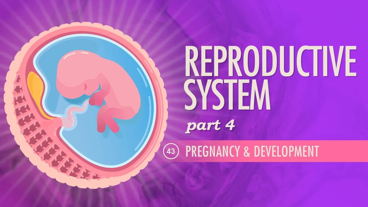
- Why this video: This video offers a highly visual, fast-paced overview of how the uterus expands to fill the abdominal cavity, displacing maternal organs. It clearly explains the development of the placenta and umbilical cord, setting a strong anatomical baseline for non-obstetric clinicians.
- Knowledge Checkpoint:
- Understand how the uterus expands from its non-pregnant state to fill the entire abdominal cavity, altering landmark anatomy.
- Explain the basic function of the placenta as a selective barrier and temporary organ of exchange.
- Identify the structural relationship between the maternal uterine wall, the placenta, and the umbilical cord.

- Why this video: This lecture highlights key physiological adaptations in pregnancy, particularly the cardiovascular shifts. For ER and EMS providers, recognizing that a 50% increase in cardiac output and a 15 bpm heart rate increase are normal is essential for accurate vital sign assessment.
- Knowledge Checkpoint:
- Recall normal cardiovascular adaptations, including blood volume expansion and baseline heart rate elevation.
- Explain how respiratory and renal parameters shift to meet maternal-fetal metabolic demands.
- Define the baseline hematological changes, such as physiological anemia of pregnancy.

- Why this video: This detailed academic breakdown reviews advanced maternal physiological adaptations. It explains why hematocrit and hemoglobin concentrations drop relative to overall plasma expansion—crucial information when interpreting laboratory blood panels in the ER.
- Knowledge Checkpoint:
- Detail the mismatch between red blood cell volume expansion and plasma volume expansion that leads to physiological hemodilution.
- Explain why pregnant patients are in a baseline hypercoagulable state.
- Discuss how these maternal-fetal changes affect standard diagnostic lab values in emergency presentations.
Module 2: Triage, Assessment, and Trauma in the Pregnant Patient
Assessment of the pregnant patient must be rapid, methodical, and tailored to altered maternal physiology. This module covers focused obstetric triage, the management of maternal cardiac arrest using Left Uterine Displacement (LUD), and the resuscitation of the pregnant trauma patient.
Recommended Videos
- Why this video: This comprehensive, trauma-specific emergency medicine lecture addresses critical gaps highlighted in curriculum reviews. It teaches clinicians how to adjust trauma resuscitations for pregnant patients, emphasizing that maternal stabilization is the best way to stabilize the fetus.
- Knowledge Checkpoint:
- Explain why a pregnant patient can lose up to 30-35% of blood volume before showing signs of shock, masking fetal distress.
- Outline the primary and secondary survey adjustments needed for a pregnant trauma patient.
- Recognize signs of placental abruption following blunt abdominal trauma.
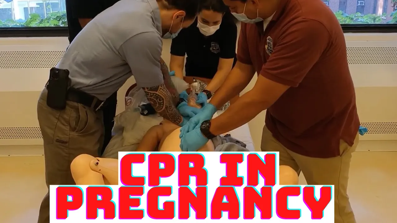
- Why this video: This video covers a crucial safety gap: managing maternal cardiac arrest. It demonstrates the mechanics of manual Left Uterine Displacement (LUD) to relieve aortocaval compression during chest compressions, ensuring adequate blood return to the heart.
- Knowledge Checkpoint:
- Explain the anatomical rationale for Left Uterine Displacement (LUD) in pregnant patients beyond 20 weeks gestation.
- Demonstrate the physical technique of performing continuous manual LUD during CPR without compromising chest compression depth or rate.
- Understand how relief of aortocaval compression improves the efficacy of chest compressions during maternal cardiac arrest.

- Why this video: Designed for first responders, this video covers pre-hospital assessment, common causes of trauma in pregnancy (such as motor vehicle collisions), and how altered center of gravity and loose joints increase fall risks.
- Knowledge Checkpoint:
- Identify the primary mechanisms of maternal trauma (e.g., motor vehicle accidents, falls).
- Recognize early signs of premature labor or uterine irritability following minor maternal trauma.
- Structure a pre-hospital secondary assessment for a pregnant patient involved in an accident.
🔍 Independent Study Suggestion: While the videos above cover clinical triage and trauma extensively, the Obstetric Triage Education literature highlights specific formalized triage scales (like the Obstetric Triage Acuity Scale - OTAS). To supplement your learning, research how your local EMS agency or hospital emergency department incorporates OTAS or similar tools to systematically prioritize pregnant patients over non-pregnant patients with similar complaints.
Module 3: Normal Labor and Emergency Delivery
When transport is not possible, emergency department and EMS personnel must be prepared to assist with a normal vaginal delivery. This module details the clinical stages of labor and guides you step-by-step through assisting an uncomplicated delivery in the field.
Recommended Videos

- Why this video: This highly acclaimed 3D medical animation illustrates the three stages of normal vaginal delivery. It provides clear, accurate visuals of cervical dilation, fetal rotation through the birth canal, and the delivery of the placenta.
- Knowledge Checkpoint:
- Identify the clinical parameters of the three distinct stages of normal labor.
- Define "crowning" and understand the rotational movements of the fetal head during descent.
- Describe the physiological detachment and delivery of the placenta.
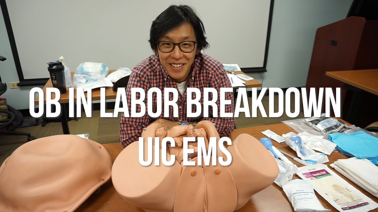
- Why this video: This focused EMT training resource demonstrates how to set up an emergency delivery field and handle immediate delivery maneuvers, such as checking for and resolving a nuchal cord.
- Knowledge Checkpoint:
- State the steps for preparing a clean or sterile perineal field in a pre-hospital environment.
- Explain how to check for a nuchal cord as the head emerges, and how to safely slip it over the baby's head.
- List the essential tools required in a standard emergency OB kit (clamps, scissors, bulb syringe).

- Why this video: This structured clinical simulation demonstrates the precise physical support required during a vaginal delivery, including how to configure the bed, support the perineum to prevent tearing, and guide delivery of the shoulders.
- Knowledge Checkpoint:
- Demonstrate how to apply gentle, controlled counter-pressure to the crowning fetal head to prevent rapid, traumatic expulsion.
- Describe the upward and downward traction maneuvers used to facilitate delivery of the anterior and posterior shoulders.
- Complete a thorough visual assessment of the perineum post-delivery.
Module 4: Hypertensive Crises and Postpartum Hemorrhage
This module focuses on the rapid identification and stabilization of two of the most common causes of maternal mortality: eclampsia (characterized by tonic-clonic seizures) and postpartum hemorrhage (PPH).
Recommended Videos

- Why this video: This comprehensive clinical lecture details the pathophysiology, multi-system signs, and nursing management of preeclampsia and eclampsia. It provides clear diagnostic parameters and outlines Magnesium Sulfate administration protocols.
- Knowledge Checkpoint:
- Define the diagnostic thresholds for gestational hypertension, preeclampsia, and severe preeclampsia.
- Recognize severe features of preeclampsia, including severe headache, visual disturbances, and epigastric pain.
- State the primary therapeutic dosage and monitoring parameters for Magnesium Sulfate administration.
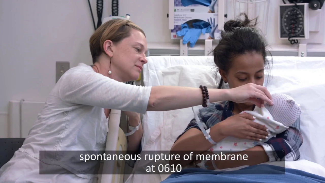
- Why this video: This realistic simulation walks through a postpartum hemorrhage crisis. It shows the critical teamwork required for immediate diagnosis, uterine massage, bladder catheterization, and the coordination of uterotonic medications.
- Knowledge Checkpoint:
- Define postpartum hemorrhage volumetrically (e.g., loss of >500ml after vaginal delivery).
- Describe and demonstrate the physical technique of fundal massage to stimulate uterine contraction (reversing uterine atony).
- Outline the collaborative steps in a PPH protocol (IV access, fluid resuscitation, medication administration).

- Why this video: This video focuses on advanced management of postpartum hemorrhage in emergency and critical care settings. It covers pharmacological interventions (e.g., Oxytocin, Misoprostol, Tranexamic Acid) and advanced mechanical interventions.
- Knowledge Checkpoint:
- Understand the "4 Ts" of PPH etiology: Tone, Tissue, Trauma, and Thrombin.
- Identify the primary uterotonics used in the emergency setting, their routes of administration, and contraindications.
- Describe the clinical indications for advanced mechanical stabilization (e.g., intrauterine balloon tamponade or bimanual compression) in refractory bleeding.
Module 5: Critical Delivery Complications and Maneuvers
This module covers high-risk, low-frequency obstetric complications where rapid, decisive physical maneuvers are required to save maternal and neonatal lives: shoulder dystocia, breech presentations, and prolapsed umbilical cords.
Recommended Videos

- Why this video: This video demonstrates the essential maneuvers to resolve shoulder dystocia. It highlights the McRoberts maneuver (hyperflexing the mother's legs to her abdomen) and the application of suprapubic pressure.
- Knowledge Checkpoint:
- Identify "turtle sign" as a key diagnostic indicator of shoulder dystocia.
- Demonstrate the McRoberts maneuver and explain how it alters pelvic anatomy to release the impacted shoulder.
- Distinguish between suprapubic pressure (indicated) and fundal pressure (strongly contraindicated, as it worsens impaction and risks uterine rupture).
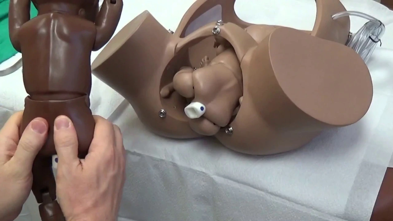
- Why this video: This video provides a systematic, highly educational guide to breech delivery. It covers breech classifications and details safe, hands-off descent techniques followed by controlled assist maneuvers.
- Knowledge Checkpoint:
- Differentiate between frank, complete, and footling breech presentations.
- Explain the "hands-off" principle during early breech descent to avoid stimulating premature fetal respiratory efforts.
- Describe the Løvset maneuver for delivering the arms and the Mauriceau-Smellie-Veit (MSV) maneuver for delivering the head.
- Why this video: This video addresses a key gap: active management of umbilical cord prolapse. It covers the mechanical relief of cord compression using a gloved, manual elevation technique alongside gravity-assisted maternal positioning.
- Knowledge Checkpoint:
- Recognize umbilical cord prolapse during physical examination or upon sudden fetal bradycardia after rupture of membranes.
- Demonstrate how to place a sterile gloved hand in the vagina to apply upward pressure on the fetal presenting part, relieving cord compression.
- Identify maternal rescue positions (knee-chest or Trendelenburg) that use gravity to keep the fetus off the prolapsed cord during urgent transport.
Module 6: Immediate Newborn Care and Resuscitation
A critical component of any emergency delivery is care of the newborn. This module covers standard care for the vigorous infant and step-by-step implementation of the Neonatal Resuscitation Program (NRP) algorithm for the compromised neonate.
Recommended Videos

- Why this video: This exceptionally clear, step-by-step tutorial breaks down the Neonatal Resuscitation Program (NRP) guidelines. It explains how to transition from the initial steps of drying and stimulating to positive pressure ventilation (PPV) when needed.
- Knowledge Checkpoint:
- State the three initial questions to assess a newborn at birth: Term gestation? Good muscle tone? Breathing or crying?
- Demonstrate the correct sequence of immediate care: warm, position airway, clear secretions (if obstructed), dry, and stimulate.
- Identify the clinical indications for starting Positive Pressure Ventilation (PPV) within the first 60 seconds of life ("The Golden Minute").

- Why this video: This video details the equipment setup, ventilation techniques, and clinical decision-making needed when a newborn does not breathe spontaneously, presenting the concepts in a highly visual, accessible format.
- Knowledge Checkpoint:
- Describe how to choose the correct neonatal mask size and achieve an effective seal for ventilation.
- Understand the ventilation rate (40 to 60 breaths per minute) and how to evaluate adequacy via chest rise and heart rate.
- Outline when to progress from ventilation to chest compressions based on heart rate thresholds.

- Why this video: This practical clinical simulation demonstrates how to position the newborn's neck in the "sniffing" position and deliver inflation breaths using a bag-valve-mask.
- Knowledge Checkpoint:
- Position a neonate's head in the neutral or "sniffing" position to ensure airway patency, avoiding hyperextension or hyperflexion.
- Perform the MR. SOPA airway troubleshooting steps if chest rise is inadequate during PPV.
- Determine APGAR scores at 1 and 5 minutes post-delivery.
Course Map
This flowchart maps the recommended learning progression and shows how earlier concepts build into advanced clinical management.
Key People Index
- Dr. Sakshi Arora Hans (Featured in Module 1)
- Context: Obstetrician, author, and medical educator specializing in high-yield physiological and clinical concepts for medical exams and clinical practice.
- Dr. Raj Sherman (Featured in Module 2)
- Context: Canadian Emergency Physician and clinical leader; provides instruction on the triage priority models used to categorize emergency patient presentations.
- Joy James (Featured in Module 6)
- Context: Senior midwifery lecturer and clinical simulation educator, specializing in hands-on clinical training for neonatal resuscitation.
Final Self-Assessment
Complete this comprehensive self-assessment checklist after finishing all modules:
- Maternal Physiology: Can you identify the normal alterations in a pregnant patient's vital signs, including baseline heart rate increases and systemic blood pressure drops?
- Maternal Resuscitation: Can you perform manual Left Uterine Displacement (LUD) to relieve aortocaval compression during a cardiac arrest scenario?
- Trauma Management: Do you understand why a pregnant trauma patient can experience significant internal blood loss before demonstrating classic signs of hypovolemic shock?
- Uncomplicated Delivery: Are you prepared to guide a normal vaginal delivery, including checking for a nuchal cord and supporting the perineum?
- Postpartum Hemorrhage (PPH): Can you identify the symptoms of severe PPH, perform a proper fundal massage, and outline the standard uterotonic medications?
- Preeclampsia/Eclampsia: Can you identify the severe features of preeclampsia and state the correct administration and monitoring steps for Magnesium Sulfate?
- Shoulder Dystocia: Can you perform the McRoberts maneuver and apply suprapubic pressure? Do you understand why fundal pressure is contraindicated?
- Breech Presentations: Do you understand the "hands-off" descent principle and how to safely assist a breech delivery?
- Cord Prolapse: Can you describe the manual elevation technique used to relieve umbilical cord compression during urgent transfer?
- Neonatal Resuscitation (NRP): Can you perform the initial resuscitation steps (warm, position, dry, stimulate) and identify when to initiate positive pressure ventilation (PPV)?