Interpreting PFTs: Spirometry & DLCO
Learning Goal: Mastering Pulmonary Function Test (PFT) Interpretation: Spirometry, Lung Volumes, and Diffusing Capacity. This course equips healthcare students and clinical practitioners with a systematic, evidence-based workflow to confidently evaluate spirometric measurements, static lung volumes, and the diffusing capacity of carbon monoxide (DLCO).
- Prerequisites: Basic human anatomy and systemic circulatory physiology.
- Estimated Study Time: 12 Hours
Module 1: Respiratory Anatomy and Physiology Foundations
This module establishes a deep physiological foundation in respiration mechanics and micro-alveolar gas exchange. To understand dynamic and static lung tests, you must first master how the diaphragm and intercostal muscles generate pressure gradients, and how the five distinct anatomical layers of the blood-gas barrier regulate diffusion.
Recommended Videos

Why this video
This video bypasses basic high-school biology to deliver high-yield medical anatomy. It specifically highlights the microcirculation surrounding the alveoli, detailing how a massive 70m² capillary network maximizes surface area for exchange, laying the physical groundwork for understanding what DLCO actually measures.
Knowledge Checkpoint
- Sketch the structural relationship between alveolar walls and pulmonary capillaries.
- Explain how a reduction in functional alveolar surface area impairs gas diffusion.
- Identify where gas exchange starts and ends along the tracheobronchial tree.
Why this video
Understanding PFTs requires mastering active inhalation and passive exhalation. This detailed lecture covers the precise roles of the diaphragm (responsible for 75% of volume changes) and the pump-handle/bucket-handle mechanics of the external intercostal muscles.
Knowledge Checkpoint
- Distinguish between the muscular work required for normal quiet respiration versus forced spirometry.
- Describe the thoracic pressure changes that drive air movement into and out of the lungs.

Why this video
This video perfectly bridges anatomy and basic ventilation dynamics. It provides a clear, high-yield overview of how pulmonary perfusion matches ventilation at the capillary level, establishing why gas transport is heavily dependent on active thoracic mechanics.
Knowledge Checkpoint
- Define ventilation-perfusion (V/Q) coupling in your own words.
- Explain how chest wall compliance affects alveolar recruitment during deep inhalation.
Module 2: Spirometry, Flow-Volume Loops, and Bronchodilator Reversibility
This module covers dynamic spirometry: assessing the speed and volume of exhalation. You will learn to differentiate obstructive and restrictive defects using the FEV1/FVC ratio and interpret the shapes of flow-volume loops. Crucially, you will master the clinical criteria for determining significant bronchodilator reversibility.
Recommended Videos
Why this video
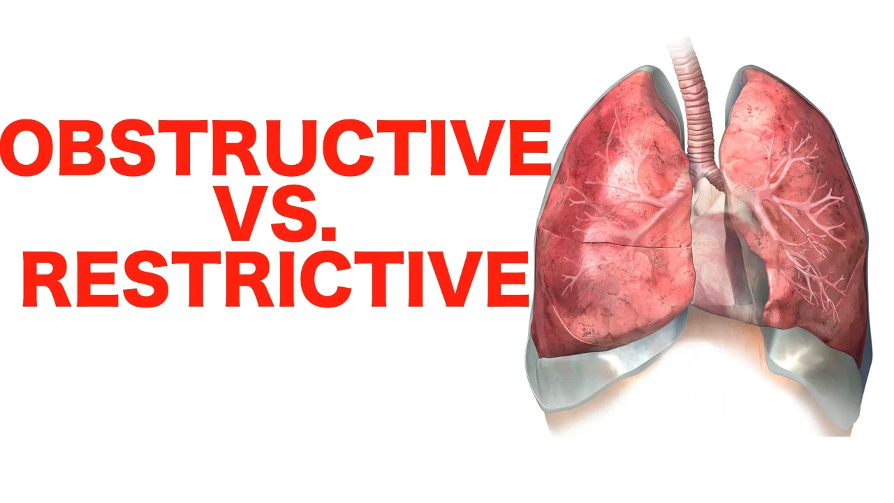
A classic medical review that simplifies the foundational paradigm of PFTs: Obstructive vs. Restrictive patterns. It clearly illustrates why obstructive diseases (like asthma and COPD) lower the FEV1/FVC ratio below 0.70, while restrictive diseases keep the ratio normal or even elevated.
Knowledge Checkpoint
- Define FEV1 and FVC, and explain why their ratio falls in obstructive diseases.
- Identify three common etiologies of obstructive lung disease and three of restrictive disease.
- Explain why a restrictive disease pattern can present with a normal FEV1/FVC ratio.

Why this video
This video is exceptional for visual learners. It demonstrates how to plot and read a flow-volume loop, comparing a normal "child's sailboat" drawing to the scooped-out expiratory limb of obstruction, and the miniature, shrunken loop of restriction.
Knowledge Checkpoint
- Draw a normal flow-volume loop, identifying the peak expiratory flow (PEF) and the residual volume (RV) position on the x-axis.
- Explain why "scooping" occurs on the expiratory curve in patients with asthma or emphysema.
- Describe the visual characteristics of a flow-volume loop in a patient with a fixed upper airway obstruction.

Why this video
This video addresses a critical feedback gap: defining clinical bronchodilator reversibility. It clearly outlines the quantitative criteria required on spirometry to confirm a positive therapeutic response to a short-acting beta-agonist (SABA).
Knowledge Checkpoint
- State the exact percentage and absolute volume improvements in FEV1 or FVC required to diagnose "positive bronchodilator responsiveness."
- Explain how demonstrating reversibility helps differentiate asthma from irreversible COPD in a clinical workup.
Module 3: Lung Volumes and Plethysmography
While spirometry measures dynamic air flow, confirming true restrictive lung disease requires measuring static lung volumes. This module explains the concepts of Total Lung Capacity (TLC), Residual Volume (RV), and Functional Residual Capacity (FRC), and contrasts gas dilution techniques with the gold standard: body plethysmography.
Recommended Videos
Why this video
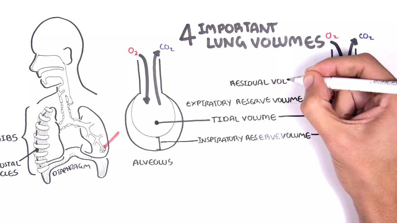
Armando Hasudungan’s beautifully hand-drawn illustrations make learning static lung volumes intuitive. The video cleanly breaks down how individual lung volumes (TV, IRV, ERV, RV) combine to form clinical capacities (VC, FRC, TLC).
Knowledge Checkpoint
- Define the four basic lung volumes and the four lung capacities.
- Identify which lung volumes cannot be measured by simple spirometry and explain why.
- Calculate TLC using Vital Capacity (VC) and Residual Volume (RV).
Why this video
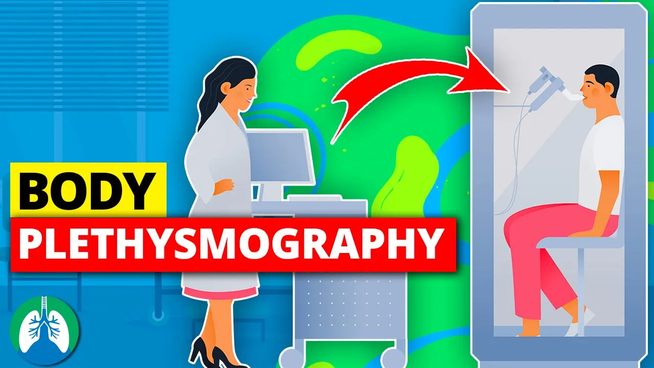
This video provides an excellent clinical overview of what patients experience inside the airtight "body box" and how changes in cabinet pressure are used to calculate the air trapped in the thorax.
Knowledge Checkpoint
- Explain the physical setup of body plethysmography.
- Describe how thoracic gas volume is measured when the airway shutter is closed.

Why this video
This highly analytical lecture compares helium dilution with body plethysmography. It explains how Boyle's Law () governs plethysmographic measurements, making it a critical video for understanding why helium dilution can underestimate lung volumes in patients with severe air trapping.
Knowledge Checkpoint
- State the formula for Boyle's Law and explain how it relates to plethysmography.
- Contrast how helium dilution and body plethysmography measure FRC.
- Explain why helium dilution fails to accurately measure lung volume in a patient with large bullae or severe emphysema.
Module 4: Diffusing Capacity of the Lungs (DLCO)
DLCO evaluates the functional integrity of the alveolar-capillary membrane. This module covers the physics of gas transfer, the single-breath carbon monoxide technique, and how to clinically interpret elevated, normal, or reduced DLCO values across a variety of cardiorespiratory disorders.
Recommended Videos

Why this video
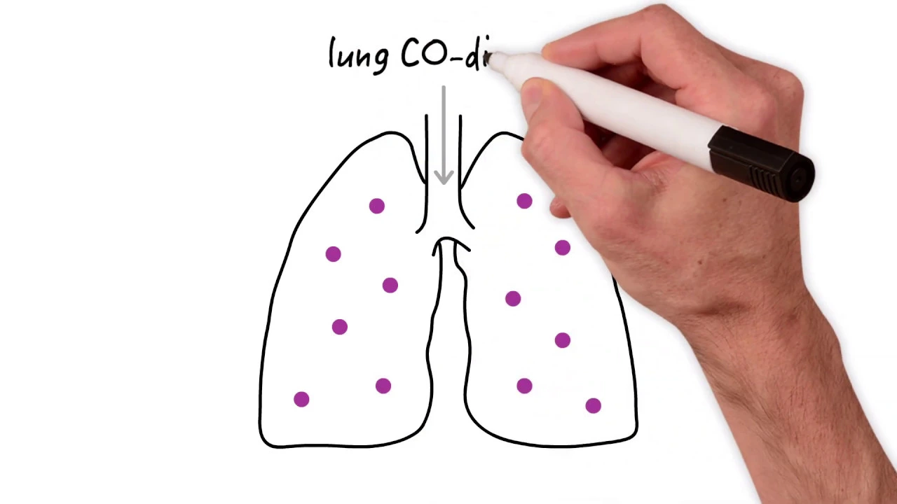
StrongMed provides a concise and high-yield presentation of the DLCO testing mechanism. It explains why carbon monoxide is used as a surrogate gas (due to its massive affinity for hemoglobin) and outlines how the test is executed clinically.
Knowledge Checkpoint
- Explain why Carbon Monoxide (CO) is the ideal gas for measuring diffusing capacity.
- Describe the clinical steps a patient performs during a single-breath DLCO test (breath-hold duration, etc.).
- List two factors that can artifactually lower a patient's DLCO reading (e.g., anemia).
Why this video
MedMastery excels at clinical application. This video demonstrates how to use DLCO to distinguish between diseases with identical spirometric patterns. For instance, it shows how DLCO differentiates intrinsic restrictive parenchymal disease (low DLCO) from extrinsic chest wall restriction (normal DLCO).
Knowledge Checkpoint
- Differentiate the DLCO profiles of emphysema vs. asthma.
- Explain why DLCO is reduced in idiopathic pulmonary fibrosis (IPF) but remains normal in chest wall kyphoscoliosis.
- Name a clinical condition where DLCO can actually be elevated above normal limits.
Why this video
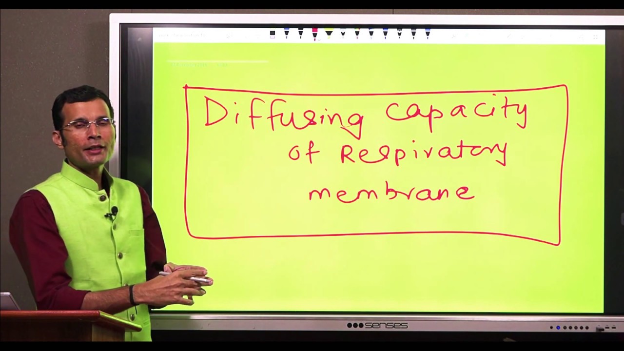
This in-depth lecture breaks down the physical barriers to diffusion. It analyzes the five physical layers of the respiratory membrane and uses Fick’s Law of Diffusion to show how membrane thickness, surface area, and pressure gradients directly determine the measured DLCO.
Knowledge Checkpoint
- List the five anatomical layers of the blood-gas barrier in order from alveolus to capillary lumen.
- Use Fick's Law of Diffusion to explain how pulmonary edema reduces diffusing capacity.
Module 5: Step-by-Step Pulmonary Function Test Interpretation and Clinical Cases
This module synthesizes everything you have learned. Using structured, algorithmic workflows, you will interpret real-world, complete Pulmonary Function Test reports. You will learn to recognize mixed defects, avoid physical fitness "PFT" search noise, and apply clinical logic to complex patient presentations.
Recommended Videos
Why this video
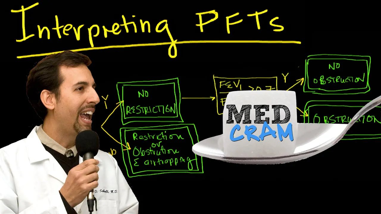
Dr. Roger Seheult of MedCram provides the gold-standard step-by-step PFT interpretation algorithm. It teaches you exactly where to look first on a clinical report (FEV1/FVC ratio), how to evaluate FVC, and how to use TLC to rule in or out true restriction.
      [ Look at FEV1 / FVC Ratio ]
             /            \
     < 70%  /              \  >= 70%
           v                v
 [ OBSTRUCTIVE ]         [ Look at FVC ]
                         /             \
                >= 80%  /               \  < 80%
                       v                 v
                   [ NORMAL ]     [ SUSPECT RESTRICTION ]
                                         |
                                         v
                                  [ Measure TLC ]
                                  /             \
                         < 80%   /               \  >= 80%
                                v                 v
                         [ RESTRICTIVE ]    [ NORMAL VARIANT ]
Knowledge Checkpoint
- Reproduce the basic MedCram step-by-step PFT interpretation flowchart from memory.
- Explain why a low FVC does not automatically confirm restrictive lung disease.
- Determine the next diagnostic step if a patient has a normal FEV1/FVC ratio but an FVC of 65% predicted.

Why this video
This video directly addresses the gap regarding mixed pulmonary defects. It explains how a patient can simultaneously suffer from airway obstruction (e.g., severe COPD) and mechanical restriction (e.g., morbid obesity), and shows how their PFT parameters present on a clinical report.
Knowledge Checkpoint
- Define the PFT criteria for a "mixed" obstructive and restrictive defect.
- Explain how a combined pathology (e.g., COPD + severe chest wall deformity) alters both the FEV1/FVC ratio and TLC.
Why this video
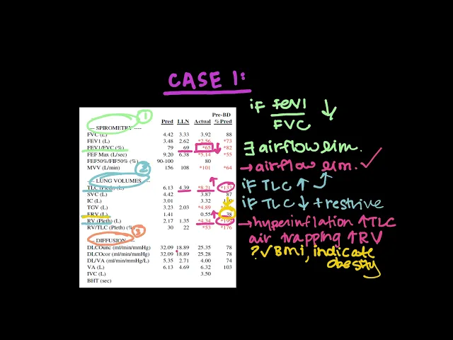
This clinical case session brings the entire curriculum together. Watching a pulmonologist work through actual patient PFT reports teaches you to integrate clinical history, spirometry numbers, plethysmography-derived lung volumes, and DLCO values.
Knowledge Checkpoint
- Work through a clinical case step-by-step: read the clinical scenario, apply the algorithm, and arrive at the correct diagnosis before the video reveals it.
- Explain how the patient's symptoms (e.g., dyspnea on exertion) correlate with their objective PFT findings.
Course Map
Key People Index
- Dr. Roger Seheult (MedCram): A double board-certified Pulmonologist and Critical Care specialist. Renowned for creating clean, highly logical diagnostic flowcharts that simplify complex physiological assessments.
- Dr. Eric Strong (StrongMed): Clinical Associate Professor of Medicine. Celebrated for his highly structured, step-by-step videos on cardiovascular and pulmonary diagnostics.
- Armand Hasudungan: Renowned online medical illustrator and educator. Famous for using hand-drawn, real-time anatomy videos to make complex physiology easily accessible.
- Dr. Vivek (Vivek's Physiology): Academic physiology professor who specializes in high-level physical-chemical gas laws and micro-layer cellular diffusion equations.
Final Self-Assessment
Test your clinical competency. Before completing this curriculum, ensure you can check off every clinical skill below:
- Explain why a patient with severe emphysema can have an elevated Total Lung Capacity (TLC) but a severely reduced Vital Capacity (VC).
- Correctly interpret a spirometry report to identify an obstructive defect based on a FEV1/FVC ratio (or below the Lower Limit of Normal).
- Determine if a patient has asthma reversibility using the clinical standard: improvement and absolute increase in FEV1/FVC post-bronchodilator.
- Identify a restrictive pattern on spirometry (FVC , normal ratio) and explain why a body box plethysmography test is required to confirm the diagnosis (TLC ).
- Differentiate between intrinsic restriction (IPF) and extrinsic restriction (obesity/neuromuscular disease) using DLCO values.
- Explain how Boyle's Law is applied inside a plethysmograph to calculate FRC and RV.
- List three independent clinical factors that can artificially lower DLCO without representing true lung tissue destruction (e.g., anemia, smoking, Valsalva maneuver).
- Identify the classic visual presentation of a "scooped out" flow-volume loop, a "shrunken" restrictive loop, and a "variable/fixed" upper airway obstruction plateau.
- Apply the step-by-step MedCram clinical algorithm to systematically evaluate any clinical Pulmonary Function Test report without skipping intermediate steps.
- Describe the physiologic changes in a combined/mixed defect (such as COPD combined with chest-wall restriction) and detail how it alters FEV1/FVC, TLC, and DLCO.