







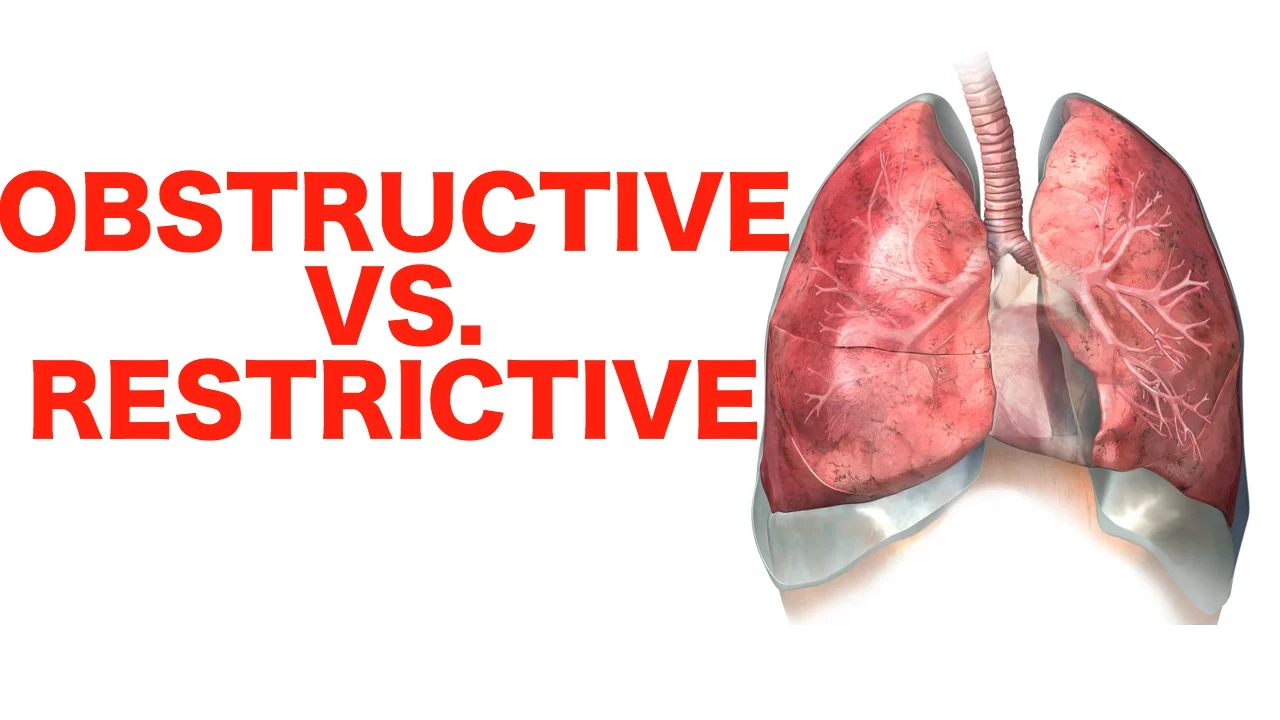


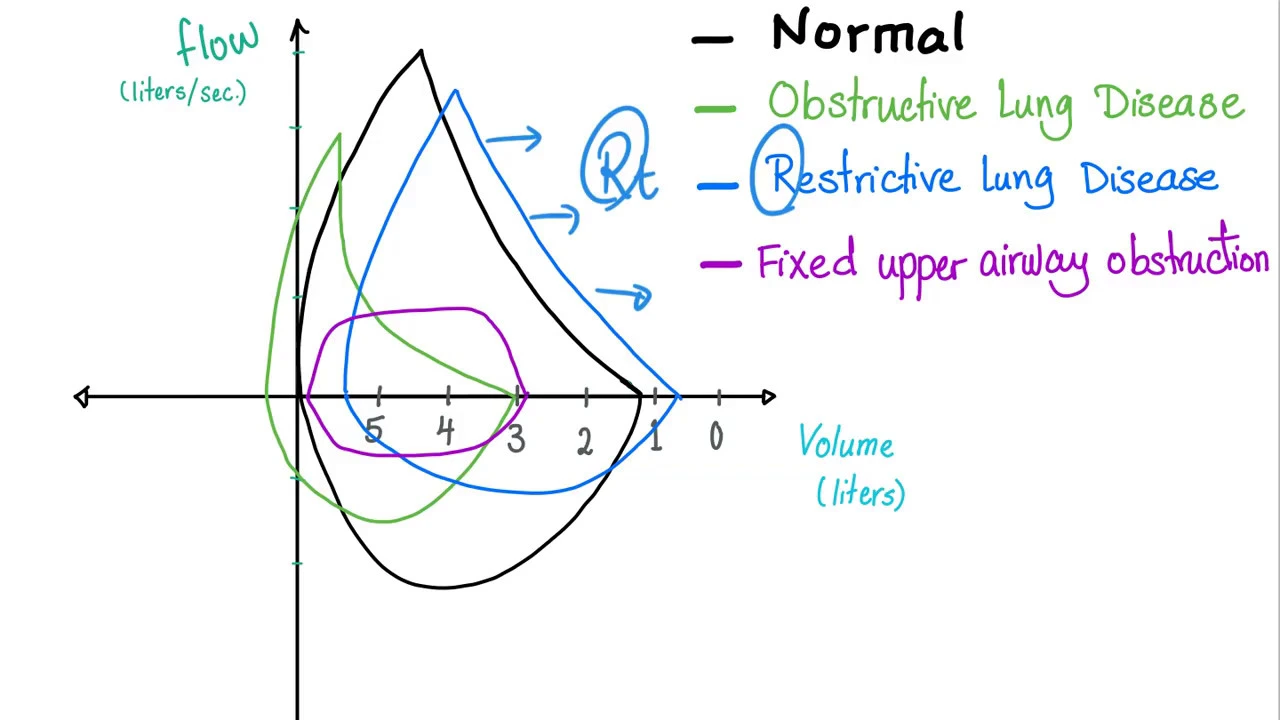









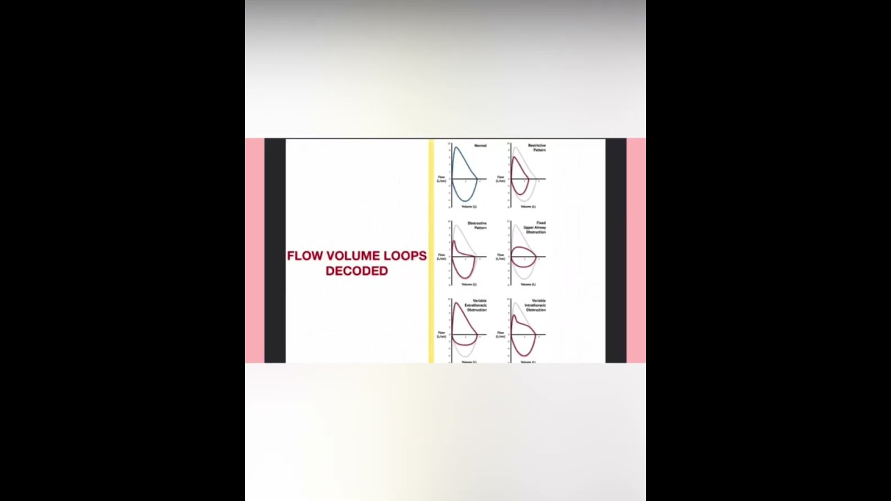
Flow-volume loops plot airflow velocity against lung volume during forced breathing maneuvers, with normal loops showing a characteristic pattern where inspiration starts at residual volume (~1.3L) and expiration shows a scooping curve; obstructive lung diseases (like emphysema) cause a leftward shift with increased lung volumes and characteristic scooping of the expiratory limb due to airway resistance and elastic recoil loss, while restrictive lung diseases (like pulmonary fibrosis) cause a rightward shift with decreased lung volumes and preserved loop shape due to reduced lung compliance and stiffness.
Flow-Volume Loops Explained: Obstructive vs Restrictive Patterns
Added:Basic pulmonary anatomy and mechanics of ventilation, including lung compliance and airway resistance.
Standard lung volumes and capacities, specifically Total Lung Capacity (TLC), Residual Volume (RV), and Vital Capacity (VC).
Spirometry basics, including Forced Expiratory Volume in 1 second (FEV1), Forced Vital Capacity (FVC), and the FEV1/FVC ratio.
Pathophysiological definitions and general characteristics of obstructive lung diseases (e.g., asthma, COPD) versus restrictive lung diseases (e.g., pulmonary fibrosis).
Localization of upper airway obstructions (fixed, variable extrathoracic, and variable intrathoracic lesions) based on flattening of the inspiratory or expiratory limbs.
Using bronchodilator reversibility testing to differentiate asthma from irreversible COPD on flow-volume loops.
Advanced pulmonary function tests (PFTs), such as body plethysmography for measuring absolute lung volumes and diffusing capacity of the lung for carbon monoxide (DLCO).
Clinical application of flow-volume loop interpretation in diagnosing and managing complex respiratory disorders in USMLE-style clinical vignettes.
603 views14likes18:32Original Release: 2026-01-17
[music] [music] Welcome back guys. Now let's continue with the topic of flow volume loops.
Flow means how the air is flowing with how much velocity the air is entering into the lungs or exiting out of the lungs and volume how much air is going and how fastly it is going. Okay, flow means the velocity. Flow here literally meaning the velocity means how much speed with which it is going. And volume means how much air is going. So we are going to compare here two things. The flow and the volume during inspiration and expiration. Okay. Now let's look at the flow volume loop. Here is the flow volume loop. And whenever you see a graph, the first thing that you have to see is what is given on the x-axis and y-axis. Look on the x-axis.
What is given the volume changes? Okay, volume how the volume is changing from 0 ml 1 2 3 4 5 6 7 8 something like that.
Okay, the volume is shown on the x-axis.
Now what is shown on the y-axis? See on the y-axis, okay, on the y-axis we are talking about the the flow changes means the velocity changes starting from here 0 liter, 1 liter, 4 liter, 6 8 10 something like that. Okay, here also 2 4 6 8 something like that. Okay, the volume um is shown on the x axis and the flow is shown on the y axis means 0 1 2 3 4 something like that. Okay, don't confuse why there are two zeros. Okay, because one is regarding the volume.
Okay, one is regarding the volume other is regarding the velocity. Now here always look on the bottom side we are representing the inspiration. We are talking about the inspiration. On the above side we are discussing about the expiration. Okay, top we are discussing expiration on the bottom side we are discussing about the inspiration. So now let's take a Hindley individual and now I'm asking this individual to take inspiration. First I'm asking him to take the inspiration. Now he's starting the inspiration and he's doing it as fast as possible. So look he's starting his inspiration at 1.3 L. He's not starting at zero. Look he's not starting at zero. He's starting at which liters guys? 1.3 L. Why? Because you already know there is residual volume. Okay, there is residual volume. So residual volume is also present here, right? So that's why he's not starting his inspiration from 0 ml. He's starting from 1.3 L. And from 1.3 L, see he's starting to take the air in. When when he's trying to do the inspiration, what will happen to the velocity by which the air is entering into the lungs? See? Now I'm doing the inspiration. when I'm doing the inspiration yes the volume is increasing but I'm talking about the velocity how the air is going into the lungs see the velocity is gradually increasing means from 0 L velocity okay velocity gradually increasing to 1 liter 2 liter 3 liter 4 liter something like that means the flow is increasing the air entering into the lung with high velocity okay now you try to do it okay practically you try to do it and check when you are doing the inspiration Initially the air is going into the lung with high velocity but at the end of the inspiration concentrate the air that enters into the lung with low velocity.
The air is entering into the lung with less velocity. You try to do it practically.
See at the end the air is not able to enter into the lung. Okay. So initially the air is going into the lung with a high velocity and at the end the air is entering into the lung with low velocity. So here I'm talking about see the velocity is increasing but at the end of the inspiration the velocity is gradually decreasing and coming back to zero. Okay let me give you an example.
See guys u you have done with your MBBS.
Okay now you are doing your internship.
Okay your MBBS life completed. Now you are doing your internship. See when you are going to internship you are going to go in a nice vehicle right? You are not going to take some local taxi or you are not going to go in some local transport.
So you are going to have your own nice bike or nice car. So when you are going to start from your home, tell me when you are going to start from your home, you are going to start your car. So initially do you think the car will go with high velocity? No. Your mom is watching you, your father is watching you. Okay. So how you will start? You will start the car and you will slowly start from your home. So once you take the turn, okay, your street turn. Now what you will start to do? You will start to increase the velocity, right?
You will start to go fast now. Okay? So in the same way initially see the velocity is increasing initially starting from zero. See starting from zero velocity starting from zero velocity gradually the velocity increased increased increased okay but what happened at the end see once you are reaching to the hospital you tell me what what will happen once you are reaching to your destination hospital you'll start to decrease your speed okay in the same way at the end of the inspiration the velocity decreases and coming back to zero. So final thing which I want you to know here is on the bottom side we are discussing about this inspiratory loop here this is the inspiratory loop exactly same what about the expiration see you are doing the expiration initially you you can take out the air with high velocity initially see you can take the air out with high velocity but at the end of expiration the velocity gradually comes back and coming to zero again coming to zero again okay so initially you can have high velocity later the velocity decreases and coming back to zero okay I'm talking about the velocity velocity see initially increases and later again coming back to zero again coming back to zero so how the volume is changing and how the flow is changing see inspiration you are starting from 1.3 L and how much maximum inspiration you have taken the maximum inspiration that you have taken let's say 6 L you have taken 6 L of air okay starting from 1.3 total and capacity 6 L and how the flow Flow is changing. So initially the flow increases. Initially the flow increases later the flow decreases. Okay. Here initially flow increases later decreases. Expiration also same thing.
Initially the flow increases later flow decreases. So how the volume is changing from 1.3 to 6. 6 to 1.3. How the velocity is changing? How the flow is changing initially? Increasing decreasing increasing decreasing. So that's it guys. So how question is going to come? is image based question. Okay, image based question. Most of the time they won't ask this normal flow volume loop. Most of the time they won't ask about this normal flow volume loop. Then how the question will come? They'll ask about the diseases. How this flow volume loop is going to change in obstructive lung diseases and restrictive lung diseases. How it's going to change? For that you have to have a basic understanding of obstructive luck disease. So in short form we can write it as OLD. Okay. OLD obstructive lung disease. The classical example of obstructive lung disease is emphyma.
Okay. Emphyimma who will have this emphyma that there are two possibilities. Um one the emphyma can be seen in smokers.
Okay. Chronic smokers or emphyma can be seen in people who are having alpha 1 antitripen deficiency. Okay. Alpha 1 antitin deficiency. Okay. Now what is the main problem here in FISMA? Already I have explained you in the basics itself. The main problem in FISMA is there is damage to okay or elastin fiber damage. Elastin protein damage.
Okay. So we all know lungs are made up of what? Elastin. So that elastin protein because of the excessive elast activity there is damage to this elastin protein gone. Today now there is no more elastin. Now tell me whenever there is no more elastin I have told you elastin protein and the collagen are the responsible for the elastic recoil. Okay the elastin protein is responsible for what? It is responsible for the elastic recoiling of the lung or the collapsing of the lung. But right now there is no elastic recoil. Elastin is gone. So elastic recoil property decreases.
Elastic recoil the collapsing nature decreases. So now tell me what will happen. Sir the alvolus your lung have to collapse. Your lung have to collapse during the expression. During expression the alvulus has to collapse so that the air will come out. Alvulus has to collapse so that the air will come out.
But right now the collapsing the recoil elastic recoil decreases. Now tell me whenever the elastic recoil decreases do you think the expiration can happen easily? No. So there will be problem with expiration. So always always remember in obstructive lung diseases are in fisma empa which problem?
Expiratory problem E for E. In emphyma expiratory problem, emphyma expiratory problem. So everything e guys emphyma elastin gone. Expression gone. Okay.
Next step. So what's going to happen now? Now the air will start to trap inside the lungs. Because expression problem, right? The expression is not happening. The air is not coming out of the lungs. So there is air trapping within the lungs. So now because of this air trapping, what do you think will happen to the size of the lungs? Now because of this air trapping, what will happen to the size of the lungs? Now these lungs are going to be voluminous.
These are big lungs, let's say. Okay, big lungs.
The lung size is going to be very very big. Big lungs. And what do you think will happen to the lung capacities? What do you think will happen to the lung capacities? Big lung. So lung volumes, lung volume and capacities will be definitely more. The lung volumes and capacities increases.
Okay. So that's why in your pathology, in your second year pathology, you have studied. Okay. In emphyma, the patient is going to have a barrel-shaped chest because of this big big size lungs.
Okay, the size of the lungs is very big, right? So the thoracic diameters, okay, the antroposterial diameters of the thorax is going to be affected because of this large lungs. So if you look at the chest of these patients that the patients are going to have a barrel-shaped chest, okay, in Ephesimma, this is going to be barrel barrel shape chest. Okay, barrel-shaped chest. And whenever you do the physical examination, whenever you try to do the percussion, okay, whenever you are trying to do the percussion, how it's going to be, there is a lot of air. So when you are tapping on the air, it will give a lot of re resonance. So there's going to be hyper resonance.
Okay, there's going to be hyper resonance, not dullness. More more sound is going to come. Okay, they're just looking like a drums now. Okay, done. So this what we have completed? We have completed the basics about the obstructive lung disease. Obstructive disease classical example is emphyma.
The problem is with the damage to the elastin protein, elastic recoil decreases. So there's going to be large volumeous lungs, a trapping, expiratory problem. Okay, lung volumes and capacities increases. Now let's see what exactly is a problem with the restrictive lung disease that is RLD.
Okay, R L D that is restrictive lung disease. RLD the classical example of RLD is pulmonary fibrosis okay pulmonary fibrosis now what is the problem with the pulmonary fibrosis let me tell you it's very simple right pulmonary means lung fibrosis means hardening or stiffening because of excessive collagen deposition let's say today there is excessive collagen that is getting deposited okay in the alvulas there is excessive excessive collagen that is getting deposited excessive collagenization excessive collagen getting deposited Now I'm asking you whenever there is lot of collagen there is these are fibroic lungs right now they are stiff for fibroic lungs. First question number one if they are stiff fibroic lungs are they going to be big in size or small in size? They're stiff very stiff hard lungs. So these are comparatively small lungs. Okay try to understand they are small lungs.
Okay small lungs not big lungs definitely. And what do you think will happen to the compliance property? These are very hard lungs stiff lungs. So do you think you can stretch the alv easily? No, you cannot stretch the lung easily. So during inspiration, what do you want? We want to stretch the alvulus. During inspiration, we have to stretch the alvulus so that air will come in. But here in this condition, you cannot stretch the alvulus. So it's a hard lung, less compliance. The compliance is affected. So it's mainly the inspiratory problem. So these are non-compliant alvi. Okay, non-compliant alvi. And what happened to the alvolar filling in alvi there is a decreased filling guys. Okay, alvi are not getting filled because they are stiff. Okay, they are not able to get the enough amount of air. So it's mainly the inspiratory problem. The problem is with the inspiration. It's a inspiratory problem. So small lungs, stiff lungs, less compliance, hard lungs. So can I say something like this? Whenever the the lungs are very small, stiff, they cannot expand. So what will happen to the lung volumes and capacities?
Definitely less. So lung volumes and capacities will be definitely decrease. So lung volumes and capacities value will be less. Now okay with this basic understanding now let's see how the flow volume loop is going to change in these diseases. Okay guys first look at this diagram. Okay now this is the image imagine this is the image based question which was asked in your exam. First the patient is suffering with which disease.
Okay this patient is suffering with which disease I'm asking you how to answer it. See in this patient he's beginning his inspiration from where a normal individual have to begin his inspiration from 1.3 L. So that this is a normal residual volume. But now right now my patient he's beginning his inspiration from 2.5 L which means what happens to his residual volume. See there is a lot of residual volume. So residual volume increases. He's having a lot of residual volume. Okay. And see at the end of the inspiration how much air he's taking in. See he's taking air a lot. He's taking a lot of air. A normal individual will take somewhere around 6 lit. But now right now our guy is let's say taking 7 lit or 7.5 L. So what happened to his total lung capacity?
This is the total lung capacity right?
The total lung capacity in a normal individual is around 6 lit but he is having around 7 lit. Total lung capacity increases. So starting point is always let's say residual volume. Okay. And ending point is always total lung capacity. Now tell me in this condition the total lung capacity increases residual volume increases okay increases increases. So now tell me what is this disease? The disease in which the residual volume increasing and as well as the total lung capacity increasing.
What is this disease? This is your obstructive okay lung disease.
Okay obstructive lung disease. So now you tell me obstructive lung diseases have I already told you she cannot get the air out. Air is not coming out air trapping large lungs big lungs okay that is the reason why lung volumes and capacities are increasing I have explained you what is the disease in which the lung volumes and capacities increases it's emphyma obstructive lung disease okay so this is how to identify now tell me how the graph is shifting see the graph it is more shifting towards the the graph is shifting more towards which side left side see it's shifting towards the left side okay see residual volume it's shifting total lung capacity also shifting so there's going to be a left shift shift.
Okay. In obstructive lung diseases, there is a left shift. Left shift of what? Flow volume loop. There is left shift of flow volume loop.
Okay.
Next guys, look at this disease. I'm asking you what is this patient suffering with? Always look at the starting point, inspiratory point.
Normal person was supposed to start his inspiration. He's going to start at 1.3 L. But right now my patient he's starting his inspiration at say at 0.8 lit. Okay. At 0.8 lit which means what happened to the residual volume? Yes. Residual volume decrease. And what happened to total lung capacity? Look the total lung capacity is also decreased. Not 6 lit.
It is less than 5 lit. Let's say 4.8 L only. So total lung capacity also decreases. Total lung capacity decreases and residual volume decreases. So what is this disease? The diseases where the lung volumes and capacities decreases that is a restrictive lung disease guys.
So here answer is restrictive lung disease.
Have I already taught you? In restrictive lung diseases the problem is he cannot get the air in. He cannot breathe normally. The inspiration is going to be less. So less inspiration automatically less expiration. Okay.
Small lungs, hard lungs, stiff lungs, less volume, some capacities. But tell me where the graph graph is shifting.
See the residual volume is shifting towards the right side as well as the total lung capacity is also shifting towards the right side from 6 lit now it is shifting towards the more right side.
So always in restrictive lung diseases there's going to be right shift R for R.
Okay. In restrictive lung diseases, there's going to be right shift of low volume loops. Low volume loop. Okay.
Next step, sir, with just a glance.
Okay. I don't want to go into too much details. Just in exam, just within a glance, within a second, I want to say whether the person is suffering with abstract disease or restrict disease.
See, how can you do that? Okay, the pattern look here in the expiratory loop. This is expiratory loop, right? In a person who is suffering with empyma or abstract disease, there's going to be a scooping pattern or coving pattern.
Okay, there's like a scoop, right? you have taken a scoop out of the ice cream.
So whenever you are seeing this bending or curve uh cving okay bending or scooping this is definitely an obstructive lung disease but here in restrictive lends no such curve or bent is going to be seen okay so here there's going to be coving pattern coving here there is no coving okay this is the one thing just with the second you can say whether it is abstract so with this finally what we have completed we have completed what is the normal flow volume loop and how the flow volume loop is going to be uh different how it's going to be affected in abstractive diseases as well as restrictive diseases. Again I'm telling you this is the key. Okay, this table is the key for understanding the pathology.
What is the problem with obstructive expiratory problem? What is the problem with the restrictive lung disease?
Inspiratory problem. Obstructive lung disease recoiling problem. In uh restrictive lung diseases it is the inspiratory problem or the compliance problem. In one place the lungs are not collapsing in the other place the lungs are not expanding. The alvi are not expanding. Okay. So with this the topic of low volume will be completed. Hope the video is helpful. Thank you.
Up Next

79:03
Lung Volumes, Capacities & Gas Exchange | Respiratory Physiology
@DrMattDrMike
16.1K views•2020-07-06

32:45
Memory and Long-Term Potentiation | Physiology | MBBS Exam Prep
@doctutorialsmbbs
307 views•2025-08-27
22:48
Electron Transport Chain & Oxidative Phosphorylation | Biochemistry Animation
@doctorbhanuprakash
186.8K views•2018-11-13

3:05
Stages of Labor and Vaginal Birth | Childbirth Animation
@nucleusmedicalmedia
52.1M views•2017-08-18
Related Study Plans & Knowledge Roadmaps
Structured learning paths in Medicine
medicine
Interpreting PFTs: Spirometry & DLCO
01Respiratory Anatomy and Physiology Foundations
02Spirometry: FVC, FEV1, and Flow-Volume Loops
03Lung Volumes and Plethysmography
+ 2 more learning modules
12h5 modules
Explore Roadmap
medicine
Abnormal LFTs: Diagnostic Workup & Pathology
01Liver Anatomy and Basic Physiology
02Introduction to Liver Function Tests (LFTs)
03Hepatocellular vs. Cholestatic Patterns
+ 3 more learning modules
12h6 modules
Explore Roadmap
medicine
Analyzing Blood Smears: RBC, WBC & Platelets
01Introduction to Blood Components & Smear Preparation
02Normal Morphology of Blood Cells
03Erythrocyte Pathologies & Morphological Variants
+ 2 more learning modules
12h5 modules
Explore Roadmap
medicine
Mechanical Ventilation: Waveforms & Alarms
01Anatomy and Physiology of Breathing
02Introduction to Mechanical Ventilation & Core Modes
03Ventilator Settings and Waveform Interpretation
+ 2 more learning modules
12h5 modules
Explore Roadmap