Mastering Peripheral Blood Smear Interpretation: Diagnosing Erythrocyte, Leukocyte, and Platelet Pathologies
Learning Goal: Master the complete diagnostic pipeline of peripheral blood smear analysis. By the end of this curriculum, you will be able to prepare and stain a high-quality human blood slide, recognize normal cellular morphology under oil immersion, systematically identify microcytic, normocytic, and macrocytic red blood cell pathologies, distinguish benign reactive leukocyte changes from malignant leukemia lineages, and confidently rule out technical artifacts like EDTA-induced platelet clumping.
- Prerequisites: Basic knowledge of human circulatory physiology.
- Estimated Total Study Time: 12 Hours
Module 1: Introduction to Blood Components & Smear Preparation
This module introduces the cellular architecture of whole blood and details the precise physical techniques required to prepare, stain, and fix a diagnostic-grade peripheral blood film. You will learn the mechanics of smear creation—controlling the spreader slide angle—and the chemical foundation of Wright-Giemsa staining.
Recommended Videos
Why this video
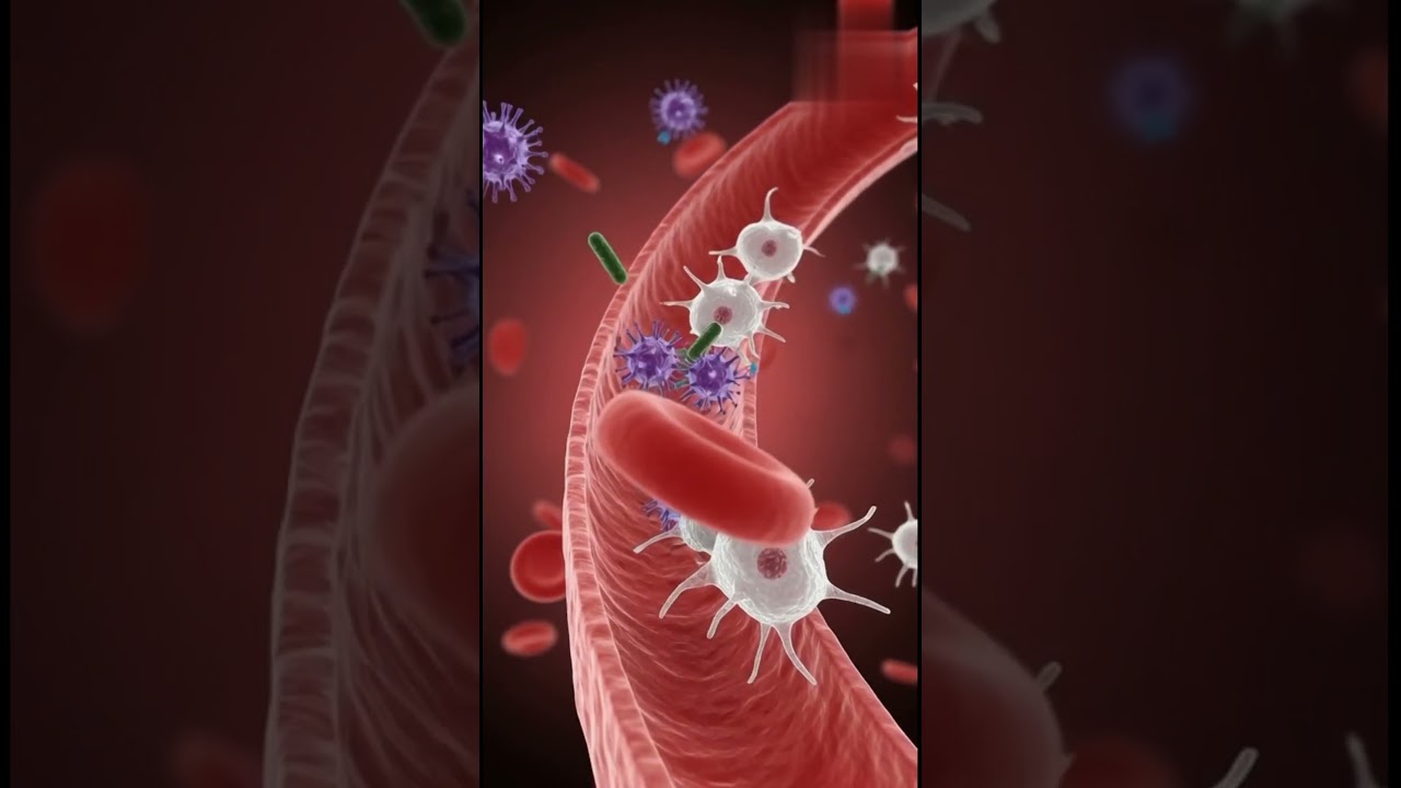
This animated introduction establishes the baseline physiological landscape of blood. It visualizes blood separation into plasma and the three primary formed elements (erythrocytes, leukocytes, and platelets), laying the groundwork for how these components appear when physically spread on glass.
Knowledge Checkpoint
- Differentiate between the liquid component of blood (plasma) and the cellular/formed elements.
- Define the primary role of hemoglobin in erythrocytes.
- State the foundational protective roles of leukocytes and platelets.

Why this video
This professional medical laboratory demonstration details the physical technique required to pull a high-quality blood film. It provides an explicit step-by-step visual on cleaning factory film from slides, managing specimen volumes, and achieving a perfect "feathered edge."
Knowledge Checkpoint
- Explain why standard factory glass slides must be wiped clean before smear preparation.
- Demonstrate the "draw-back and push-forward" technique using a spreader slide at a 30–45 degree angle.
- Describe the appearance of a perfect feathered edge and identify why too steep or shallow an angle compromises smear thickness.

Why this video
Staining is necessary because blood cells are transparent under standard light microscopy. This video walks through the complete chemical theory and manual protocol of the Wright-Giemsa stain, explaining how basic and acidic cellular structures interact with methylene blue and eosin dyes.
Knowledge Checkpoint
- Explain the stain chemistry of Wright-Giemsa, highlighting how basic cellular components bind to acidic eosin (red/pink) and acidic components bind to basic methylene blue (blue/purple).
- Outline the physical workflow steps of fixing the dried slide and timing the buffer stage.
- Identify signs of poor staining, such as excessive blue tinting or precipitate artifacts.
Module 2: Normal Morphology of Blood Cells
Before diagnosing disease, you must recognize health. This module focuses on identifying normal, non-pathological red blood cells, platelets, and the five main classes of white blood cells (neutrophils, lymphocytes, monocytes, eosinophils, and basophils) under high-power oil immersion.
Recommended Videos
Why this video
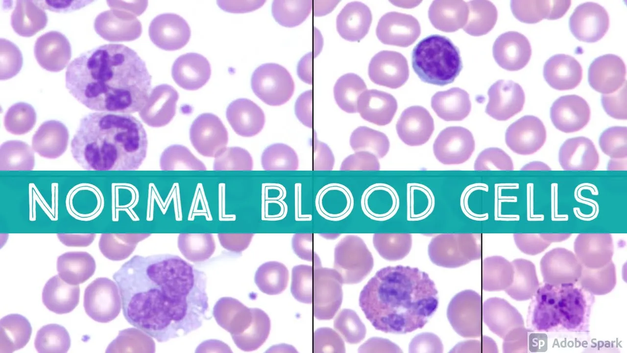
Led by a board-certified pathologist, this high-yield tutorial provides close-up microscopic reviews of every healthy blood cell type. It provides the essential visual vocabulary for comparing normal red blood cell size (relative to a small lymphocyte nucleus) and normal platelet distribution.
Knowledge Checkpoint
- State the normal proportion of central pallor in a healthy, resting erythrocyte (should be approximately 30–45% of the cell diameter).
- Identify the nuclear lobation pattern of a mature segmented neutrophil (3 to 5 lobes linked by thin chromatin threads).
- Distinguish eosinophils from basophils based on granule color (bright orange-red vs. dark blue-black).

Why this video
Distinguishing lymphocytes from monocytes is a classic clinical pain point. This video provides a structured comparative methodology, analyzing cell size, nuclear shapes (round vs. kidney/folded), and cytoplasmic textures (scanty pale blue vs. abundant ground-glass gray).
Knowledge Checkpoint
- Compare the chromatin pattern of a lymphocyte (highly condensed, dark, blocky) to a monocyte (lacy, loose, lighter purple).
- Identify cytoplasmic vacuoles and pseudopods as key features pointing toward a monocyte.
- Utilize the size of surrounding red blood cells as a structural ruler to measure lymphocyte and monocyte diameters.
Why this video
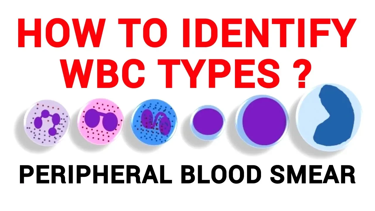
This clinical physiology lab presentation provides a systematic diagnostic key to categorize leukocytes into granulocytes and agranulocytes. It reinforces morphology with side-by-side comparative microscope footage.
Knowledge Checkpoint
- Classify neutrophils, eosinophils, and basophils as granulocytes, and lymphocytes and monocytes as agranulocytes.
- Identify the unique "spectacle-shaped" bilobed nucleus characteristic of eosinophils.
- Explain why basophilic granules often obscure the outline of their underlying nucleus.
Module 3: Erythrocyte Pathologies & Morphological Variants
This module covers clinical red blood cell morphology. You will learn to categorize anemias microarchitecturally, moving beyond broad indices like MCV to identify physical variants such as target cells, spherocytes, elliptocytes, and fragmented schistocytes.
Recommended Videos
Why this video
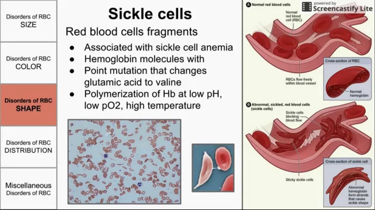
This comprehensive lecture links abnormal morphology to exact underlying pathophysiologies. It covers anisocytosis (size variations), poikilocytosis (shape variations), and intracellular inclusions, explaining why membrane defects or hemoglobinopathies yield distinct microscopic shapes.
Knowledge Checkpoint
- Define anisocytosis and poikilocytosis, correlating them with elevated Red Cell Distribution Width (RDW).
- Explain the pathogenesis of schistocytes (fragmentation due to physical shearing across microvascular fibrin strands).
- Differentiate between microcytosis (MCV <80, iron deficiency/thalassemia) and macrocytosis (MCV >100, B12/folate deficiency) using visual criteria.

Why this video
This medical lecture bridges pure morphology with clinical algorithms. It demonstrates how to utilize specific peripheral smear findings (like spherocytes, elliptocytes, or target cells) to narrow down the differential diagnosis of hemolytic and non-hemolytic anemias.
Knowledge Checkpoint
- Recognize spherocytes (cells lacking central pallor due to membrane loss) and associate them with hereditary spherocytosis or warm autoimmune hemolytic anemia.
- Identify target cells (codocytes) and understand their clinical association with asplenia, hemoglobin C, and thalassemias.
- Explain the diagnostic significance of finding more than 5 target cells per high-power field (HPF).

Why this video
A rapid, high-intensity slide review focusing on key pathognomonic shapes. It provides excellent clear-field microscopy captures of helmet cells, schistocytes, and target cells side-by-side, cementing your visual pattern recognition.
Knowledge Checkpoint
- Visually identify schistocytes (broken, triangular, or helmet-shaped RBCs).
- Link schistocytes on a smear to critical medical emergencies (TTP, HUS, and DIC).
- Describe the "bull's-eye" mechanism that causes target cells to stain with a dense center, clear ring, and dark outer border.
Module 4: Leukocyte Pathologies: Reactive and Malignant
This module covers white blood cell pathology. You will learn to distinguish benign, reactive leukocytic changes—such as left shifts, toxic granulation, and reactive lymphocytosis—from acute and chronic hematologic malignancies.
Recommended Videos
Why this video
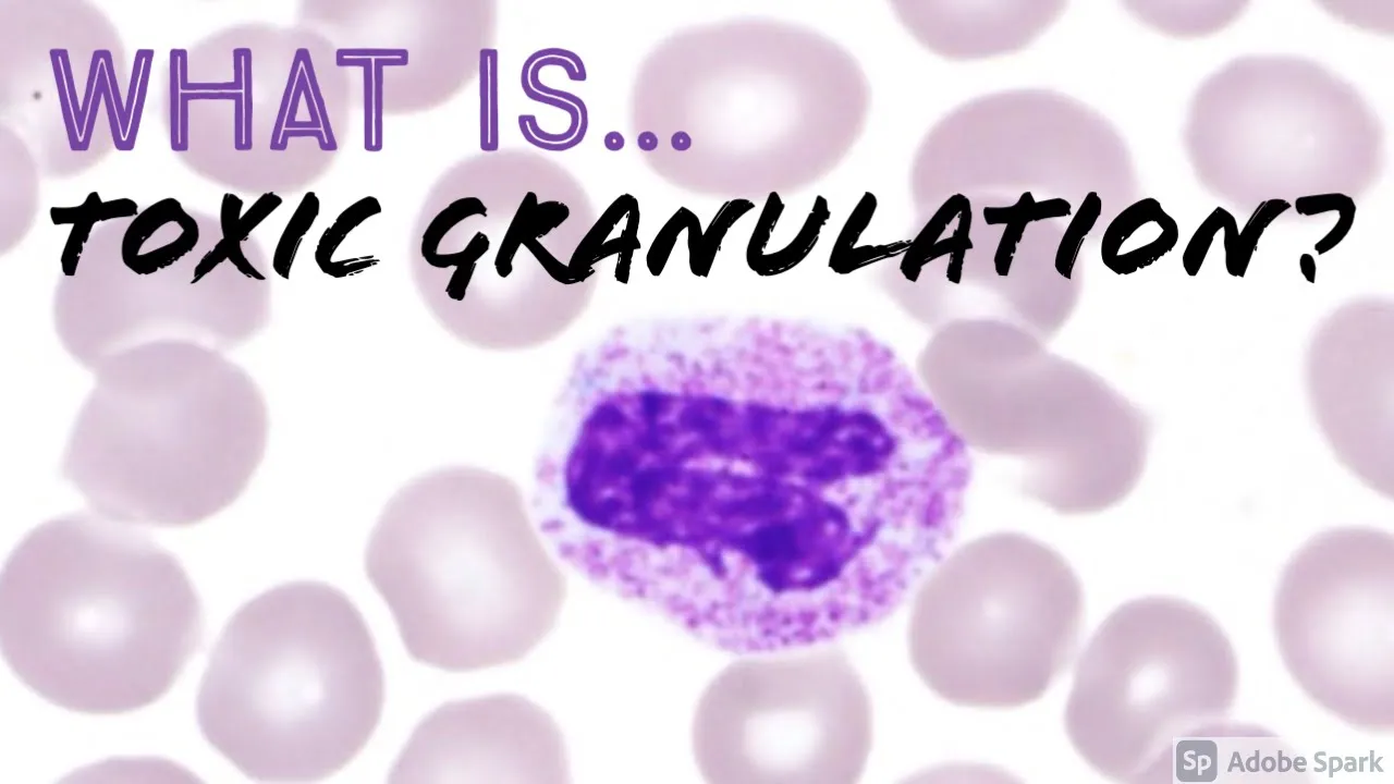
Addressing a critical feedback gap regarding benign reactive changes, this focused hematopathology video outlines toxic granulation. It explains how severe systemic inflammation or cytokine stimulation causes neutrophils to retain coarse, dark blue-purple azurophilic granules.
Knowledge Checkpoint
- Define toxic granulation and describe its visual presentation (coarse, prominent purple-black granules in neutrophil cytoplasm).
- Explain the physiological trigger for toxic granulation (bone marrow stimulation during severe infection or inflammation).
- Distinguish toxic granules from normal neutrophil granules and basophilic granules.

Why this video
This masterclass addresses the complex task of differentiating reactive lymphocytes from neoplastic lymphoid cells. It covers how antigenically stimulated cells (reactive "Downey" cells) expand their cytoplasm to mold around neighboring RBCs, compared to the rigid, monomorphic populations seen in malignancies.
Knowledge Checkpoint
- Identify reactive lymphocytes by their key visual markers: increased overall size, variable nuclear shape, and deep blue/basophilic cytoplasmic borders that "scallop" around adjacent red blood cells.
- Contrast the polymorphous, varied presentation of reactive lymphocytosis (typical of Epstein-Barr virus/mononucleosis) with the monotonous, uniform cell populations characteristic of neoplastic processes.
- Explain the diagnostic risk of confusing large immunoblasts with malignant blast cells.

Why this video
This practical clinical guide presents a fast, diagnostic flowchart to classify leukemias (AML, ALL, CML, and CLL) under microscopy based on three core questions: cellular maturity, lineage (myeloid vs. lymphoid), and cell count.
Knowledge Checkpoint
- Recognize blast cells (large cells with high nuclear-to-cytoplasmic ratio, fine chromatin, and distinct nucleoli) indicative of acute leukemias (AML/ALL).
- Differentiate Chronic Myeloid Leukemia (CML) by its hallmark "spectrum of myeloid maturation" (blasts, promyelocytes, myelocytes, metamyelocytes, bands, and mature neutrophils simultaneously present).
- Identify Chronic Lymphocytic Leukemia (CLL) by the presence of mature but fragile small lymphocytes and characteristic "smudge cells."
💡 Curriculum Note on Gaps: While the video pool covers toxic granulation and reactive vs. malignant lymphocytes, detailed videos on Döhle bodies (pale blue-gray, peripheral cytoplasmic inclusions of ribosomal RNA in neutrophils during toxic states) are limited. Ensure you search for clinical images of Döhle bodies and memorize their association with leukemoid reactions and severe infections alongside left shifts.
Module 5: Platelet Disorders & Systematic Smear Analysis
The final module integrates your lineage-specific knowledge into a professional, systematic microscope scanning workflow. It also covers platelet pathology, focusing on how to spot laboratory artifacts like EDTA-induced clumping to prevent false diagnoses of thrombocytopenia.
Recommended Videos

Why this video
This video explains the clinical difference between true thrombocytopenia and pseudothrombocytopenia—an in-vitro laboratory artifact caused by EDTA anticoagulant tubes which triggers antibodies to clump platelets together, leading automated analyzers to output a falsely low count.
Knowledge Checkpoint
- Explain the mechanism of EDTA-induced platelet clumping (pseudothrombocytopenia).
- Describe the corrective laboratory action when platelet clumping is suspected (redraw blood in a sodium citrate/blue-top tube and re-run).
- Identify giant platelets on a peripheral smear and understand their clinical association with increased platelet turnover.

Why this video
This video outlines the standard operating procedure for a complete, professional microscopic blood smear review. It emphasizes structural navigation: scanning the physical slide borders first, locating the ideal "zone of morphology," and building a systematic reporting pipeline.
Knowledge Checkpoint
- List the three parts of a blood smear (head, body, tail) and identify exactly where the "zone of morphology" or monolayer lies.
- Explain why the feathered edge (tail) must be scanned first at low power (to check for platelet clumping, microfilaria, or large abnormal cells).
- Describe the systematic "battlement" scanning pattern used to perform a manual differential count without repeating fields.

Why this video
This video provides a practical walkthrough of the complete analytical stage of blood smear reading. It covers how to use low power (10x) for quality assessment, transitioning to high-dry (40x) for estimate counts, and completing analysis under 100x oil immersion.
Knowledge Checkpoint
- Detail the objective lens sequence (10x -> 40x -> 100x oil) and what diagnostic information is extracted at each magnification level.
- Calculate a manual platelet estimate from a 100x oil immersion field (Average number of platelets per 100x field multiplied by 15,000 or 20,000).
- Formulate a structured diagnostic report detailing red cell morphology, white cell differential, and platelet adequacy.
Course Map
Key People Index
- Dr. Jeanette Ramos
Pathologist and Hematopathology Educator
Featured in the clinical training videos from@JMGardnerMD. Known for high-yield, clear-field microscopic walkthroughs identifying key morphological variants, toxic changes, and normal cell baselines. - Dr. Preeti Sharma
Pathology Board Review Lecturer
Featured in@UnacademyLiveNEETPGand@PrepLaddermedpg. Highly regarded for breaking down pathognomonic cell structures (schistocytes, target cells) into memorable, high-yield diagnostic patterns. - Dr. Hardik Mistry
Physiology Professor
Featured in@drhardikmistry. Specialized in demonstrating physical laboratory competencies and systematic microscopic leukocyte identification workflows for medical and lab science students.
Final Self-Assessment
Complete this comprehensive self-assessment to verify your mastery of peripheral blood smear analysis.
- Slide Preparation: Can you consistently prepare a wedge peripheral blood smear showing a distinct head, body, and an even, wave-free feathered edge?
- Wright-Giemsa Staining: Can you troubleshoot stain abnormalities (e.g., recognizing that an overly red slide indicates an overly acidic buffer or insufficient staining time)?
- Red Cell Sizing: Can you visually differentiate a microcytic erythrocyte from a macrocytic erythrocyte by comparing its diameter to the nucleus of a nearby small, resting lymphocyte?
- Spherocyte Recognition: Can you identify spherocytes under oil immersion and explain why they lack central pallor?
- Microangiopathic Hemolytic Anemia (MAHA): Can you identify schistocytes (helmet/triangle shapes) and list three microvascular conditions associated with their presence?
- White Cell Lineage Differentiation: Can you distinguish a normal monocyte from a normal large lymphocyte based on nuclear chromatin condensation and ground-glass cytoplasm?
- Left Shift Identification: Can you identify an increase in band neutrophils and intermediate myelocytic precursors, and link this pattern to bone marrow response to acute infection?
- Benign vs. Malignant Lymphocytes: Can you distinguish reactive lymphocytes (showing cytoplasmic molding around adjacent erythrocytes) from malignant blast cells (showing prominent nucleoli and high nuclear-to-cytoplasmic ratios)?
- Smudge Cells: Can you identify smudge cells on a smear and associate them with the fragile neoplastic cells of Chronic Lymphocytic Leukemia (CLL)?
- Pseudothrombocytopenia: Can you recognize platelet clumping under 10x or 40x magnification and explain why it causes a false-low automated platelet count?
- Manual Platelet Estimation: Can you perform a manual platelet estimate on 100x oil immersion and verify if it matches the automated analyzer output?
- Systematic Reporting: Can you execute a structured clinical report detailing erythrocyte variations (anisocytosis/poikilocytosis), leukocyte differential counts, and platelet adequacy in the correct monolayer zone?