

















Red blood cell morphological abnormalities include changes in size (macrocytosis with MCV >100 indicating B12/folate deficiency, microcytosis with MCV <80 indicating iron deficiency/thalassemia/lead poisoning), color (hypochromasia with central pallor >1/3 diameter indicating low hemoglobin, polychromasia showing immature reticulocytes), and shape (target cells in liver disease/thalassemia, spherocytes in hereditary spherocytosis/autoimmune hemolysis, schistocytes in microangiopathic hemolytic anemia, sickle cells in sickle cell anemia from HbS polymerization, echinocytes in renal disease, acanthocytes in liver disease, teardrop cells in myelophthisis), as well as distribution abnormalities like rouleaux formation and agglutination, which help diagnose various hematological conditions.
Red Blood Cell Morphology Abnormalities Explained
Added:Okay, so this is a video on morphological abnormalities found in red blood cells. Now, just before we begin real quickly, these abnormalities are pretty crucial to identifying and diagnosing many hematological diseases.
After we do a blood smear, where we smear a bit of blood and look at it under the microscope, we can use these morphological abnormalities to diagnose several diseases. And uh just in general, we're going to be talking about changes in red blood cell size, color, shape, and distribution. So if we look at these four images on the front here, the leftmost one has red blood cells of different sizes. You can see that some are big, some are small, some are medium sized. Um we're going to talk about what that's called and what that might be indicative of. We can also change in red blood cell color. Sometimes they appear blue under the microscope. Um that's that second image there. The shape can change as in cickle cell anemia. You can see that sickle-shaped cell in that third image. And of course the distribution of red blood cells can change. And sometimes we see weird patterns like that weird row of cells in the leftmost image. And we'll talk about that one too.
So first we have macrocytosis.
Macroytosis means large red blood cells.
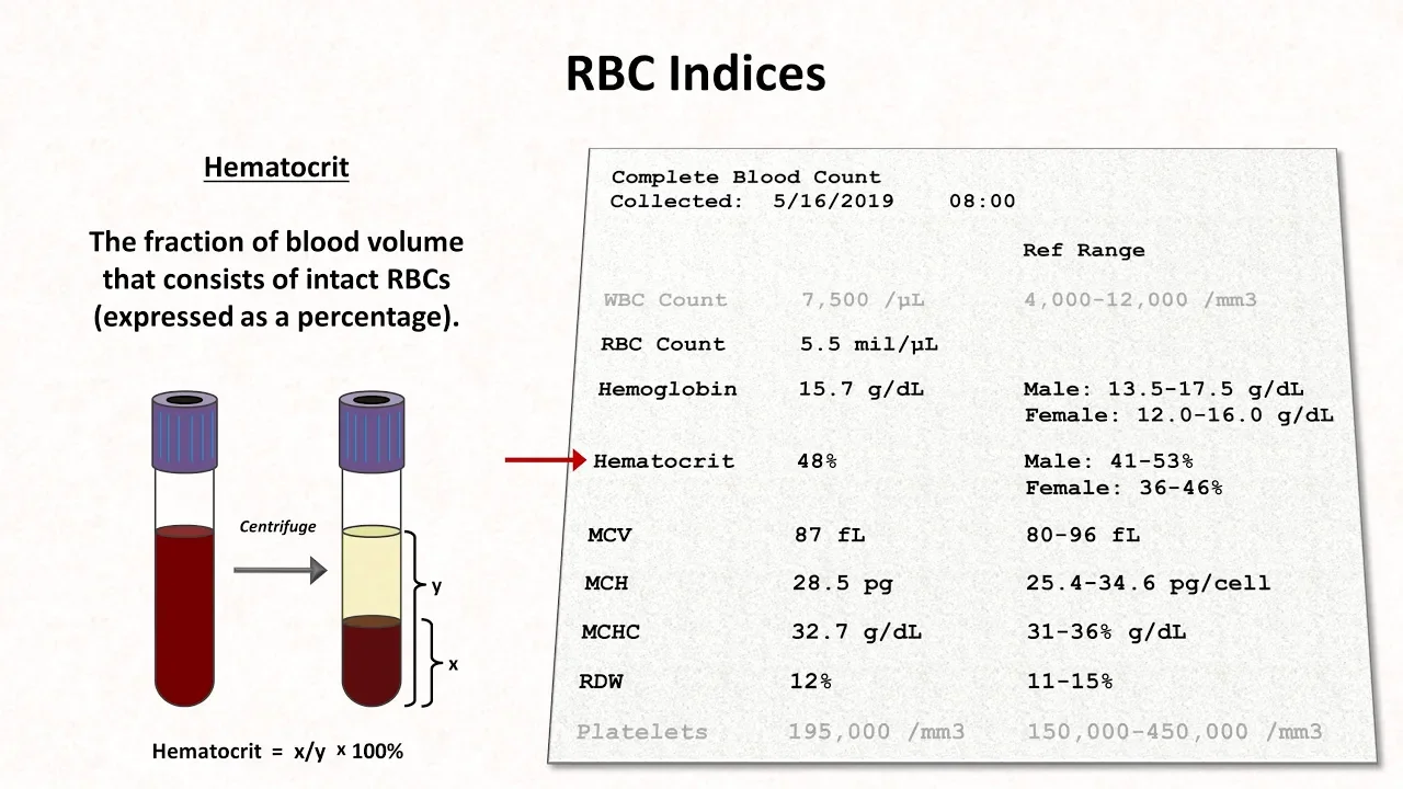
Now we measure the size of red blood cells with a lab test called mean corpuscular volume. uh MCV for short.
When this mean corpuscular volume is above about 100, we say we have macrocytosis and usually this is indicative of a disease where DNA synthesis is altered such as B12 deficiency and uh folate deficiency.
Both of those molecules are involved in either replicating DNA or the division of cells and so the cells keep growing and growing and growing and they never divide. That's why we see large red blood cells in macrocytosis.
The opposite of macroytosis is microytosis which of course means small red blood cells. This is also measured with MCV the mean corpuscular volume and it's associated with several diseases such as iron deficiency, lead poisoning and thalismia. Thalosmia is a kind of a fancy word. It means a problem making hemoglobin specifically the globin part of hemoglobin. So uh the globin is is like a globular protein that that red blood cells make that create hemoglobin which is the oxygen carrier. When you have a deficiency in the enzymes or in the in the process that makes the globin proteins you have athalosmia that causes you to make less hemoglobin and it results in smaller red blood cells. You can essentially think of red blood cells as like sacs or carriers of hemoglobin that that essentially kind of protect hemoglobin throughout the bloodstream.
So if you have less hemoglobin such as in iron deficiency and thalosmas, you're going to have smaller red blood cells.
And when you have a wide range of red blood cell sizes, it's called anocytosis.
The uh the measure for this is the red cell distribution with RDW for short.
red cell distribution with and this is kind of a standard deviation of all the red blood cell sizes. So that you you could you can see in the smear that there is a high RDW because some of the smells are really really small, some of the cells are really really big um and they just kind of vary in size. It's when you have a large range of red blood cell sizes. That's anocytosis.
Let's jump into disorders of red blood cell color. Now normally let's go back to this anocytosis slide real quick on a blood smear the actual microscope image you see red blood cells and they have like a little palar inside a little white region on the inside we call that palar now this white region comes from the shape of red blood cells you could see in the left um the the 3D model there that the red cell is a bonave discshaped so it's kind of like a disc but both sides of the disc are concave meaning they they kind cave in on both sides. That caved in region shows up as white on the blood smear. So if we go back to uh discoloration of red blood cells, we know that in normal cells, the red blood cell is supposed to have about 1/3 of its diameter be shaded white.
Have a palar that is about 1/3 the total cell diameter. That's in normal sizes.
Sorry, in normal red blood cells. When that palar is too big, we call it hypochromatic cells. Um, when that pellar is larger than one/ird of the diameter of the cell, like these six cells on the bottom here, [snorts] we call that hypochromatia.
Hypochromasia means too little color.
Um, and it describes red blood cells that have too little hemoglobin. It's measured with a uh a lab value called mean corpuscular hemoglobin. And it essentially if this was a real blood smear it would look like the same size cells but a larger white region inside.
It has less color. It is less reddish pink than the normal red blood cells.
That's hypocchromasia.
Polychromasia means multiple colors.
It's when we see red blood cells of multiple or unusual colors. Uh here's red blood cells that are shaded grayish blue which is what's most commonly seen.
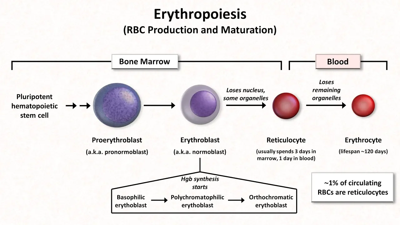
And these grayish blue red blood cells are often reticulytes which is the precursor to red blood cells essentially immature red blood cells. So when we have polychromasia we usually see these grayish blue reticular sites which are immature red blood cells.
All right let's jump into disorders of red blood cell shape. Now first before we begin this term poilocytosis means that you have a lot of red blood cells that vary in shape. A few slides back we had anocytosis which meant red blood cells that vary in size.
Poilocytosis means red blood cells that vary in shape. We see a blood cell here or a blood smear here that shows red blood cells that are of very different shapes.
We can see in the zoomed in image we have some teardrop shaped cells. That bottom one right there looks like a sickle cell. We have red cells that look like spheres. Some are kind of ovalshaped and some still look normal.
So poiloytosis means red blood cells that vary widely in shape. And we're going to kind of go through these different shapes and um talk about what the shapes look like and what might be causing them.
So first we have target cells. Target cells are called target cells because well they look like targets kind of kind of easy uh to remember this one. They're also called cottoytes if you need a another another word to memorize. And it's related to liver disease, thalismas, a misshapen or an abnormal form of hemoglobin called hemoglobin C.
And we also see target cells after the spleen is removed post spleenctomy. Now, target cells look like targets on a normal blood sphere because they have they're like more wavy than normal red blood cells. We know normal red blood cells, if you look at them from the side, they look like this this top series of circles here. It's like a by concave disc where the where each side of the disc kind of dips in once. In a target cell, we have each side of the disc dipping in twice. So, we kind of see how that would form a target if we were to turn that cross-section over and look at it as we do in the blood.
Next, here we have spherosites, which are exactly what the name implies. They are red blood cells that are shaped like spheres instead of, of course, the usual by concave disc shape of the red blood cells. I put a normal cross-section at the bottom middle here of a normal red blood cell. You can see that both ends of the disc kind of dip in. They kind of cave in. This is not the case in a spherosite at the bottom right here where the where the cells are around.
They're more spherical. On the blood smear on the left, this looks like a loss of central palar. So that white region in the middle of the cells is gone in spherosittes. Uh this is associated with several diseases such as her hereditary spherosytosis and autoimmune hemolysis. These are spherosytes. They're spherical red blood cells instead of the normal by concave disc shape of red blood cells.
[snorts] Next, we have shistytes, which are essentially red blood cell fragments that look like the red blood cells were torn apart. Look, if you were to pull on a red blood cell until it broke, it looks like or that's what a shistyite would look like. And that's kind of what happens to form shistytes. Uh, oftentimes when a when a red blood cell goes by an arterial wall that has a plaque on it, either a calcium plaque or a fatty plaque, the red blood cell can shear if there's a sharp edge on that plaque. When you shear a red blood cell, you're going to see a fragment that has sharp edges as if you just tore it apart. One disease that's associated with the formation of shistotes is microangopathic hemolytic anemia. And that forms red blood cell fragments that look like red blood cells were just ripped apart with sharp edges called shistytes.
[snorts] We have a big one here, sickle cells, which of course are most often associated with sickle cell anemia. A normal red blood cell, as we said several times, looks like a bonave disc.
If you can imagine a polymer like a long stick of uh a long stick shaped molecule forming inside that biconave disc, it's going to stretch that normally round red blood cell until it looks like a sickle.
As you can see in that bottom middle image there, the polymer that forms is made of hemoglobin molecules. And there are a few uh kind of factors, a few environmental changes that can cause hemoglobin to sickle like that. Uh this includes low pH, low oxygen concentrations, and high temperatures.
Um the the abnormal hemoglobin that causes sickling is actually a single point mutation in the gene that makes hemoglobin. And when hemoglobin forms these long polymers that kind of stretch out and bend that cell, it looks like a sickle. sickle is an old farming tool.
You could look up what that looks like and see that it kind of does look like that um that that sickle. [snorts] Now, this sickle-shaped cell does not bend in the microvasculare as well as a normal red blood cell does. And you can see that in the right image there. This is the basis behind some sickle cell crises. And this can be incredibly painful when these cickle cells block the tubes that are your blood vessels and prevent blood from getting to certain parts of the body. So cickle cell anemia is a very wellstied um disease. We we know quite a bit about it and it causes pain in people that have this mutation when it blocks blood flow to uh to to parts of the body.
Next we have ekinosytes. These are also called burr cells. I don't know if I'm pronouncing that right but burr cells is easier to pronounce. Uh these have projections. These are like little spiky projections around the cells. And um in burr cells these projections are regular. So they're about the same size all the way around the cell. Uh burr cells are associated with renal disease.
Similar to bur cells are spur cells. And the fancy word for these are aanthsytes.
Kanthytes differ from bur cells in that their projections are irregular. They are not evenly spaced. They are not necessarily the same size. And uh spur cells are associated with liver disease.
So let's go back one more time. Burr cells have regular projections associated with renal with kidney problems. Spur cells are irregular projections associated with liver disease.
Next [snorts] we have teardrop cells.
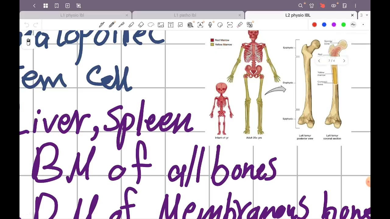
And this is pretty easy to remember because they look like teardrops. These are formed when the bone marrow is infiltrated with something that should not be there. So something like scar tissue or maybe cancerous cells or lymphosytes or maybe even bacteria goes into the bone marrow grows in the bone marrow and of course bone marrow is where all these red blood cells are produced. It's the site of urethropoesis. So if there's something invading the bone marrow it's going to malform the shape of the red blood cells that come out and make teardrop cells.
Next, we have a couple diseases associated with a red blood cell distribution. Um, first we have this French word, rulo or ru. Um, it's it's literally like a linear stack of red blood cells. Ruo means like a roll in in French, like like a stack of coins. Um, and you can kind of see this formation on the blood smear. The red blood cells are stacked in a line like that. Now, why does this happen? Usually red blood cells have a surface charge on their outer membranes that keep them from sticking together. This means that they have a similar surface charge that kind of keeps them separate. That's why in all the previous blood smears, none of the red cells were were touching.
Usually [snorts] when you have too many antibodies, too many imunoglobulins that neutralize these charges, the red blood cells can attract each other with charge charge interactions that were usually repelling each other and then they form these long stacks, these rulo.
Next we have aglutination which is similar to ru but much less orderly. This is when a bunch of red blood cells form big aggregates big stacks and come together and touch and overlap and are all over the place next to each other. They collect in clumps and it looks very disorderly. It's not in a straight line like in ruo. It's a big pile, a big aggregate of red blood cells. This occurs when red blood cells are coated with IGM which is a type of is a type of antibbody. It's a pentimer.
So it's a large type of antibody that has five binding sites and it's large enough to bridge the red blood cells together to cause this aggregation um seen in aglutinated red blood cells.
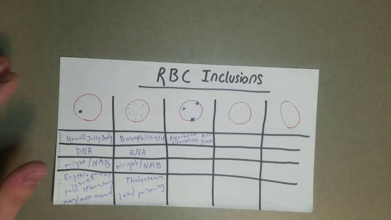
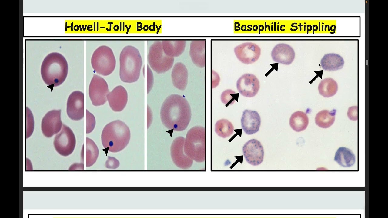
[snorts] Now we have one miscellaneous condition of red blood cells that I figured I'd throw in there. This is the howell jolly bodies. How well jolly bodies are they on the blood smear they look like small purple dots that um that are inside the red blood cells. It's usually one howell jolly body per red blood cell. This uh you start seeing this after the spleen is removed from the body or if the spleen is not functioning properly. So if you have like a spleenic infarction which means the spleen is small and and not working or if your spleen is otherwise just nonfunctional you're going to have how jolly bodies. Now what are these little purple dots? They're actually remnants of nucleic acids. They are nucleic acids that normally would have been um collected in the spleen and reintegrated into the body, but instead they were released and they form these these aggregates, these insoluble uh bodies that that collect in the red blood cells. These are nucleic acids that form howell jolly bodies.
Up Next

Leukemia Diagnosis on Peripheral Blood Smear: 3-Question Method
@pathology360
395 views•2024-03-29

Graphic Medicine: Comics for Collaborative Healthcare Communication
@nationalpatientadvocate
189 views•2023-12-04

Neuroanatomy: Central and Peripheral Nervous System Divisions Explained
@AKLECTURES
136.2K views•2014-09-20

Stages of Labor and Vaginal Birth | Childbirth Animation
@nucleusmedicalmedia
52.1M views•2017-08-18
Related Study Plans & Knowledge Roadmaps
Structured learning paths in Medicine