















![Hematologie - Leucemie Myeloide Chronique (LMC) [FMPC]](https://i.ytimg.com/vi/a9YarX5HIpQ/hqdefault.jpg)
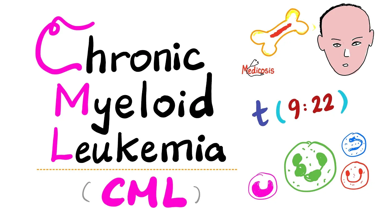

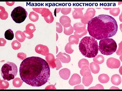




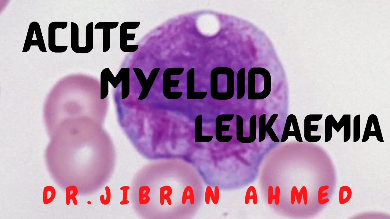
To diagnose leukemia types (ALL, AML, CML, CLL) on peripheral blood smear within 30 seconds, systematically answer three questions: (1) Are the cells mature or immature? Mature lymphocytes with smudge cells suggest CLL; (2) Is there a mixture of immature and mature cells with very high WBC count? This indicates CML, where myeloblasts proliferate but retain maturation capacity, creating a 'family reunion' picture of various cell stages; (3) If predominantly immature blastic cells are present, check for Auer rods—presence indicates AML (myeloid lineage), absence indicates ALL (lymphoid lineage). This approach is based on the fundamental principle that CML shows maturation while acute leukemias show blocked differentiation.
Leukemia Diagnosis on Peripheral Blood Smear: 3-Question Method
Added:Understanding of normal hematopoiesis, specifically the divergence of the myeloid and lymphoid cell lineages.
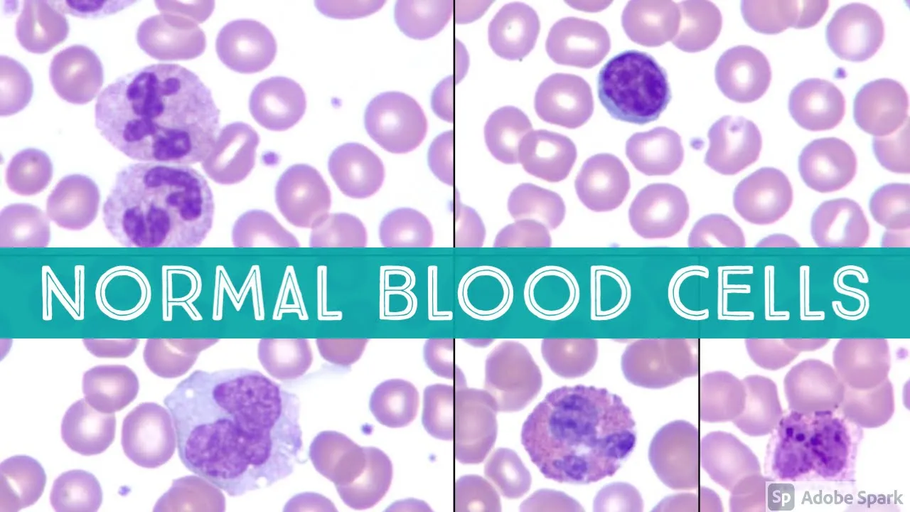
Familiarity with normal peripheral blood smear morphology, including the identification of mature white blood cells like neutrophils, lymphocytes, and monocytes.
Broad definitions of 'acute' versus 'chronic' hematologic malignancies in terms of cell maturation and clinical onset.
Basic cytological terminology used to describe abnormal blood cells, such as 'blasts', 'smudge cells', and 'Auer rods'.
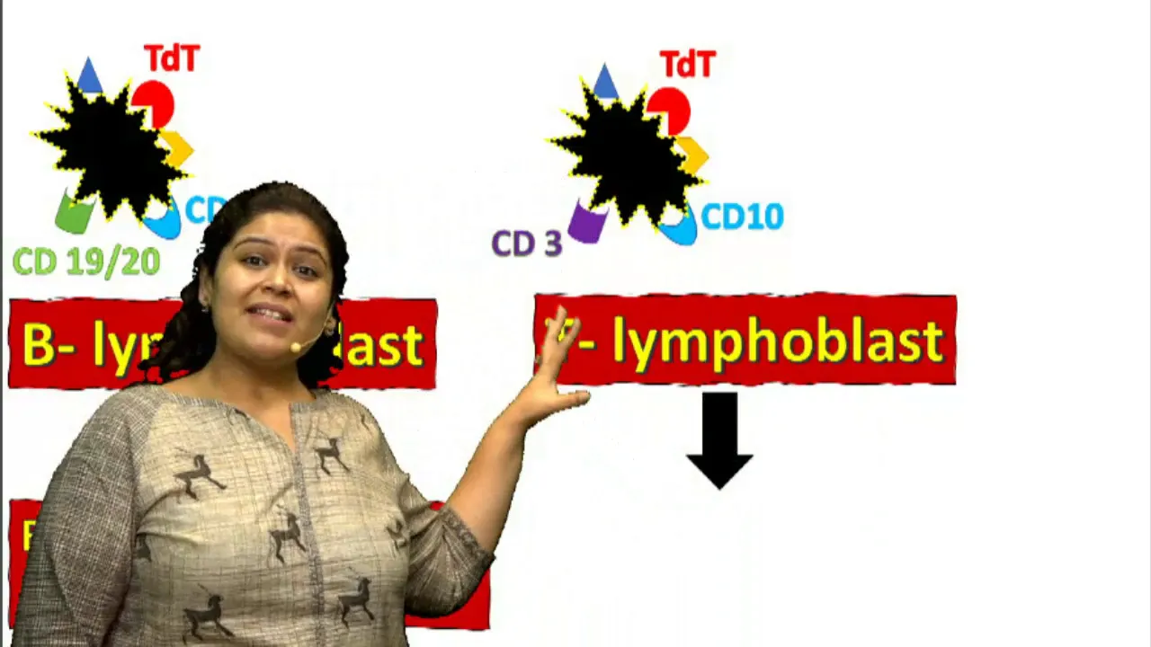
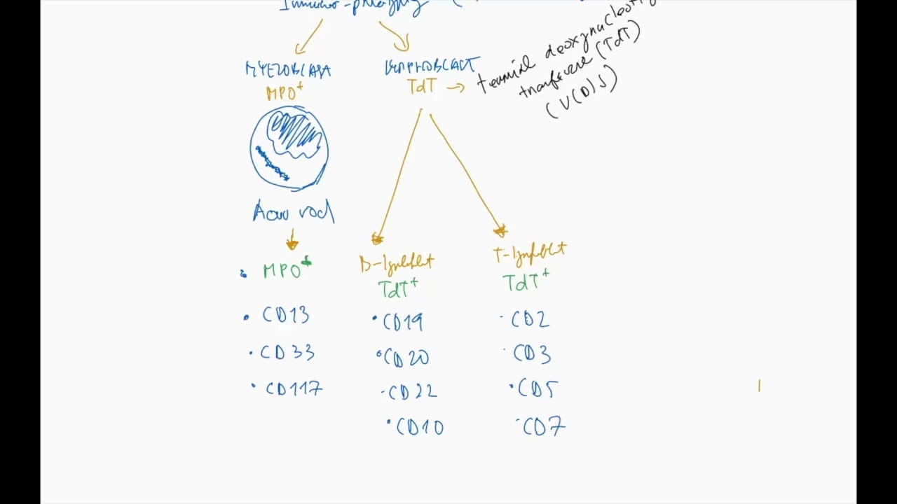
Use of flow cytometry and immunophenotyping (CD markers) to definitively characterize and confirm leukemia subtypes.
The role of cytogenetics and molecular genetics (such as FISH or PCR) in detecting key mutations like the BCR-ABL translocation in CML.
Interpretation of bone marrow biopsy and aspirate findings to complement peripheral blood smear analysis.
Familiarity with international classification systems (such as the WHO classification of myeloid and lymphoid neoplasms) and clinical staging systems.
395 views31likes9:59Original Release: 2024-03-29
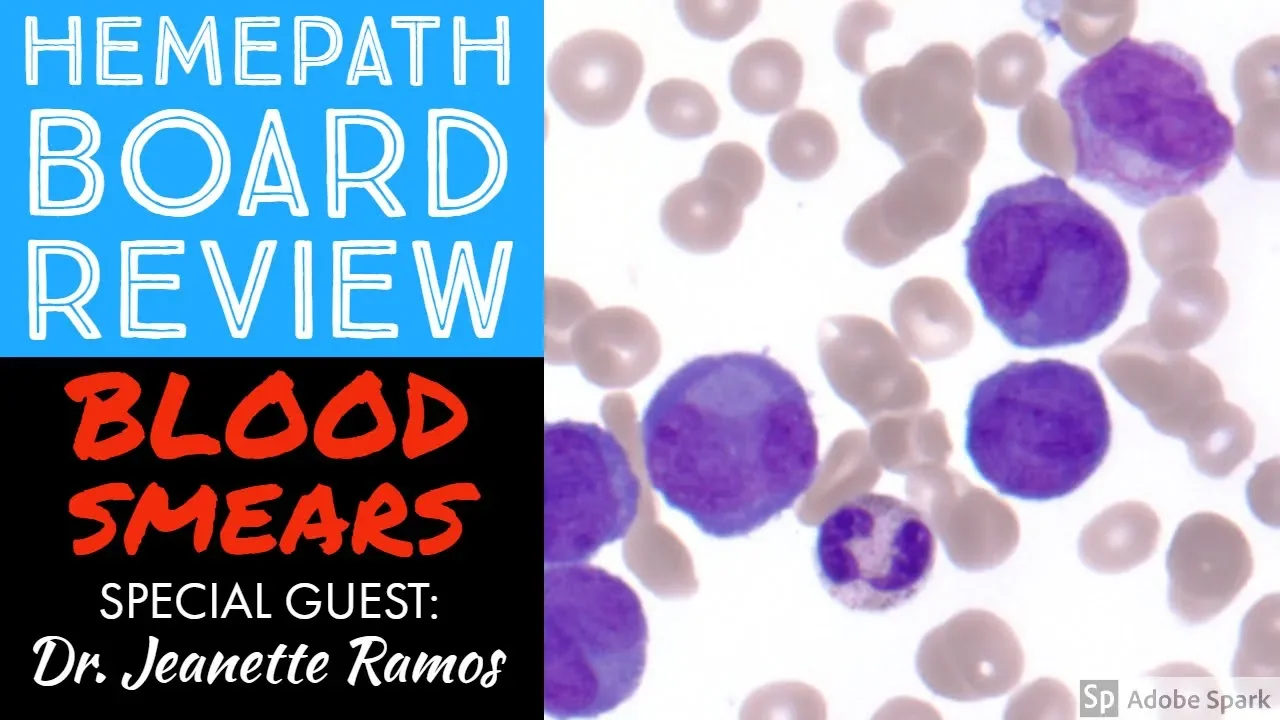
how to train your brain to figure it out that what kind of leukemia is this is it Al or AML or CL or CML under 30 seconds on the peripheral blood smear on the questions which you have received how to reach out to the accurate response that I'm going to teach you right now the number one you need to basically ask three questions in this process and if you accurately answer this three question you get the accurate answer and none of this is basically based on cramming or memorization of any fact this all based on validated scientifically proven tools and concepts of the leukemias if you apply them it becomes very easy for you question number one if you see a peripheral blood smear and the options are L ml CL CML the first question that you need to ask yourself that what I'm seeing on the peripheral blood smear are they all mature looking cell or mature appearing cell or I'm seeing immature cells also if the answer is I'm only seeing mature looking cell I'm not seeing any bizarre looking large plastic cells it would be most likely CL chronic lymphocytic ukia Plus in clll as you are all aware you'll be seeing lot of ruptured lymphocytes also on the peripheral blood smear those are called SMUD cells or parachute cells or basket cells plus some other buzzwords would be for cl that typically it would be seen in the adults elderly population 60 plus plus the the tumor cells the peripheral blood they can show soccer ball like chromatin pattern or cracked mud like chromatin pattern this is another buzz word for them and definitely the diagnosis of C is done by doing flow cytometry that this cells should be positive for cd5 cd23 cd200 all these characteristic markers so this is now in this list of CLL CML Al AML clll is ruled out now I have three options Al AML and the CML so next question if while looking at the peripheral blood you need to ask yourself that as I'm seeing all the cell population are all the cell population on the peripheral blood smear on the peripheral blood this patient are there entirely immature blastic cell bizar looking large cell or there is a kind of mixture of immature looking plastic cells and mature looking cells you need to answer this question and if you are getting on the peripheral blood smear that you are getting a mixture of immature looking cells like your getting a lot of myoblast prosite myosite and also getting mature looking neutrophil this is a characteristic feature of peripheral blood SP for CML chronic M leukemia and chronic my leukemia also getting too many WBC there would be very striking leucocytosis very high WBC count now what is this is also not based on cramming actually this is based on a simple principle that CML versus AML or acute leukemia which I usually teach earlier also on the Bas concept of hypothesis of taale of Two Cities the basic fundamental principle of CML versus acute leukemia B it ml or L is that in CML there is a proliferation of myoid cells are occurring in the bone marrow precursor cells but the myo precursor cells like you know that they usually mature myoid precursor cells in this stage myoblast prosite myosite metam myosite ban for mature neutrophil in CML what is occurring that the myoblast are proliferating but the myoblast are not trapped in that myoblast stage they are maturing down the line they are becoming those myoblast they're becoming proside miloy metam band from at neutrophil but what is the difference fundamental difference with ML is that in AML what is happening the myoblast are proliferating but they have lost the capacity of maturation or differentiation further down the line the myoblast cannot become Pro myoside metam myosite band from mature neutrophil like they cannot become mature neutr usually when I teach this I usually tell It's A Tail of Two Cities you can think that this is a city a and City B in City a what is happening that lot of newborns are taking birth in City b a lot of newborns are taking birth in City a the newborns are basically trapped in that newborn stage they cannot become the schoolgoing kid adolescent mature adult but in City B the newborns are taking but but the newborns can become mature adults they can become schoolgoing kid mature adults so 10 years down the line what you would be seeing the difference between two cities population that in City a and City B both the cases pop population is increased but CTA is entirely filled with newborn babies which is acute leukemia and CTB AML in that case acute Miler leukemia and CML what is happening that the newborn babies you are also getting newborn babies you're also getting mature adults and all all kind of population and the whole population is increased with a mixture of population because there is no blockage of maturation or differenciation of the blast cells Malo blast in the same so now I'm coming back to that question if you see that on peripheral blood smear that I am get seeing mixture of immature s I'm seeing some Malo blast Pro I'm also seeing mature looking neutrophil with a very high WBC count this is a feature typical peripheral blood SM picture of chronic Myer liuk chronic M when I teach I usually tell them that they look like a uh Family Image you know family reunion image there is a baby there there is a grandfather is also there grandmother is also there there is a new the uncle is also there the parents father mothers are also there so I have myoblast I have mature looking neutrophil grandfather I have the I have the inprocess Milo blast Pro myosite all the cells are also there that is the typical picture of the CML compared to an AML you see entirely all the picture is made of newborn babies so if you get that the mixture population mature and maturing cells IM mature cells this is a CML feature plus other buzz word would be CML typically would be seeing 50 plus around typical presentation and the characteristic very characteristic feature they would be describing that massive splen omally or they can say that there's a dragging sensation in the left side of the abdomen this is due to increased spinning SI enlargement this would be the typical feature of this Plus on peripheral blood spere and same you can also get osop increased and Bops increased these are the other features so now in the list clll CML Al AML CL CML has been crossed out now I if I look at the Al and AML are remaining now if I look at the peripheral BSM if I see that entirely the population population is mostly is blastic population IM immature looking bizarre looking cells this is you have to think about acute deuk because acute deia's Hallmark feature is blockage of differentiation of blastic cells blastic cells are multiplying but blastic cells has lost the capacity of maturation that is the Hallmark feature of acute my leukemia acute leukemia by the way if it is for myoblastic proliferation is occuring where myoplast lost the capacity of differentiation then it is AML if it is lymphoblast or proliferating but they have lost the capacity of differentiation to mature lymphoblast lymoc side that is Al and that's why in W classification you see that they are characterized by more than 20% blasters in the bone marrow or in the peripheral blood because blast cells are replicating but they have lost the capacity of maturation and defens so this is now I have so the third question that I'm going to ask you that I see entirely on the pop peripheral blood SM a lot of blastic cells let's say in the question they mentioned that there is 37% Blaster present and the Blaster showing presence of our rods the blast are showing voluminous cytoplasm and lot of nucleoli so basically they're talking about feature of myo blast so if you have more than or equal to 20% Milo blast on the peripheral blood smar obviously it would become acute Milo leukemia that is the point you need to remember so peripheral pme typically to make your life simple at the basic level they give you our rods feature if you see rods on a blast it is basically indicative of feature of myoid lineage myoid blast but in lympho blast this hour rods would be absent so the question number three that you need to ask you that in both AML and L I'm seeing lot of newborn babies are there but if they on the forhead of newborn babies there is our Rod there is a sticker there of our Rod that would be typically AML but that sticker would be absent in the AL that is how you need to approach this is the question number three that you need to ask to to reach out that this is AML or Al so so this is the way actually you need to think act think that how you should reach out that this is AML uh cl cl AML and obviously other features would be easy for you like if you look at age that could be an ad buz word for you l typically we get that common in the first decade of Life 2 to 5 years but in AML M you can be seen mature adults typically this is typical for that TL L can also occer in the adolesents but Al is typically disease of the the Pediatric population this is the most common cancer in the Children Al So based on this principle I think it becomes very easy for you and obviously there will be markers markers I would discuss in a separate session the markers would be helpful you to differentiate between the LL and ml but this is on the basis of the peripheral blood smear if you approach in this way that are all look cells looking mature lymphocytes with some smart cells think about cell are there is a mixture of mature and immature cell maturing cell with lot of lucites wbcs on per blood think about CML if you're seeing entirely immature population think about acute leukemia they showing our rods it would be acute my li then not showing our rods it would be acute lymas this is a simplified approach actually how can you figure it out on that 30 seconds I would continue with this in the further sessions also thank you so much
Up Next
11:23
Petechiae vs Purpura vs Ecchymosis: Causes, Size & Clinical Signs
@MedicosisPerfectionalis
62.9K views•2019-11-13

48:47
Graphic Medicine: Comics for Collaborative Healthcare Communication
@nationalpatientadvocate
189 views•2023-12-04

10:28
Neuroanatomy: Central and Peripheral Nervous System Divisions Explained
@AKLECTURES
136.2K views•2014-09-20

3:05
Stages of Labor and Vaginal Birth | Childbirth Animation
@nucleusmedicalmedia
52.1M views•2017-08-18
Related Study Plans & Knowledge Roadmaps
Structured learning paths in Medicine
medicine
Abnormal LFTs: Diagnostic Workup & Pathology
01Liver Anatomy and Basic Physiology
02Introduction to Liver Function Tests (LFTs)
03Hepatocellular vs. Cholestatic Patterns
+ 3 more learning modules
12h6 modules
Explore Roadmap
medicine
Analyzing Blood Smears: RBC, WBC & Platelets
01Introduction to Blood Components & Smear Preparation
02Normal Morphology of Blood Cells
03Erythrocyte Pathologies & Morphological Variants
+ 2 more learning modules
12h5 modules
Explore Roadmap
medicine
Mechanical Ventilation: Waveforms & Alarms
01Anatomy and Physiology of Breathing
02Introduction to Mechanical Ventilation & Core Modes
03Ventilator Settings and Waveform Interpretation
+ 2 more learning modules
12h5 modules
Explore Roadmap
medicine
Neuro Exam: Localization & Stroke Syndromes
01Foundations of Neuroanatomy and Vascular Supply
02The Neurological Examination: Step-by-Step
03Principles of Neurological Localization
+ 3 more learning modules
18h6 modules
Explore Roadmap