












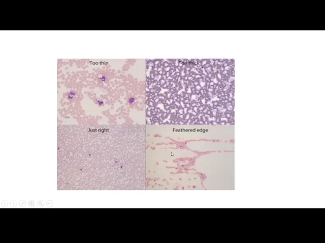




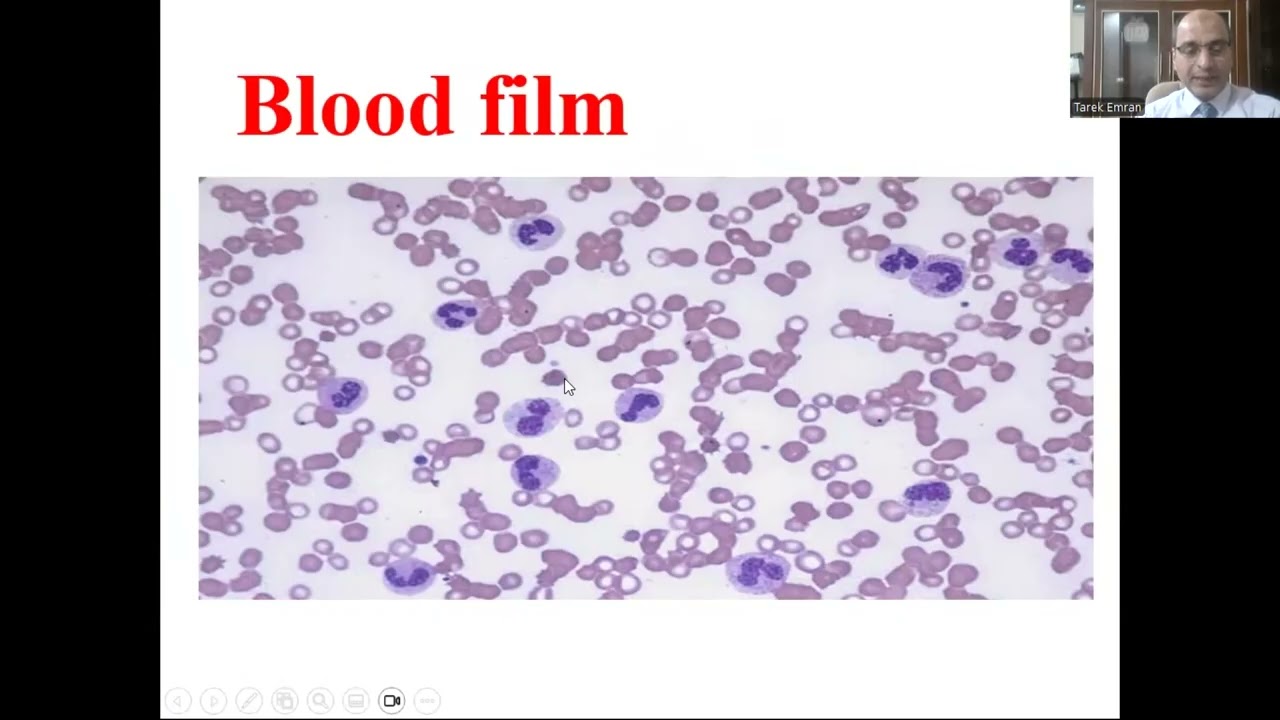



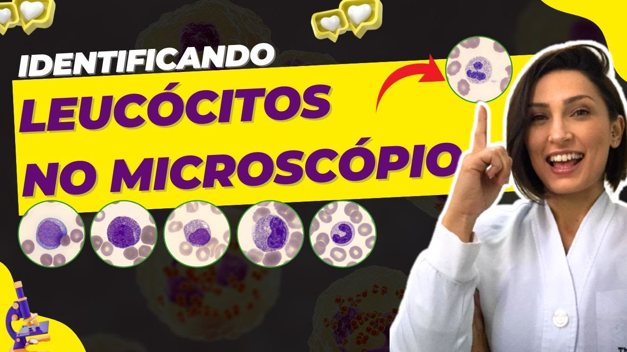
White blood cells (WBCs) are classified into granulocytes (neutrophils, eosinophils, basophils) and agranulocytes (lymphocytes, monocytes), identified through systematic morphological analysis: compare cell size relative to RBCs (7.2 μm), assess granule presence/absence, and examine nuclear characteristics; neutrophils (50-70% of WBCs) have 1-6 nuclear lobes with fine granules, eosinophils (1-4%) show bi-lobed spectacle-shaped nuclei with brick-red granules, basophils (<1%) have S-shaped nuclei with purple/blue granules, lymphocytes (20-40%) display single large oval nuclei with no granules (small lymphocytes = RBC size, large lymphocytes = double RBC size), and monocytes (2-8%) feature kidney-shaped nuclei with pale cytoplasm.
WBC Identification in Peripheral Blood Smear | Hematology Lab Guide
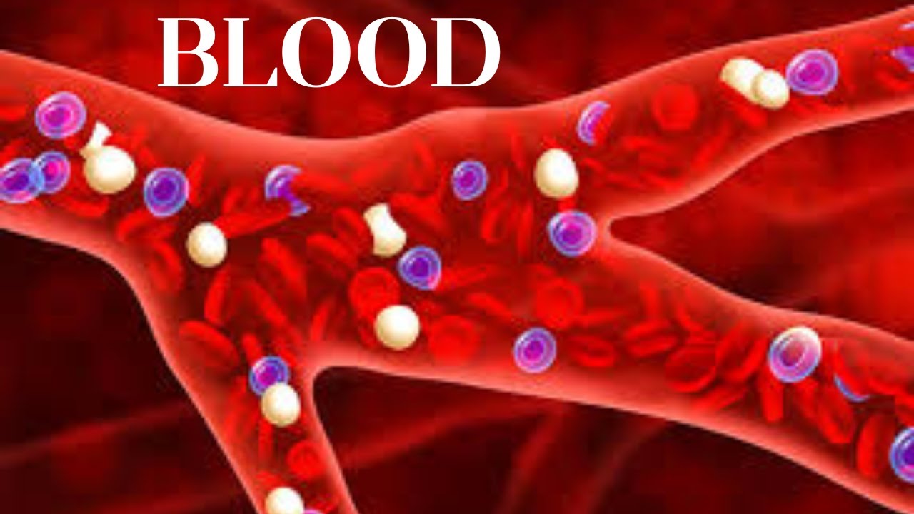
Added:Basic components of blood (plasma, red blood cells, platelets, and white blood cells) and their primary functions.
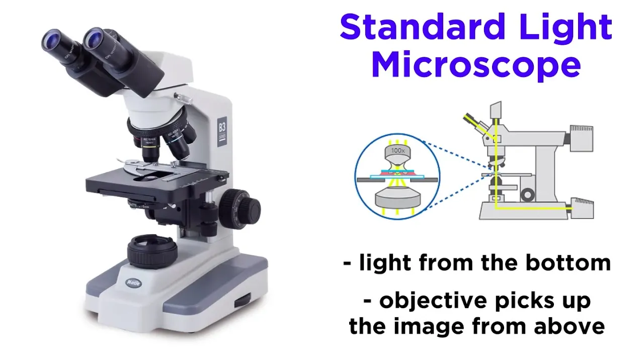
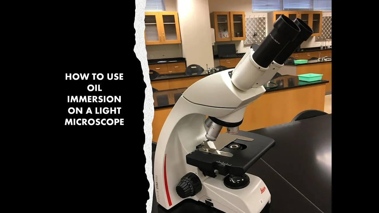
The principles of light microscopy, specifically how to use the oil immersion lens (100x magnification) required for cell morphology.
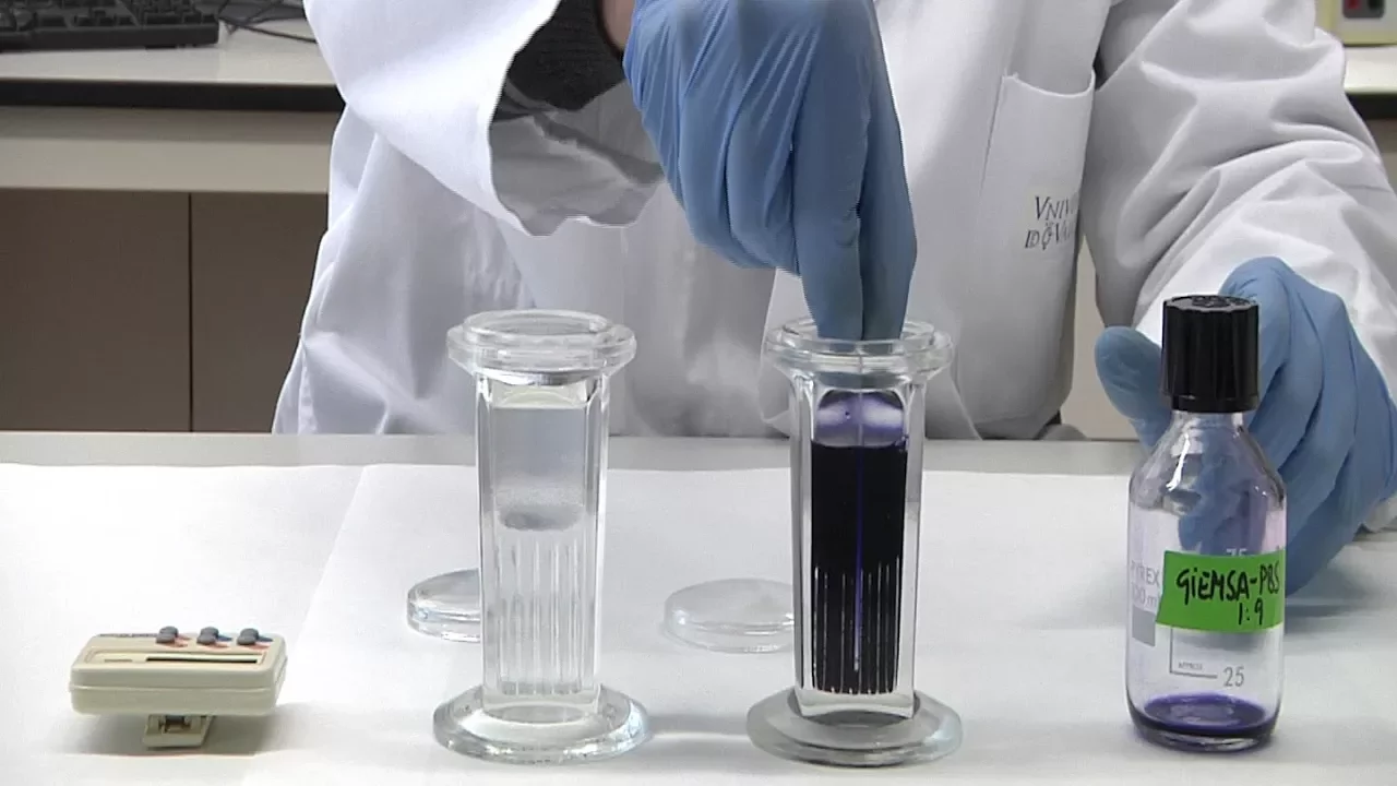
Understanding of standard blood smear preparation and staining techniques (such as Wright-Giemsa stain) and how stains affect cellular structures.
The classification of white blood cells into granulocytes (neutrophils, eosinophils, basophils) and agranulocytes (lymphocytes, monocytes).
How to perform a manual 100-cell differential count to calculate the relative percentages of each WBC type.
Recognition of abnormal WBC morphology, such as hypersegmented neutrophils, reactive lymphocytes, toxic granulation, and Döhle bodies.
Clinical correlation of abnormal WBC counts (e.g., identifying bacterial infection via neutrophilia/left shift, or allergic reactions via eosinophilia).
Identification of immature WBC precursors (blasts, myelocytes) associated with hematological malignancies like leukemia.
178.1K views4Klikes13:03Original Release: 2019-10-18
hello everyone in this video I am going to explain how to identify WBC types in peripheral blood smear white blood cells or leukocytes or in short form it is known as WBC there are two types of WBC granulocytes and a granulocytes neutrophils eosinophils and basophils these are the granulocytes wears lymphocyte and monocyte these are the granulocytes now how to identify white blood cell in peripheral blood smear first of all look size of the cell as compared to size of RBC here we are going to compare different types of WBC as comparable to size of the RBC the size of the RBC normally it is 7.2 micrometer so first of all you have to look the size of cell as compared to size of RBC now you have to find out whether the size almost equal to size of RBC little larger or double to size of RBC very large as compared to size of RBC now you have to find out granules whether granules are present or absent so if the size of the cell is almost equal to size of the RBC granules absent and nucleus it is the oval same then this cell it is known as small lymphocyte now if you find a cell which is little larger or double to size of the RBC see whether Grendel's are present or absent if granules are present now check for the number of lobes of the nucleus or the focus over the nucleus if granules are present little larger or double to the size of RBC as well as one to six lobes of the nucleus then this cell it is known as neutrophil inside this neutrophil you will find 1 to 6 lobes of the nucleus granules are praising a little larger or double to size of the RBC alright now the second if you find two lobes of the nucleus and there in a spectacle shape then this cell it is usable field so in the use you know field there are two loads of the nucleus and nucleus arranged in such a way that it looks like spectacle in same granules are praising and larger or double to the size of pharmacy so that is the Yoshino field now if you find a cell which is little larger double to the size of our PC granules present and two lobes of the nucleus and lobes of the nucleus are arranged in such a way as seen with granules on the nucleus so this is the base often in basal field you will find two lobes of the nucleus both lobes are arranged in a safe manner with granules over the nucleus granules are present and little laser or double to the size of our business so that cell is basal film now if you find a cell which is little larger or double to the size of our PC granules are absent now find out the nucleus oval shape of the nucleus so this cell it is the large lymphocyte so which are the specification of the large lymphocyte little larger or double to size of the RBC first thing second thing granules are absent the nucleus is oval shape so this cell it is the large lymphocyte now if you find a cell which is very large as compared to size of the RBC and granules are absent now check for the nucleus if you find that the nucleus is kidney shape then this cell it is the monocyte so specification of the monocyte the cell size is very large as compared to size of the RBC granules are absent kidney shaped nucleus so this cell it is the monocyte this neutrophils eosinophils and basophils these are the granulocytes where more lymphocyte large lymphocyte and monocyte these are the a granulocytes i hope from this flowchart you are able to understand and identify the type of WBC's very easily now I am going to explain the characteristic features and specification of each cell in detail first start with the neutrophil neutrophil it is a granulocytes out of hundred WBC we are able to find 50 to 70 of the neutrophil or in other words in normal condition we are able to find 50 to 70 percentage of the neutrophil out of all WBC cell size 10 to 14 micrometer nucleus multi load one to six lobes of the nucleus and the nucleus color is purple laser the number of the loops of the nucleus the neutrophils are more juvenile and more the number of lobes of the nucleus the neutrophil are more semi or in other words the lace the number of the lobes of the nucleus the neutrophils are younger ones and higher the number of the lobes of the nucleus the neutrophils are elder or senior citizen cytoplasm slight bluish in color granules fine sand like particles red brown or purplish in color function phagocytosis mediate febrile response or due to related to fever producing response so here whatever the cell you are seeing it is the neutrophil you are able to see the number of lobes of the nucleus 1 2 3 & 4 it is a granular cells so brain duals are present and the granules are the red or brown or purple is in color now these are the actual photographs of the neutrophil which I took in the hematology lab during the practical of peripheral blood smear now look at this neutrophil there are four lobes of the nucleus look at this there are three lobes of the nucleus look at this there are one two three four and five lobes of the nucleus so all these cells have the neutrophil and these are the granulocytes now you see no film out of hundred WBC's we are able to fire one two four percentage of the eosinophils or in some other words the concentration of use you know fill out of all the blue pc is one to four percentage cell size ten to fourteen micrometer nucleus by loop spectacle shape purple in color cytoplasm use you know philic light pink in color granules large coarse brick red in color and do not cover the nucleus function mild phagocytosis limiting allergic manifestations and provide mucosal community now this cell which you are seeing it is the ii you know fill it is a granular site so these all are the granules the granules are brick red in color and do not cover the nucleus and the nucleus is by load spectacle in shape and it is the purple in color so this is the photograph of actual yoshino field which I found in the hematology lab look the spectacle shape by love nucleus and these all are the granules if you find this type of cell then it is absolutely yoshino film now the piece of film the concentration of P so fill is less than one percentage very less and they are not able to found on routine basis on a peripheral blood smear cell size ten to fourteen micrometer nucleus by load purple in color arranging a ship cytoplasm basophilic or blue in appearance granules large coarse purple or blue in color and overlying the nucleus function mild phagocytosis and allergic manifestations so here whatever the cell you are seeing it is the basal field it is a by load and the lobes are joined in such a way that it looks like a as in shape and granules are purple or blue and it covers the nucleus or it overlies the nucleus alright so this cell it is the basal field till date I am NOT able to see any type of the basal field so I am sorry I don't have the real photograph of the basophils now lymphocyte the concentration of lymphocyte or the differential count of the lymphocyte is 20 to 40 percentage both small lymphocyte and large lymphocyte now cell size small lymphocytes 7 to 10 micrometer equal to the size of the RBC large lymphocyte 10 to 14 micrometer double to size of the RBC nucleus single very big oval or round in shape central in position occupying hood of the cell cytoplasm scanty sky blue in color less amount than the nucleus granules are epson so lymphocyte is a a granulocytes a function produce antibodies so here whatever the cell you are saying that is the small lymphocyte and here whatever the cell you are saying it is the large lymphocyte see the nucleus it is a single very big oval or round in shape and it is occupied central in position and occupied almost full of the cell or in some other words 2/3 portion of the cell has been occupied with this nucleus the same thing in the small deposit and large lymphocyte so I have real images of the small and large lymphocyte so here this is the small input side and here this cell it is the last lymphocyte find out as compared to size of the RBC so all these are the RBC this is the last lymphocyte look at this same if you are able to see the size almost larger and upper to that of the size of the RBC then that is the large lymphocyte look at this cell you are also finding that the size is almost larger to that of the size of the RBC so these all are the this both are the large lymphocyte and this one smaller one it is the small Impa site now the monocyte differential count of the monocyte is 2 to 8 percent that means how 200 WBC we are able to find two to eight of the monocytes on a normal basis or in some other we are able to find two to eight percentage of the monocyte out of all WBC's cell size ten to eighteen micrometer or even more than it nucleus single kidney say pale in color peripherally position cytoplasm pale blue amount more than the nucleus granules are absent so that monocyte is a or granulocyte function phagocytosis work as tissue macrophages so this is the blood vessels and this is the monocyte once the monocyte is circulating inside the blood vessel the cell is known as monocyte but once it is coming out of the blood vessel and enter into the tissue the cell it is known as macrophages all right okay now dear whatever the cell you are saying it is the monocyte single kidney say pale in color nucleus and it is peripheral in position so look at this this is the kidneys about the nucleus and it is situated on the peripheral part of the cell so here I have few images of the real monocyte now look at these both pictures you are clearly able to find kidney shape of the nucleus and it is situated on the peripheral part of the cell if we are going to compare the size of the monocyte with this size of the RBC then you are able to see that it is either twice or thrice bigger than the size of the RPC alright so this cell it is known as mono site you
Up Next

33:46
RBC Morphology & Shapes | Pathology | NEET PG 2022 | Dr. Preeti Sharma
@UnacademyLiveNEETPG
13.7K views•2022-03-05

48:47
Graphic Medicine: Comics for Collaborative Healthcare Communication
@nationalpatientadvocate
189 views•2023-12-04

10:02
9th to 12th Cranial Nerve Examination | Clinical Lab | Physiology
@drhardikmistry
123.5K views•2019-07-21

3:05
Stages of Labor and Vaginal Birth | Childbirth Animation
@nucleusmedicalmedia
52.1M views•2017-08-18
Related Study Plans & Knowledge Roadmaps
Structured learning paths in Medicine
medicine
Abnormal LFTs: Diagnostic Workup & Pathology
01Liver Anatomy and Basic Physiology
02Introduction to Liver Function Tests (LFTs)
03Hepatocellular vs. Cholestatic Patterns
+ 3 more learning modules
12h6 modules
Explore Roadmap
medicine
Analyzing Blood Smears: RBC, WBC & Platelets
01Introduction to Blood Components & Smear Preparation
02Normal Morphology of Blood Cells
03Erythrocyte Pathologies & Morphological Variants
+ 2 more learning modules
12h5 modules
Explore Roadmap
medicine
Mechanical Ventilation: Waveforms & Alarms
01Anatomy and Physiology of Breathing
02Introduction to Mechanical Ventilation & Core Modes
03Ventilator Settings and Waveform Interpretation
+ 2 more learning modules
12h5 modules
Explore Roadmap
medicine
Neuro Exam: Localization & Stroke Syndromes
01Foundations of Neuroanatomy and Vascular Supply
02The Neurological Examination: Step-by-Step
03Principles of Neurological Localization
+ 3 more learning modules
18h6 modules
Explore Roadmap