
![BIOLOGÍA - Célula Eucariota Parte 01 [CICLO FREE]](https://i.ytimg.com/vi/mTbeSPHnZ9U/maxresdefault.jpg)

![Normal Morphology of Blood Cell [ part 01] | Morphology of RBCs in Hindi](https://i.ytimg.com/vi_webp/SsnhAc3EWCw/maxresdefault.webp)









![Hematologie - Spherocytose Hereditaire [FMPC]](https://i.ytimg.com/vi/QUdIjwK8kLI/hqdefault.jpg)
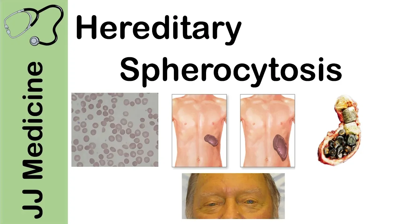


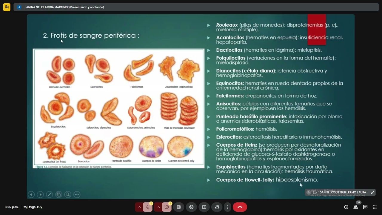








This comprehensive pathology lesson covers essential red blood cell shapes and morphologies for NEET PG exam preparation, including microcytic and hypochromic cells (smaller than 7 microns with increased central pallor), spherocytes (round, spherical cells without pallor seen in autoimmune hemolytic anemia, hereditary spherocytosis, and G6PD deficiency), reticulocytes (large blue cells indicating immature RBCs), bite cells (degmacytes) and sickle cells (drapanocytes) associated with G6PD deficiency and sickle cell anemia respectively, teardrop cells (dacrocytes) in myelofibrosis, fragmented cells (schistocytes) in microangiopathic hemolytic anemia, target cells (codocytes) in hemoglobin disorders, echinocytes (burst cells) in burns and renal failure, acanthocytes (spur cells) in abetalipoproteinemia, rouleaux formation (stack of coins) in multiple myeloma due to loss of zeta potential, agglutination in cold agglutinin disease, stomatocytes (with central slit) in stomatocytosis, and pincer cells in band 3 defects.
RBC Morphology & Shapes | Pathology | NEET PG 2022 | Dr. Preeti Sharma
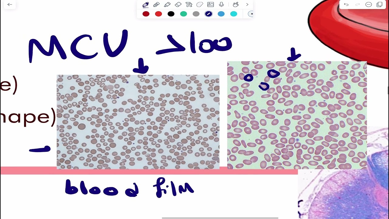
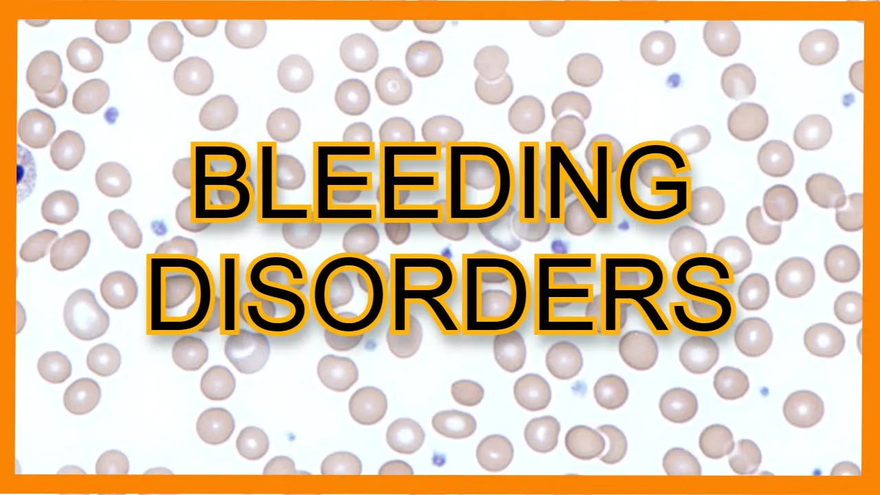
Added:hi everyone good afternoon guys i hope i'm audible and visible to all of you so welcome to this afternoon session and like i had told all of you yesterday also we are continuing the same pattern and that happens to be two lessons that we have back to back so first from two o'clock right now up till approximately 2 30 we will be having a session on this channel where we will be discussing the different red blood cell shapes and morphologies thereafter we will be meeting once again at 2 40 p.m which is a thing that we started on um the unacademy app which is known as the open house session now what do i uh many of you were a part of this open house session it's like an audio only session no video no pdfs no screen sharing it's an audio alone session more like a podcast but also a method of learning which sometimes is very good for building up your memory so yesterday if you all remember we had discussed something to do with the cd markers today we'll be having cd markers part two so that all the markers of whatever we have to know in pathology are completed for us so these are the two lessons that you have to look out for and additionally all of you know that today morning we could not have a kickstart morning session so like a bonus session i've put up one session again at 9 00 pm tonight this 9 pm session is going to be on the unacademy app it is free for everyone to attend and this is going to be based on the image based questions so this is for all students neet fmg ini cet some quick 40 45 minute session some quick image-based questions we'll be discussing and that will be on the unacademy app tonight at nine o'clock okay so um dr sayak skin tumors and salivary gland tumors are planned in the coming month in the kickstart morning session so that is where you could attend these topics right okay so i guess we are done with the all the orientation of the classes of the day let's start with the session yesterday if you all remember we did all the inclusions all these bodies written over here we did all of that and what are we going to do today all the different shapes that are written first we'll figure out those so before i begin the very first one which is the most important and i'll enlarge it even further if you guys want whenever you get a ps ps means whenever you get a peripheral smear the first thing that you have to search you want to comment on the red blood cells you want to comment on their size their shape so the first thing that you try to pick out is a lymphocyte that's the trick of solving a peripheral smear so the first thing i pick up is a lymphocyte and which part of a lymphocyte the blue color dot i pick up the nucleus of a lymphocyte because i know that a nucleus of a lymphocyte is approximately 7 microns means it is similar to the size of a red blood cell even a red blood cell is 7.2 microns right so 7 microns is what i know a lymphocyte is and i have to compare the red blood cells with the size of a lymphocyte so for example if this is the size of the lymphocyte that we have and i ask you that what do you think is the size of this red blood cell or what is the size of this red blood cell so you can very clearly see that these red blood cells are all smaller so can i label them as microcytic cells that is a very important feature that the first shape variation that you can get of a size variation i would say size being small and that is going to be microcytic so how did i get to know it's a smaller size i compared it with the size of a lymphocyte nucleus thereafter the next thing that you have to comment on is the color so if i draw a normal red blood cell this is how i draw a normal red blood cell means how much of the center will i leave as empty so remember one third pallor is a normal thing one third pallet is considered as a normal finding and if i say that no the red blood cell is looking like this means very very white it's showing you more than one third pallor you'll say ma'am this means the hemoglobin is less this means the cell is hypochromic less of color hypochromia is what i'm seeing so have a look at these guys have a look at for example this red blood cell or look at this red blood cell or any one that you pick up you don't see only central one third you see most of them are empty so i can clearly mention that they are hypochromic so what kind of a peripheral smear does this become you will say that okay this happens to be a microcytic hypochromic peripheral smear anything else that you guys see over here any any shape variation that you guys are able to appreciate i'll highlight it what is this cell that i've highlighted over here because this gives you a very good hint and a very famous question so it's known as a pencil cell this cell is referred to as the first shape variation that i'm telling you this is a pencil cell so overall now i know it's a microcytic hyperchromic blood picture with the presence of pencil cells and when i get this kind of a peripheral smear i'm very commonly doubting that this is a case of iron deficiency anemia so i think the first two three things are very clear in front of me if i have to categorize this chart you know that fine if there is a normal size if the size of the red blood cell is small it becomes microcytic if the size of the red blood cell would have been more it would have been macrocytic further anything less in color means how much of pallor is more you say one-third pallas is other more than one-third pallar so you can see how the center is becoming empty empty empty so all of these come under hypochromia cells so first uh size and hypochromia understood now let's go on to the next slide so what do you see over here first let me ask you what is the shape of the cells that i have encircled the ones that i am encircling any comment on those you will say ma'am they are completely round they are completely spherical and they don't have any pallet they are full of the hemoglobin can you see there is no pallet that i am seeing they are full of hemoglobin so firstly because they are completely round we call them correct everyone's right these cells are referred to as the spherocytes these cells are spherocytes so first thing that the examiner will ask you is that how do you define spherocytes are they i've understood they are round they are spherical but are they smaller than the red blood cells or are they bigger than the red blood cells normal red blood cells so i like to add a word over here i like to call them micro spherocytes means that their size is lesser than a regular red blood cell they are smaller in size and what are the three conditions the three most important conditions in which i can see spherocytes what is the most common cause of pserocytosis and that is auto immune hemolytic anemia that's the most common cause other than autoimmune hemolytic anemia the very famous hereditary spherocytosis i'm sure everyone thought of as the first thing and thirdly even g6pd deficiency can show us zero side so please remember as i see most of you are answering cerosites are seen in three main conditions out of which the most common cause is autoimmune hemolytic anemia then hereditary spherocytosis and g6pd deficiency but other than that but other than this steroid part of it did you also see some other population if i open this for the house today i ask all of you any other cell population that you see out here anyone who wants to give me an answer other than the spherical population yes a quick 30 seconds i give to everyone to think over it any other cell that you wish to point out and if you see teardrop no teardrop is my name teardrop i'll be showing you in a while then you'll know the difference anything else see uh the size maybe all the cells are very small but then can you see some cell which is very very big can you pick up these two jumbo cells out here yes and i want you to analyze two things jumbo cells in these two jumbo cells that i have highlighted out here first thing you'll say that ma'am their size is more i agree with you that's why we are calling them jumbo cells and also look at their color does everyone agree that these two jumbo cells have a little bit of a bluish color than the other red blood cells other red blood cells are reddish but these cells are not only bigger but they are giving me a little bit of a bluish tinge so how have i learned these cells please remember when i'm seeing cells which are big and blue the red blood cells which are big and blue are known as the reticulocytes yes what is the other name for a reticulocyte reticulocyte is also known as a polychromatophile the two names of a reticulocyte are firstly reticulocyte and second it is a poly chromatophil and how will i identify it apart from the regular cells it will be big and it's going to be blue big and blue cells are polychromatophils i hope uh that is okay with everyone so this mayor taught me two things first is the spherocytes and the second thing that it taught me was the big and blue cells that is the reticulocyte okay having said that now let's move on to the third image um is it crystal violet stainless whatever i have taught you is a normal romanovsky stain but if you ask theory questions from that point of view then reticulocyte is not stained with crystal violet reticulocyte is stained with new methylene glue reticulocyte is stained with new methylene blue but that's not the question here over here i'm showing you all normal romanovsky stained slides okay meanwhile i started off with the third one and yes all of you are right i think this is the most classical one the three arrows because the cell looks as if it's bitten off so firstly all of you are right these are definitely bite cells but byte cells have another name to them and white cells are also known as digma sites byte cells also known as digma site and which disease do we see this in we see this in g6pd deficiency g6pt deficiency yesterday i had taught you some bodies also in g6pd deficiency which all of you were very confident about yesterday so now we've completed everything when i talk about g6pd deficiency we see two things number one happens to be the bodies that we saw that was the heinz bodies i hope everyone remembers we've studied it yesterday and number two the cells that we see are known as the bite cells or the degmar sites so the first there are a lot of d's over here that i'm going to teach you today this is the first d the d for decmar site is a byte cell because now comes the second d what is what are all these cells that you're seeing over here what shape is this what rbc shape very classical once you see this shape i think we are never going to forget it it looks like a sickle because these cells are said to be the sickle cells and no one's going to ask you the name of this because they know it's very evident it's a sickle cell but they'll ask you the other name for it and the other name for a sickle cell is a drapanocite it's a drypanocyte and no points for guessing where will you see a sickle cell you will see it in a patient of sickle cell anemia so yes that's the next d that you learn the drapanosite is the sickle cell there's a third d also that comes in red blood cells and this is the third d abby sometime back someone told me ma'am tear drop cell hotel see a teardrop cell should look like a teardrop like a drop of water like a drop of tear right and this is how a teardrop cell will actually look like so see can you see this cell over here it's looking like a teardrop this cell over here this cell over here so there'll be one pointed extension coming out of it that is what you will call as you know a drop of water or a drop of tear that is a teardrop and that's the third d that is known as dacrosite that is known as dacrosite so three ds that i've taught you degmaside drapanocyte dacrocyte first tell me this dacrocyte or teardrop cell which condition is it seen in everyone's right this is seen in myelofibrosis a very important question that you get in the exam i'll tell you the concept behind it when they say myelofibrosis i'll draw this bone marrow out here when they say milo fibrosis they mean in the milo in the marrow bone marrow there is fibrosis so in the bone marrow there is a lot of fibrosis that is there which means that when the red blood cells are trying to come out when the red blood cells are trying to come out they've got a proper mesh work of fiber they have to force and find their way through they have these fibers stopping it so in that attempt of struggling and coming out of the marrow their shape changes they become these tear drop in shape so the reason for teardrop is the resistance that they face the fibrotic tissue creates resistance and their shape changes so finally when i try to put all the three d's together these are the three d's that you have to know which they will ask you and you don't have to get confused the first d that you see over here i know it's a byte cell but the first d that you should know by itself how will you learn it so bite should remind you of dog bites so you can even learn it as dogma site dogma site is which cell dogma site is the bite cell so decmar site dog bite coming to this one this is the this is the dacrosite dacrocyte basically means tear drop cell i think everyone knows that when i in ophthal when we use the word dakro we use it for the lacrimal gland like dacroadinitis so dacrover is for lacrimal gland right the primal gland which produces tears so dacrocyte is the word that i will be using for the teardrop set and the third d that we have the one that you don't need a mnemonic for now that is the drapano side and rapanocyte is nothing but a sickle cell are we all clear with this the three ds degma dogma bite cell dacro tears so teardrop cell and drapanocide sickle cell so all of this is done let's move on to the three d's which are most confusing i hope those are over now let's move on to the next one here all the weird rbcs are encircled and you say ma'am i can't come to a conclusion all of them are of a different shape this one is looking a little pointed this one is looking like a triangle this one is looking like a helmet so all weird weird kind of shapes are put up over here so can i say these are fragmented red blood cells these are fragmented red blood cells means they are broken they are fragmented into pieces so first thing is it can fragment and it can change into any shape it can look irregular like this also it can look like a triangle also it may look like this it may look like a helmet so if they are talking about helmet cells triangular cells they're talking about the same thing so all these they have grouped under one term called fragmented red blood cells which are also known as schistocytes so schistocytes is the name given to fragmented red blood cells so next question that they'll ask you is where do you see schistocytes you see it in micro angiopathic hemolytic anemia where do we see schistocytes micro angiopathic hemolytic anemia that's a very famous question if we have fmg students attending this uh this is a question that has come in your previous year also that fragmented red blood cells are a part of which anemia they are a part of miha microangiopathic hemolytic anemia which many of you are calling as hus ttp they are types of mihaolina hemolytic uremic syndrome ttp dic all of them come under the microangiopathic hemolytic anemias only so they are just examples of the same thing perfect so yeah what is this this is a slide where i want all of you to give me observations because it doesn't have one cell it has lots of cells lots of varied type of cells it has so i want all of you to give me an observation on what all are you seeing yes so okay first answer i've got target cells i see three cells guys i'm asked i'm expecting three answers one is target cell i agree two more answers left two more okay echinocytes have got second answer media third answer target cells done echinocytes done echinocyte sky cousin brotherhood very similar sounding echinocyte and acanthocyte okay so three answers i've got now let's start labeling what all i'm seeing over here subsequently i'll start with the first answer that you guys gave me all these cells which look like a target all of these because i you know exactly like a target some of the hemoglobin is here some of the hemoglobin is at the center so these are known as target cells but everything has a different name right target cells are also known as codocytes target cells are also known as kodo sites so first you'll ask me my target word i've understood because you know it's looking like that dart board the target that we have why do you call it codocide because the meaning of the word kodo kodo kamath and when they saw these target cells on electron microscopy they realized they look like a bell shape these on electron microscopy have a bell shape and bell so that is why target cells are also known as codocides okay having said that now you'll ask me where all can we see target cells is it specific for anything not at all target cells can be seen in many conditions but where do you get maximum questions t for t target cells are seen in hemoglobin disorders like thalassemia like hemoglobin c disease like hemoglobin e disease and even in cases of iron deficiency anemia but again reminding you some important question here that is target cells are seen in thalassemia and thalassemia means hemoglobin disorder so along with that some other hemoglobin disorders like hemoglobin c hemoglobin e they can also also show you target cells also known as codocytes that was the first finding target cell huge second finding dick the second finding all of you told me was echinocyte so now i'm putting up the second finding in this particular picture target cell to done now i see the second finding and that is this so let me draw what is this cell showing you i see a cell which is blurred out like this it is blurred out like that you know the membrane is not defined like this it's blurred out it's irregular and blurred out so you blurred out membrane i call that a burst the blurred out membrane is basically a burst cell and please remember the other name for a burst cell is an echinocyte the other name for a burst cell is an ekinocite you know many of you were also calling it something called spur cells and acanthocyte so there's too much of confusion which is a burst cell which is an echinocyte which is a spur cell which is any canthocyte what sorry term say so remember these three alphabets will have everything to do with it the way you write a b the way you write or probably the way you write an e for an echinocyte is the same way you write a b so if you get confused was it a burst cell or was it a spur cell so remember the way i wrote an e is the same way i wrote a b so equinoxide and word cell happen to be the same thing where do i see these i see this in three conditions listen very carefully so first and foremost these cells are seen in burn patients number one second these cells are seen in patients with renal failure that is patients who have uremia and number three these are seen with prolonged edta changes means if the blood is kept in edta for a very very long time this kind of cell can come up so i think many of you did know this answer which is very good that is how do i learn this give birth cells so b say i will think of burns you will stand for uremia and e of a kynosite will remind me of adta changes is the second cell also of the smear okay because now i come to the third and the last cell of the smear that you had to know which looks as if it has spikes which looks as if it has spikes on it so spike matlab if i want to call it a spiky cell s for s i also call it a spur cell spiky cell or spur cell or a canthosite is what we need to know here spiky cell or spur cell or a canthocyte they all mean the same the meaning of spike spur and acantho what do all of them mean all of them mean that there are spikes on the surface and where are you going to see this please remember a for a acanthocite is seen in a beta lipoprotein it is seen in a beta lipoprotein so finally i think now you will not get confused that which is the acantho and spur and which is the echino and bird the combinations are clear because somewhere you know the way you write e is the way you write b so that is echinocyte and bursal and the other one is spiky spurry that is the acanthocite so if i again show you the diagram now all of you can identify the first cell that i showed you over here was a target cell the second cell which was blurry all around the blurry cell was the bursa and the third cell that i showed you over here was the spiky one that was the spur cell so i hope all three over here are also okay with everyone now let's move on to the last two three i think that we have can you identify these two see the difference between these two is the most important see what has happened here versus what has happened here so firstly let's see what has happened in the first one i see that all the red blood cells are stacked they've given you a picture for reference also all the red blood cells are showing you like a stack of coins and this stack of coins is very famously yes known as the perfect many of you have given me a right answer and this is known as the roulette formation very famous question of pathology and can you tell me which disease roulette formation the stacking of coins is seen in a plasma cell tumor that is multiple myeloma multiple myeloma shows us the presence of roulette formation it's a five star question that you get in hematology roulette formation i hope everyone is able there is a very long stack of coins but if you look carefully there's a shorter stack of coins here and there's an even shorter stack of coins here so it necessarily doesn't have to be a very long stack of coins even short short stacks of coins are being formed over here that is all known as roulette formation and you see that in a case of multiple myeloma compare it with the other picture you will say over here also the rbcs are coming together but are they coming together like a stack of coins or are they coming together like a clump so please remember clump or agglutination is what is happening moving so the difference between the two is very important to know i put it side by side i hope you are able to identify the difference if they come together like a stack of coins you will call it roulet if they come together like this clump you call it agglutination and can you now tell me which disease is going to show you this kind of an agglutination the name of the disease has the word agglutination in it that is what is special about the disease it is cad that is cold and gluten in disease cold and gluten in disease is what you need to know out here so it has the word right the name in it okay it's a disease i know that secondly it's going to show me agglutination that i can see over here and cold cold means that this condition will be seen at low temperatures what is the temperature cold at 4 degree celsius is where we see this and i'll ask you one small little theory question that which is the antibody which works at this temperature or which is the antibody which is responsible for this cold temperature or cold and gluten in disease very good and that is going to be igm so you've uh i've got two questions which i'm coming up to this uh so if i ask you that why did the rbcs make a clump why did they have gluten in it you'll say man this is a disease which happens in cold temperatures at four degrees and the antibody which is responsible for this is igm so you've got your answer for the agglutination the reason for that some of you want to know what is the reason of this stack of coins why in multiple myeloma have you got a stack of coins i'll take exactly 30 seconds to explain that not a very tough concept let us draw normal red blood cells these are the normal red blood cells that we have and what kind of a charge do normal red blood cells have all the normal red blood cells have a negative charge and we know negative charge this negative charge is known as zeta potential so can i say all the red blood cells which have this negative charge have basically something called zeta potential that is normal and my basic understanding from school days tells me that all same same charges will repel since all of them are negative they are going to repel from each other they stay away from each other but now imagine what happens in a case of multiple myeloma do we agree multiple myeloma means lots and lots of antibodies that will be circulating in the blood so can i say all the red blood cells now will have a coating of an antibody around them they will have lots of coating of antibody around them so somewhere is their negative charge hidden because all these antibodies are coating them can i say there is a loss of that negative charge or a loss of zeta potential that is going to occur so now they are no longer showing that negative charge or negative negative that has been masked that has been covered so do you think now they're going to stay away from each other or are they now because of the zeta potential being lost they'll end up coming close to each other and if they end up coming close to each other they'll make a stack of coins so guys everyone understood that the reason for the roulette formation is that in multiple myeloma there are so many antibodies which results in a loss of the zeta potential and that is the reason that the stack of coins is formed so whosoever asked me i'm sorry i couldn't see your name but i hope your doubt is solved right okay coming to the last one that we have these are very two rare ones but we should know rare things also just in case they come in the exam what are these red blood cells this is an inherited disorder autosomal dominant disorder you'll see ma'am over here if i look at the red blood cell they've got a slit in the center they've got a line in the center as i see it again like a stomata like an opening they've got a line in the center so remember in school days we used to read something like this in plants like plants have stomata so same way these red blood cells also have a stoma or a stomata or a mouth in them and these are known as stomatocytes these are known as stomatocytes if you ask me ma'am why does this happen i'll say it's an autosomal dominant condition it's a genetic disorder so a very rare question to come but i'm just showing you the rare ones also one last one last that we have is this and who's going to answer this for me the one that has been shown with an arrow if you want i can draw it also it's looking more like a mushroom more like a mushroom um okay doctor i'll come to your doubt also after this what is this mushroom-shaped cell known as this is also known as a pincer cell now that's something that could come as a future potential question yes very good this is a pincer cell this is like you are seen it's in the shape of a mushroom so you will obviously ask me which disease are you going to see this in so remember in the membrane of the red blood cell do we all know there is a protein called band 3 so please remember if there is a band 3 defect if there is a band 3 defect that is when i see a mushroom or a pincer cell that could be one of the future potential questions so have a look at this mushroom-shaped pincer cell band three defect okay dr abhinandini had a question why doesn't the stomatocyte look like the hbc disease uh hbc hemoglobin was what was uh shaded now here the center part is white that is the pallar part in hbc you are saying yourself hemoglobin c so that part had to be colored because that was hemoglobin here this part is totally white because it's the stomata it's the palette that is there so they will look definitely different okay and yes with that we also wrap up all these shapes that we had to do so i think all of you can see all of them now see have you done target cells quickly tell me one more name for it target cell target cell also looks like a codocyte right so target cell was codocide then you had spherocyte which was totally totally round spherocyte then you had something with a stomata so that is a stomatocyte then you had the sickle type so that is sickle cell other name for sickle cell was dropanosite then look at these you have these blurry blurry cells the burst cells b and e were looking the same so that was echinocyte then you had the spiky spiky cells those were the acanthocytes then you had all broken broken cells which were the schistocytes and if they are broken in the form of a helmet then you call it a helmet cell lastly when there is a teardrop cell tear sounds like dacro so that is going to be a dichrocite apart from that did you also know when the clump is formed that is an agglutination and when a stack of coins is formed that is going to be rule deformation lastly you also studied which was the cell which was big and blue that is also what i taught you the cell which was big and blue was a reticulocyte so i think the entire table for all of you yesterday we did inclusions today we've done the shapes morphology so all in all the entire rbc morphology is over i'll be sharing the pdf of both these sessions today on the telegram group but before that we'll be meeting shortly in five minutes on the an academy app and we are going to take up the open house session and finish off our cd markers that we had to do yesterday we did half of that pdf today we are going to do the rest of it and cd markers will be done and just as an update for those who joined in late there's another session at nine o'clock today which is an image based session a 40 minute session at the max and yes that will deal with ibq's as a mixed pack okay dr epstein like i said stomatocytosis is tomatocytosis hey he is genetic disorder so if you ask me where do we see a stomatocyte we see it in stomatocytosis and that is an autosomal dominant disorder okay so yes and dr soham um uh definitely the 2.0 version is different because it's got more questions it's got some lengthier questions updated information things that we've learned from the last year exams so definitely it's added on and an improved version of the previous one okay so thank you so much guys for joining in and i think in another five minutes let's catch up again open house on the unacademy app let's quickly take 15-20 minutes of cd markers and get rid of that topic also thank you so much have a great day see you in exactly
Up Next
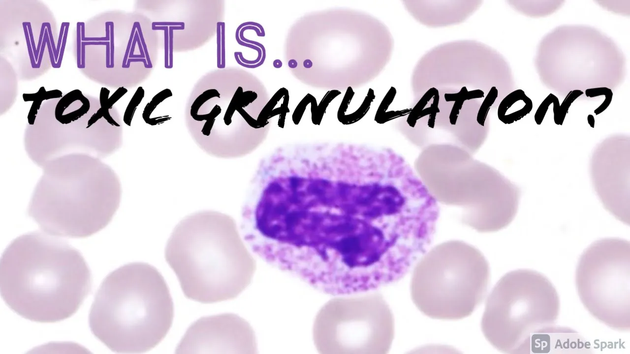
Toxic Granulation of Neutrophils: Causes and Hematopathology
@JMGardnerMD
13.3K views•2021-05-06

Graphic Medicine: Comics for Collaborative Healthcare Communication
@nationalpatientadvocate
189 views•2023-12-04

Neuroanatomy: Central and Peripheral Nervous System Divisions Explained
@AKLECTURES
136.2K views•2014-09-20

Stages of Labor and Vaginal Birth | Childbirth Animation
@nucleusmedicalmedia
52.1M views•2017-08-18
Related Study Plans & Knowledge Roadmaps
Structured learning paths in Medicine