Toxic granulation in neutrophils refers to the appearance of larger, more prominent, and coarser granules within the cytoplasm, which occurs when the bone marrow is stimulated to produce increased numbers of neutrophils. This phenomenon can be triggered by various conditions including growth factor administration (such as after bone marrow transplantation or chemotherapy-induced neutropenia), sepsis, myelodysplastic syndromes, or other situations requiring enhanced granulocyte production. The presence of toxic granulation indicates that the body is responding to stress or infection by ramping up its white blood cell production capacity.
Toxic Granulation of Neutrophils: Causes and Hematopathology
Added:Deep Dive
Normal neutrophil morphology and life cycle, including their maturation stages in the bone marrow.

Medical School Pathology, 2013 Season, Session #52: WBC-I
11.8K
Neutrophils are produced in red marrow, sequestered in spleen, and live only 2 days in peripheral blood. The marrow-to-peripheral blood ratio is 50:1. Maturation stages include: myeloblast (no granules, nucleoli), promyelocyte (starts granules), myelocyte (mature granules), metamyelocyte (indented nucleus), band/stab (two lobes), and mature neutrophil (multiple lobes with thin connections). The average number of lobes is 3.5. High turnover cells are more sensitive to radiation and neoplasia.
Click to watch →

Fases da maturação dos Neutrófilos
1.4K
Neutrophils develop through a sequential maturation process in the bone marrow, progressing from multipotent myeloid stem cells to segmented neutrophils. The stages include: (1) Myeloblast - most immature, 15-20 μm, high N:C ratio, basophilic cytoplasm, visible nucleoli; (2) Promyelocyte - larger (12-24 μm), azurophilic granules appear; (3) Myelocyte - secondary granules develop, nuclear condensation begins; (4) Metamyelocyte - first post-mitotic stage, indented nucleus; (5) Band neutrophil - 10-15% of marrow cells, S/C-shaped nucleus; (6) Segmented neutrophil - mature form with 2-5 lobes connected by filaments. Each stage shows progressive nuclear condensation and cytoplasmic specialization.
Click to watch →

Anomalías Serie Blanca
5.1K
Neutrophils mature through distinct stages: myeloblast, promyelocyte, myelocyte, metamyelocyte, band, and segmented neutrophil. Each stage has characteristic nuclear and cytoplasmic features. Normal neutrophils measure 2-20 micrometers with 2-5 lobes connected by thin filaments. Primary granules contain lysosomes and myeloperoxidase, while secondary granules constitute 80% of total granules and contain specific enzymes.
Click to watch →
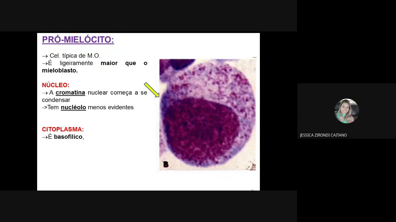
INTRODUÇÃO A LEUCOPOIESE E FORMAÇÃO DE NEUTRÓFILOS
5.9K
Neutrophils mature through a 14-day process in the bone marrow: myeloblast (large nucleus, delicate chromatin, no granules) → promyelocyte (primary granules appear) → myelocyte (secondary granulation predominates) → metamyelocyte (kidney-shaped nucleus, condensed chromatin) → band neutrophil (rod-shaped nucleus) → segmented neutrophil (2-5 lobes connected by chromatin filaments). Each stage has distinct nuclear and cytoplasmic characteristics, with chromatin progressively condensing and nuclear shape changing from round to lobulated.
Click to watch →

Ann Ramsey: EVs and Neutrophils
508
Neutrophils undergo a complete life cycle involving differentiation and maturation in the bone marrow, followed by release into the blood, migration to specific tissues, and eventual clearance. The maturation stages progress through distinct developmental phases before cells become fully functional mature neutrophils ready for deployment.
Click to watch →
Deep Dive
Fundamentals of the innate immune response, particularly the role of neutrophils in phagocytosis and acute inflammation.

Jak cukry obniżają współczynnik fagocytozy, a więc nieswoistą odpowiedź immunologiczną organizmu
26K
The immune system operates through two main responses: innate (non-specific) and adaptive (specific). Phagocytosis is a key innate immune process where cells engulf and destroy pathogens. Professional phagocytes include neutrophils (40-70% of leukocytes), monocytes, macrophages, dendritic cells, and osteoclasts. Neutrophils serve as the first line of defense, circulating in blood and immediately attacking pathogens. The phagocytosis coefficient measures immune function by quantifying how many pathogens neutrophils can engulf. Higher values indicate better immune competence.
Click to watch →

Acute Inflammation | Immunology
180.4K
Neutrophils are the most abundant and important white blood cells for acute inflammation. They arrive at the injury site within 24-48 hours and perform phagocytosis, engulfing and destroying damaged cells and bacteria. This process often results in pus formation. Neutrophils have a short lifespan and are replaced by macrophages after 48 hours, which provide longer-term defense and tissue repair functions.
Click to watch →
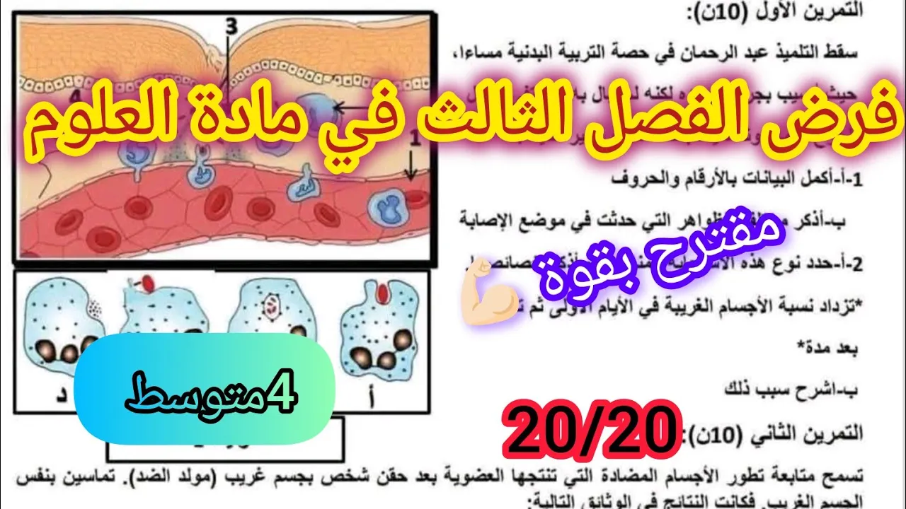
فرض الفصل الثالث في العلوم الطبيعية السنة الرابعة متوسط2026
133.6K
The innate immune response involves phagocytosis, a non-specific defense mechanism where phagocytes engulf and destroy pathogens. The process occurs in four phases: recognition/adhesion, ingestion/digestion, chemotaxis/adhesion, and excretion. Inflammation produces visible signs (swelling, pain, heat, pus) and invisible signs (bacterial proliferation, blood vessel dilation, plasma exudation). This response is immediate, rapid, non-specific, lacks immunological memory, and cannot be transferred. The number of foreign bodies increases initially to provide favorable conditions for reproduction, then decreases due to phagocytic resistance.
Click to watch →
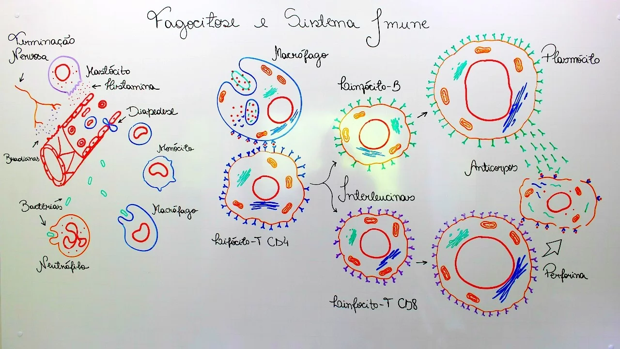
FAGOCITOSE E SISTEMA IMUNE
28.7K
The innate immune response begins when tissue damage occurs, releasing bradykinin that signals pain and alerts the brain to foreign invaders. Mastocytes release histamine, increasing blood vessel permeability and causing inflammation with heat and edema. Defense cells like macrophages and neutrophils exit vessels through diapedesis to engulf pathogens via phagocytosis. Macrophages specialize in different tissues (alveolar, Kupffer, mesangial, histiocytes, microglia, Langerhans, osteoclasts). Neutrophils, comprising 70% of phagocytes, are the primary defenders, with their death forming pus. This non-specific response provides immediate but general protection against threats.
Click to watch →
Inflammation - causes, symptoms, diagnosis, treatment, pathology
121.4K
Neutrophils are the first leukocytes recruited during the acute inflammatory process and are like hungry athletes—they immediately start phagocytosing or eating pathogens in damaged cells. Neutrophils take in a lot of pathogens quickly, kind of like a vacuum, and then undergo apoptosis, destroying themselves along with all of the pathogens they've taken in.
Click to watch →
Deep Dive
Basic hematological staining techniques (such as Wright-Giemsa stain) used to visualize peripheral blood smears.

How to do a hematology stain (Wright-Giemsa stain) Laboratory Tutorial Procedure
83.8K
The Wright-Giemsa stain is a Romanowsky stain that uses oxidized methylene blue dyes (basic, binding to acidic nuclei for blue-purple color) and Eosin Y (acidic, binding to alkaline cytoplasm for red-orange-pink color) to differentiate cellular components in peripheral blood and bone marrow specimens; the procedure involves preparing clean blood smears, fixing with methanol, staining with Wright-Giemsa stain for 60 seconds, rinsing with buffer for 60 seconds, rinsing with distilled water, and examining under microscope at 1000x magnification using oil immersion.
Click to watch →

PERIPHERAL BLOOD SMEAR | HAEMATOLOGY LAB | PHYSIOLOGY PRACTICALS
521.2K
This comprehensive section covers the complete procedure for preparing and staining peripheral blood smears. Required instruments include glass slides, pipette, lancet, distilled water, Leishman stain, and a compound microscope. The technique involves creating a thin, uniform smear by placing a blood drop on a slide and spreading it at a 45-degree angle. An ideal smear must cover the entire slide width, extend 3-4 cm, have uniform thickness, and show no air gaps. The staining process uses Leishman stain containing methylene blue (basic dye for acidic cellular components) and acetone-free methyl alcohol (for fixation without cell lysis). The procedure includes a 2-minute fixation time followed by 8-10 minutes of staining, then washing until pinkish and drying before oil immersion microscopy.
Click to watch →
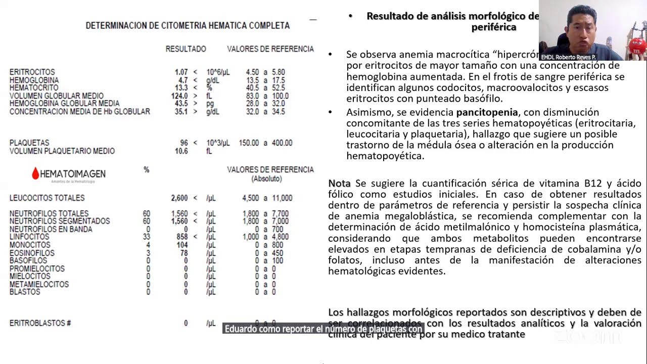
HALLAZGOS HEMATOLÓGICOS
185
Proper blood smear preparation requires specific techniques: the angle should be 45 degrees, and the spreader should be narrower than the slide. The smear should occupy approximately three-quarters of the slide, with a head, body, and tail. The monocapa (single cell layer) is the ideal zone for morphological examination. Wright-Giemsa staining uses two colorants: methylene blue (basic) and eosin (acidic). Methylene blue stains acidic substances like DNA, RNA, and mucopolysaccharides. Eosin stains basic substances like red blood cell proteins, hemoglobin, and eosinophil proteins. The Romanowsky effect (metachromasia) allows methylene blue to produce various colors depending on the substance being stained. Staining times must be established for each new batch to ensure standardization.
Click to watch →
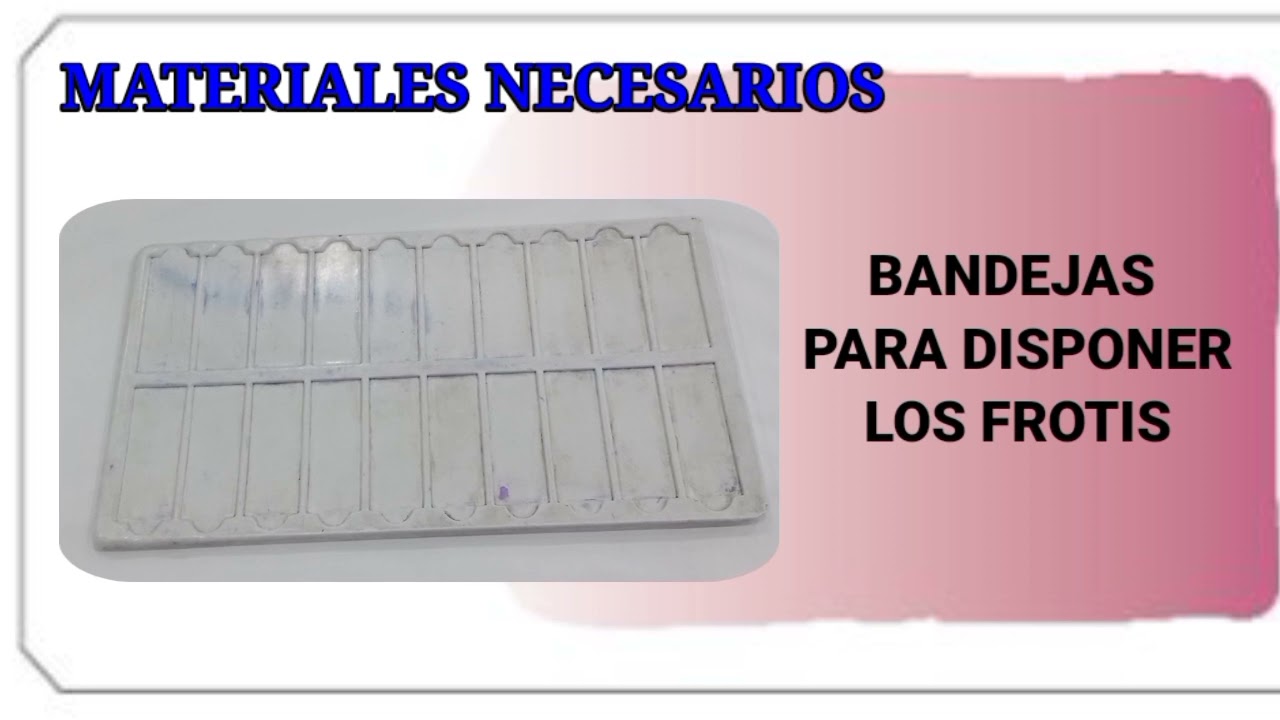
TINCIÓN MAY GRUNWALD GIEMSA, Tutorial y procedimiento simple y fácil de entender.
6.9K
The Wright-Giemsa staining technique is a differential staining method used in hematology to differentiate normal and abnormal blood cells in peripheral blood smears and bone marrow samples, as well as for histological and cytological specimens. The procedure involves three main steps: first, immerse the dried blood smear in Wright's stain for 5 minutes, then rinse with water; second, immerse in diluted Giemsa stain (1:10 dilution with buffer at pH 7.2) for 15 minutes, followed by a final water rinse; third, dry the smear completely on absorbent paper or with warm air before placing it in special trays for microscopic examination.
Click to watch →
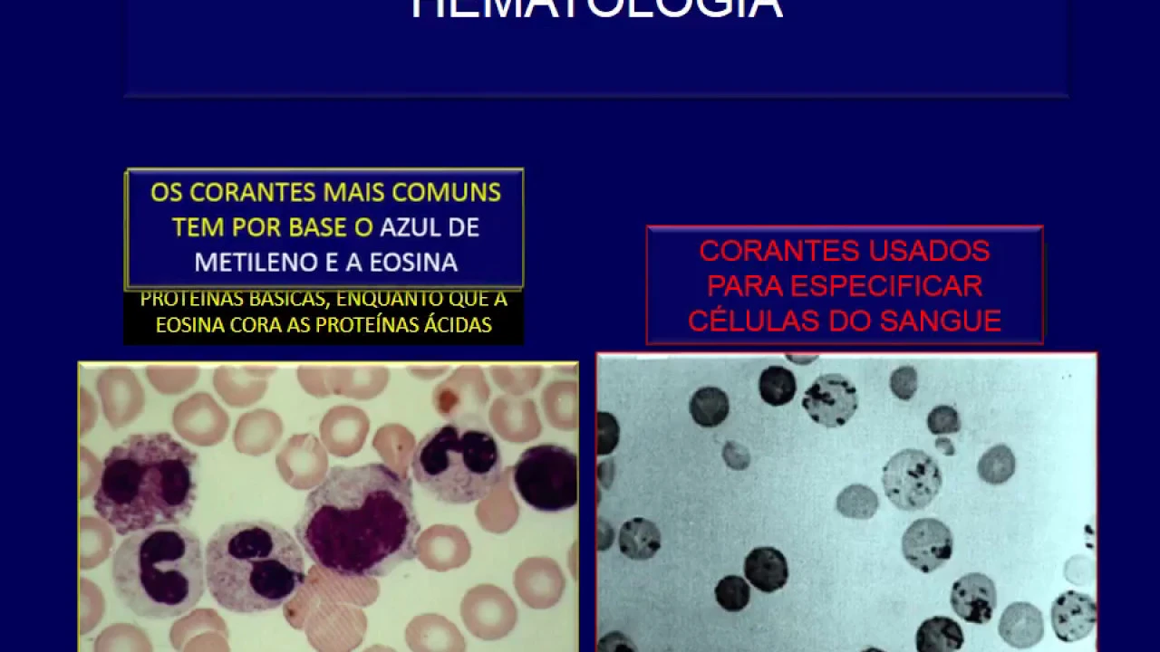
Colorações hematologicas e aplicações
11.6K
Hematological diagnostics examine red blood cells, white blood cells, and platelets using specialized stains. Two basic stain types exist: differentiating stains showing cell morphology and specifying stains for particular cells. Common stains include methylene blue (staining basic proteins/nuclei) and eosin (staining acidic proteins/cytoplasmic granules). Crystal violet brilliant specifically stains reticulocytes (containing ribosomal RNA), Heinz bodies, and hemoglobin H. Pathological findings include toxic granulation in neutrophils during severe bacterial infections, Chediak-Higashi syndrome with intensely granulated cells, macroplatelets indicating bone marrow conditions, gray platelets with aggregation defects, and polychromasia indicating reticulocytosis. Stain preparation involves weighing 2 grams of stain salt, blending with 500ml ethanol in two stages, filtering through paper towel, and storing in amber bottles at 10-25°C for 4-5 days. Blood smear preparation requires 0.1-0.3mm thickness using 2-2.6ml blood. Staining takes 3-6 minutes, followed by buffer water (pH 7-7.4) application. Proper analysis requires zigzag traversal in the central portion, as neutrophils, lymphocytes, and platelets are more abundant peripherally.
Click to watch →
Deep Dive
The concept of systemic inflammation, infection, and the physiological definition of sepsis.

Sepsis: Systemic inflammatory response syndrome (SIRS) to multiple organ dysfunction syndrome (MODS)
830K
Sepsis involves infection or inflammation in the bloodstream, causing decreased tissue perfusion, oxygenation, and hypotension. It exists on a spectrum from mild to severe. SIRS (Systemic Inflammatory Response Syndrome) is the initial stage requiring two or more of four criteria: temperature extremes (above 38°C or below 36°C), heart rate above 90, respiratory rate above 20 or PaCO2 below 32, and abnormal white blood cell counts (above 12,000 or below 4,000, or >10% band cells). These criteria indicate widespread inflammation but not necessarily infection.
Click to watch →

Điều trị nhiễm trùng da và mô mềm do vi khuẩn
26.9K
Systemic Inflammatory Response Syndrome (SIRS) is a systemic inflammatory response to infection characterized by fever, tachycardia, tachypnea, and leukocytosis. Sepsis is defined as infection accompanied by SIRS. The four diagnostic criteria for SIRS are: temperature >38°C or <36°C, heart rate >90 bpm, respiratory rate >20 breaths/min or PaCO2 <32 mmHg, and white blood cell count >12,000 or <4,000 cells/μL. Diagnosis requires at least two criteria in the presence of suspected infection.
Click to watch →

Tudo o que você precisa saber sobre sepse - Aula de Infectologia | Curso Pré-Internato
112K
Sepsis is an infection with excessive inflammatory response causing organ dysfunction, not merely fever or infection. Key distinctions: infection alone without organ dysfunction is not sepsis; bacteremia without organ dysfunction is not sepsis; multiple organ infections without systemic dysfunction are not sepsis. Sepsis-3 criteria define sepsis as infection plus two or more qSOFA criteria (altered mental status, SBP ≤100, RR ≥22). Septic shock requires refractory hypotension requiring vasopressors despite fluid resuscitation.
Click to watch →

Sepsis 1, Infection and response
65.4K
Sepsis begins with infection, which triggers acute inflammation characterized by heat, pain, redness, swelling, and loss of function. Damaged tissues release components that interact with enzymes to produce inflammatory mediators like prostaglandins. Simultaneously, damaged cells release cytokines—small protein/polypeptide messenger molecules that communicate between cells. During infection, white blood cells (particularly macrophages and monocytes) recognize foreign antigens and produce additional cytokines. These inflammatory mediators and cytokines enter body fluids and distribute systemically, potentially leading to Systemic Inflammatory Response Syndrome (SIRS). SIRS is defined as a systemic condition caused by circulating inflammatory mediators and pro-inflammatory cytokines that affect all body tissues.
Click to watch →

Sepsis
317.8K
Sepsis originates from infection triggering acute inflammation through cytokine release from damaged tissue and immune cells. Inflammatory mediators circulate systemically, causing Systemic Inflammatory Response Syndrome (SIRS). Cytokines stimulate bone marrow to produce leukocytes (leukocytosis with left shift), while bacterial toxins can cause leukopenia. Different infections produce characteristic patterns: neutrophilia suggests bacterial infection, lymphocytosis indicates viral etiology, and eosinophilia points to parasitic or allergic conditions. This cascade involves multiple organ systems including bone marrow, liver, hypothalamus, and brain, creating a complex network of defensive responses that increase the body's energy demands.
Click to watch →
Prerequisite Knowledge
- Concept 01Normal neutrophil morphology and life cycle, including their maturation stages in the bone marrow.
- Concept 02Fundamentals of the innate immune response, particularly the role of neutrophils in phagocytosis and acute inflammation.
- Concept 03Basic hematological staining techniques (such as Wright-Giemsa stain) used to visualize peripheral blood smears.
- Concept 04The concept of systemic inflammation, infection, and the physiological definition of sepsis.
Subsequent Learning
- Step 01Identification of other reactive neutrophil changes, such as Döhle bodies and cytoplasmic vacuolization.
- Step 02Differential diagnosis between toxic granulation and genetic leukocyte disorders like Alder-Reilly anomaly or Chédiak-Higashi syndrome.
- Step 03Distinguishing a leukemoid reaction (marked by toxic granulation) from Chronic Myelogenous Leukemia (CML).
- Step 04Clinical application of neutrophil morphology in monitoring patients receiving granulocyte colony-stimulating factor (G-CSF) therapy.
Band Neutrophil
Toxic Granulation
Band Neutrophil
0:00Playing Section
- 1
Identifies a band neutrophil by its U-shape and unsegmented nucleus.
- 2
Distinguishes it from a hypo-segmented neutrophil, a younger cell type.
- 3
Notes differences in granule color and prominence, indicating toxic granulation.
Subjectivity and Quantitative Alternatives to Manual Toxic Granulation Grading
While manual assessment of toxic granulation is a traditional method for detecting systemic inflammation or infection, it is highly criticized in modern hematopathology for its subjectivity, lack of standardization, and susceptibility to pre-analytical artifacts (such as staining delays or pH variations). Many clinical pathologists argue that relying on manual visual grading is outdated. Instead, they advocate for objective, automated alternatives. These include quantitative neutrophil activation markers, flow cytometric measurement of CD64 expression, and automated hematology analyzer parameters (like cell population data or neutrophil granularity index). These modern technologies provide reproducible, standardized, and rapid assessments of neutrophil activation, reducing the inter-observer variability inherent in manual smear reviews.
Deep Dive
Identification of other reactive neutrophil changes, such as Döhle bodies and cytoplasmic vacuolization.

ASCP | MLS | MLT | Nonmalignant Leukocyte Disorders
2.4K
Reactive morphologic changes in neutrophils include toxic granulation (dark blue/black granules containing acid mucosubstance enhancing microbial destruction), Döhle bodies (cytoplasmic inclusions resembling ribosomal RNA remnants), cytoplasmic vacuoles (small clear areas indicating phagocytosis or self-cleaning), hypersegmentation (nuclei with more than five lobes), and pyknosis (nuclear shrinkage and denser chromatin). Left shift indicates varying degrees of immature neutrophil release: mild shows bands and metamyelocytes; moderate includes myelocytes; marked shows promyelocytes and blasts. Cytoplasmic vacuoles measuring ~2 micrometers can result from drugs (sulfonamides, chloroquine), EDTA artifact, autoantibodies, alcoholism, or radiation. Phagocytic vacuoles up to 6 micrometers indicate bacterial or fungal invasion. Anisocytosis reflects osmotic changes or slide affinity during blood film preparation.
Click to watch →

Hematopathology-Myeloid-Toxic_Neutrophils.mp4
114
Reactive neutrophilia is characterized by three classic morphological findings in peripheral blood smear: toxic granulation (coarse, larger, basophilic granules resembling primary azurophilic granules), toxic vacuolation (clear spaces in cytoplasm representing sites of digestion), and Dohle bodies (bluish cytoplasmic inclusions representing remnants of rough endoplasmic reticulum or denatured ribosomes). These toxic changes accompany a left shift in neutrophil maturation and are associated with clinical conditions including severe infections, burns, trauma, and G-CSF administration; however, marked vacuolation may also result from blood storage artifacts, requiring careful differentiation from genuine toxic changes.
Click to watch →
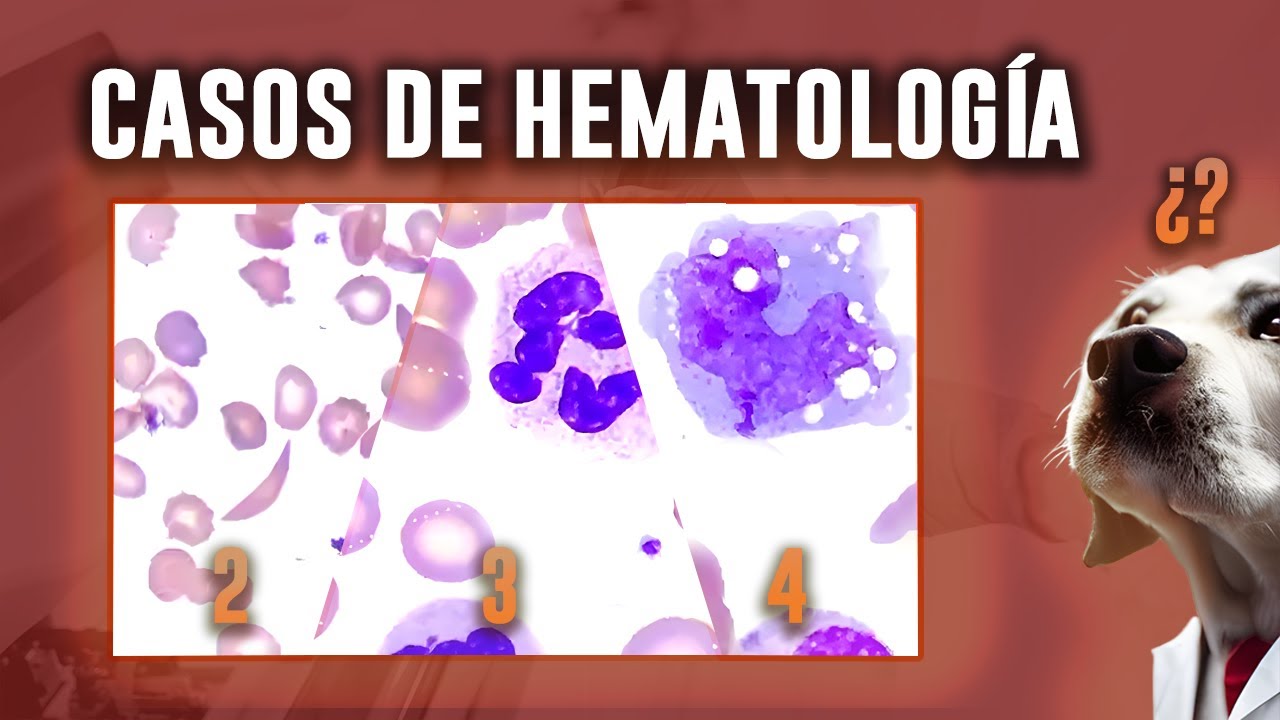
DESAFÍA tu CONOCIMIENTO CON ESTOS CASOS CLÍNICOS hematológicos (2, 3 y 4)
3.2K
Toxic changes in neutrophils include toxic granulation, Döhle bodies, and cytoplasmic vacuolization. These changes occur when neutrophils are exposed to severe infections, inflammation, or certain medications. The changes represent accelerated maturation and increased demand for neutrophils. The laboratory should recognize these morphological findings and communicate them to the clinician.
Click to watch →

The ABCs and 123s of the CBC
606
Neutrophil immaturity progresses from mature segmented neutrophils to band neutrophils (U-shaped nucleus, uniform throughout) to metamyelocytes (bean-shaped nucleus). The chromatin remains chunky in immature forms. Toxic changes include Döhle bodies (blue granular material in cytoplasm) and increased cytoplasmic basophilia (bluer cytoplasm), indicating inflammation. Döhle bodies are only seen in neutrophils, not other white blood cells. Reactive lymphocytes have more prominent blue cytoplasm and may contain granules. Atypical white blood cells are very large, very blue, and difficult to identify definitively, often requiring flow cytometry for identification.
Click to watch →
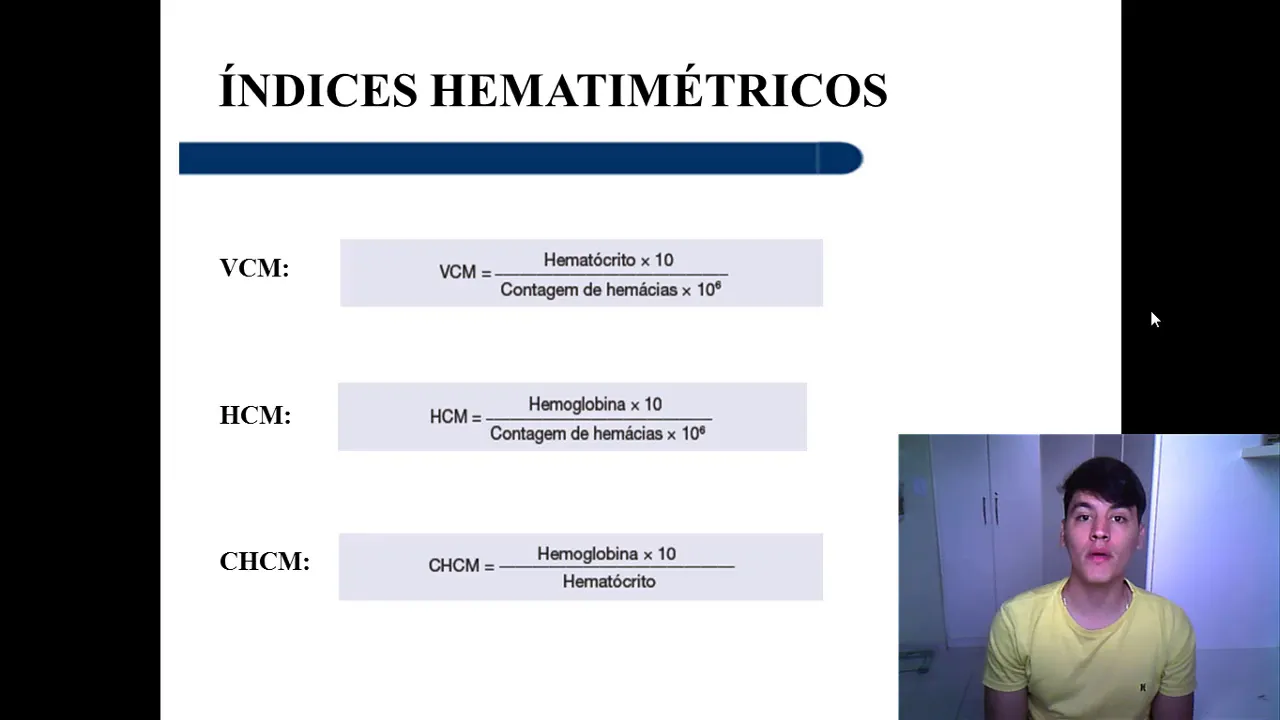
INTERPRETAÇÃO DO HEMOGRAMA
22.7K
In bacterial infections, the body responds with neutrophilia (elevated neutrophil count). The process involves infection stimulus triggering bone marrow production of more neutrophils, which are released into circulation and increase above normal reference values. In viral infections, the body responds with lymphocytosis (elevated lymphocyte count), with lymphocytes increasing above normal values while neutrophil count may decrease relative to lymphocytes. Neutrophils mature through several stages: myeloblast, promyelocyte, myelocyte, metamyelocyte, band cell, and segmented neutrophil. In severe infections, immature neutrophils (band cells) may be released prematurely, a condition called left shift. Toxic granulation appears as coarse, dark granules in neutrophils during severe infections. Toxic changes include toxic granulation, Döhle bodies (blue-gray inclusions), and cytoplasmic vacuolization, indicating the severity of the inflammatory process.
Click to watch →
Deep Dive
Differential diagnosis between toxic granulation and genetic leukocyte disorders like Alder-Reilly anomaly or Chédiak-Higashi syndrome.

طب المستنصرية | المرحلة الثالثة | PATHOLOGY | WBC DISORDERS | BENIGN DISORDER
130
Congenital neutrophil abnormalities include hypersegmented neutrophils (>5 lobes), Pelger-Huët anomaly (bilobed neutrophils), and Alder-Reilly anomaly (coarse dark granules in all cell types). Pelger-Huët anomaly is inherited and benign. Alder-Reilly anomaly can be mistaken for toxic granulation in infections. Toxic changes include toxic granulation, toxic vacuolation, and Döhle bodies. These morphological changes indicate severe infection or toxic conditions and are important for clinical diagnosis.
Click to watch →

WBC | Leucocyte abnormalities || inclusion bodies | Hematology
1K
This section covers inclusion bodies and granular abnormalities in WBCs. Döhle bodies are pale blue, acquired inclusions from ribosome polymerization during infections. May-Hegglin anomaly is genetic with dark blue inclusions, giant platelets, and thrombocytopenia. Toxic granulations are large dark blue granules in neutrophils from severe infections. Alder-Reilly anomaly is a genetic lysosomal storage disorder with large granules in all WBC types, seen in Hunter syndrome. Chediak-Higashi anomaly is a fatal inherited lysosomal transport disorder with very large purple/red granules in all WBC types, causing death before age 10.
Click to watch →

Leukocyte Abnormalities and Disorders (An overview)
68.3K
Leukocyte abnormalities encompass structural and functional deviations in white blood cells that correlate with specific hematological disorders; these include smudge cells (indicating leukemia), hypersegmented neutrophils (pernicious anemia), Pelger-Hewitt anomaly (dysfunctional nuclear segmentation), Alder-Reilly bodies (mucopolysaccharidosis), Chediak-Higashi syndrome (large granules), May-Hegglin anomaly (triad of thrombocytopenia, giant platelets, and inclusion bodies), toxic granulation (acute infection), Dohle bodies (severe infection), and Downey cells (infectious mononucleosis); leukocyte disorders are broadly categorized into quantitative (reactive/non-malignant: neutrophilia, lymphocytosis, eosinophilia, basophilia, monocytosis; malignant: myeloproliferative diseases like AML, CML, polycythemia vera, essential thrombocytosis and lymphoproliferative diseases like CLL, lymphomas) and qualitative (with morphologic abnormalities or normal morphology).
Click to watch →
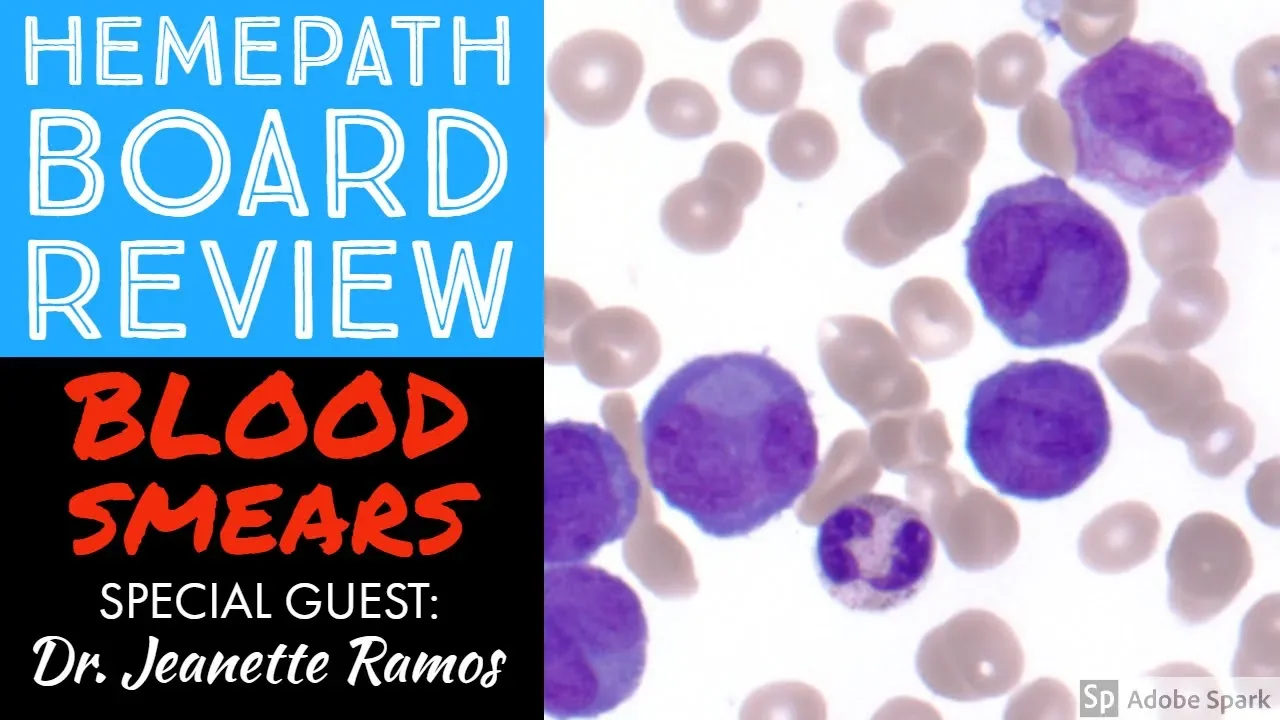
Hemepath & Hematology Board Review: Peripheral Blood Smears with Dr. Jeanette Ramos
200.2K
Toxic granulation shows larger, darker pink granules in neutrophils from infection, inflammation, or growth factor stimulation. Hypogranular neutrophils show reduced granules; Pelger-Huet anomaly is autosomal dominant with abnormal nuclear segmentation. Auer rods (rod-like granule inclusions) historically suggested AML but can appear in lymphoid leukemias. Downey cells in infectious mononucleosis show irregular nuclei, prominent nucleoli, and merge with neighbors ('Dutch skirting'). Reactive lymphocytes can mimic blasts but differ in nuclear behavior.
Click to watch →

Chapter 21 Non-malignant disorders of Granulocytes and Monocytes
3.7K
This extensive section covers morphological and inherited leukocyte disorders. Nuclear abnormalities include Pelger-Huet anomaly (autosomal dominant, 1/5000 prevalence, 2-lobed neutrophils) and hypersegmentation (>6 lobes, early megaloblastic anemia indicator). Cytoplasmic abnormalities include Döhle bodies (rough ER aggregates in severe infections/burns/drugs), toxic granulation (retained basophilic granules), and toxic vacuolation (end-stage phagocytosis). Inherited functional disorders causing recurrent infections include Aldrich-Herlin anomaly (large granules, mucopolysaccharidoses), Chediak-Higashi syndrome (giant lysosomes, defective killing), May-Hegglin anomaly (RNA inclusions, thrombocytopenia), myeloperoxidase deficiency (alternate antimicrobial pathway), and leukocyte adhesion deficiency (serious recurrent infections). The section concludes with eosinophil/basophil disorders and lysosomal storage diseases: Gaucher's disease (beta-glucosidase deficiency, Gaucher cells), Niemann-Pick disease (sphingomyelinase deficiency, foamy macrophages), and sea-blue histiocytosis (enlarged spleen, decreased platelets, sea-blue macrophages).
Click to watch →
Deep Dive
Distinguishing a leukemoid reaction (marked by toxic granulation) from Chronic Myelogenous Leukemia (CML).

MORPHOLOGICAL EVALUATION OF WBCs/PLATELETS/ORGANISM II PRACTICAL PATHOLOGY II @DR. JIBRAN AHMED
8.3K
Leukemoid reaction presents with markedly elevated WBC (>50,000/μL) and immature cells resembling CML but occurring in non-leukemic conditions. Differentiation from CML requires comprehensive analysis: underlying disease features (fever) versus hepatosplenomegaly; leukocyte count <50,000 versus usually >100,000; resolution with underlying disease versus progressive nature; evidence of infection (toxic granules, Döhle bodies, vacuoles) versus absence; immature cells up to metamyelocyte stage versus blast forms; marrow showing myeloid hyperplasia versus monoclonal blast proliferation; normal karyotype versus abnormal karyotype. This distinction is critical for appropriate patient management.
Click to watch →

Hemato-oncology | Test and Discussion | Dr. Mukesh Bhatia |
25.4K
CML and leukemoid reaction present similarly: very high WBC count (up to 60,000+), left shift with immature cells, splenomegaly. Key differentiating features: (1) LAP score: reduced in CML, normal/elevated in leukemoid reaction; (2) Philadelphia chromosome: present in CML, absent in leukemoid reaction; (3) Blast cells: few in CML, none in leukemoid reaction. The LAP score is the first test to perform, followed by Philadelphia chromosome testing for confirmation. This differentiation is crucial because CML requires targeted therapy while leukemoid reaction is reactive.
Click to watch →

LEUKEMIA(CML) vs LEUKEMOID REACTION V.IMP topic for ug/pg #neetpg #inicet #pathologyconcepts
4.9K
Leukemoid reaction is a reactive condition that mimics leukemia on peripheral smear, while CML is a neoplastic disorder. Key differences include: leukemoid reaction is caused by infections, intoxications, or paraneoplastic syndromes, whereas CML results from Philadelphia chromosome translocation (9:22) causing BCR-ABL fusion protein. Clinically, CML presents with splenomegaly while leukemoid reaction shows symptoms related to underlying cause. In peripheral smear, leukemoid reaction shows 5-15% immature cells with WBC counts up to 1 lakh, while CML shows higher immature cell percentages and counts exceeding 1 lakh. CML also shows basophilia, eosinophilia, and lack of maturation features.
Click to watch →

Saiba tudo sobre Avaliação Global do Hemograma. Com Dra. Carolina Perrone
601
Distinguishing leukemoid reaction from chronic myeloid leukemia (CML) is essential for appropriate management. Features favoring leukemoid reaction: WBC <50,000/μL, resolution after treating underlying condition, absence of basophilia, elevated leukocyte alkaline phosphatase (LAP) score, presence of toxic granulation and vacuolization, and absence of Philadelphia chromosome. Features favoring CML: WBC >50,000/μL, constitutional symptoms (fever, weight loss, abdominal pain, splenomegaly), basophilia (>5%), normal or decreased LAP score, and presence of Philadelphia chromosome (t(9;22) fusion of BCR and ABL genes). In the clinical case presented, a patient with sepsis showed left shift with toxic granulation and vacuolization, confirming reactive leukemoid reaction. The resolution of infection should lead to hemogram normalization.
Click to watch →

Chronic Leukemia | Clinical Medicine
65.6K
Distinguishing CML from leukemoid reaction requires evaluating white blood cell count, differential, left shift pattern, and LAP score. Counts >100,000 favor CML; <50,000 favor reactive process. CML shows significant left shift with myelocyte bulge and basophilia, while reactive processes show minimal left shift with bandemia. CML has low LAP scores due to non-functional neutrophils, whereas reactive processes have elevated LAP scores. These distinctions are critical for accurate diagnosis and appropriate treatment planning.
Click to watch →
Deep Dive
Clinical application of neutrophil morphology in monitoring patients receiving granulocyte colony-stimulating factor (G-CSF) therapy.

Granulocyte Colony Stimulating Factor (G-CSF) Peripheral Blood Smear
456
Granulocyte Colony Stimulating Factor (G-CSF) therapy induces characteristic peripheral blood smear changes including toxic granulation (abnormally coarse, dark granules in neutrophils), left shift (increased immature granulocytes such as myelocytes and metamyelocytes), and occasionally hypogranular neutrophils with ghostly blue cytoplasm; these findings are commonly observed in oncology patients undergoing chemotherapy to stimulate neutrophil production and enable safe hospital discharge.
Click to watch →
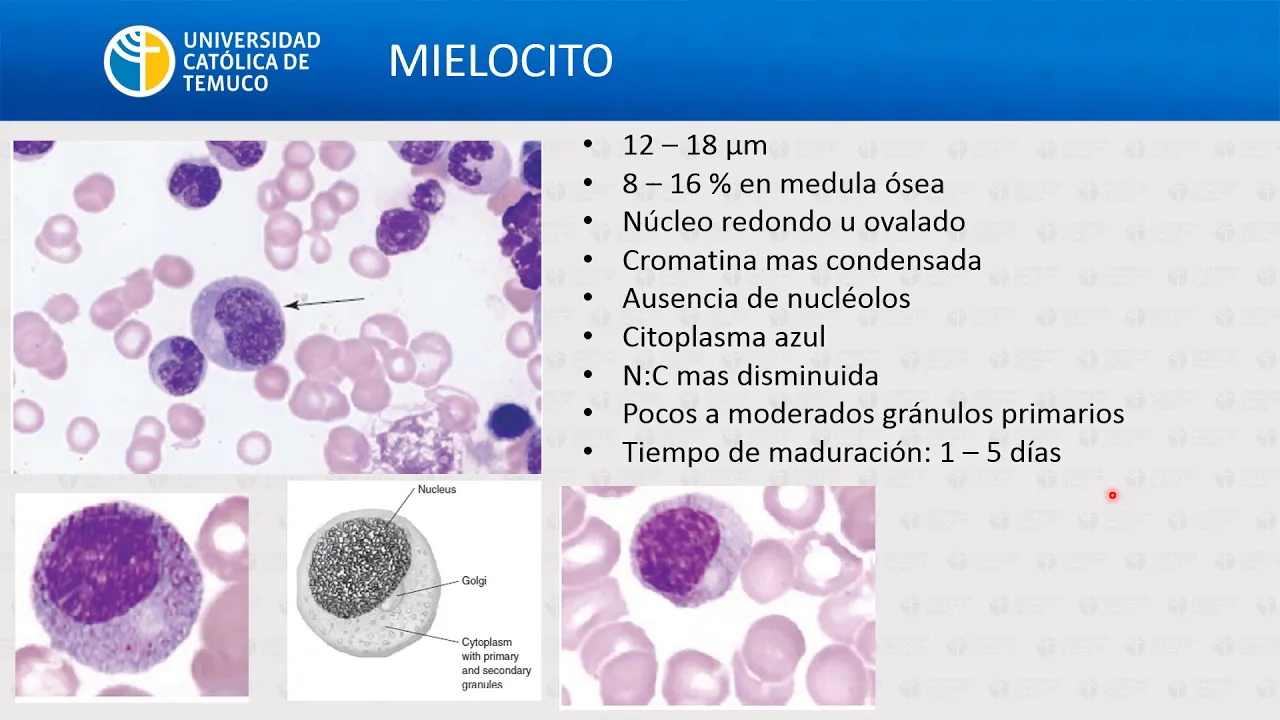
HG: Generalidades de leucocitos
14K
Neutrophils are key effectors in innate immunity, maturing through distinct stages: myeloblast (14-20 microns, lax chromatin, primary granules), promyelocyte (15-21 microns, prominent primary granules), myelocyte (12-18 microns, fewer primary granules), metamyelocyte (10-18 microns, kidney-shaped nucleus), band neutrophil (9-15 microns, horseshoe-shaped nucleus), and segmented neutrophil (9-15 microns, 2-4 lobes). Maturation takes 1-5 days, regulated by G-CSF, GM-CSF, and IL-3. Neutrophil functions include pathogen recognition, phagocytosis, and killing via reactive oxygen species (superoxide, hydrogen peroxide, hypochlorite) and antimicrobial proteins (defensins, lactoferrin, lysozyme). Extravasation involves rolling (selectins), firm adhesion (integrins), and transmigration.
Click to watch →
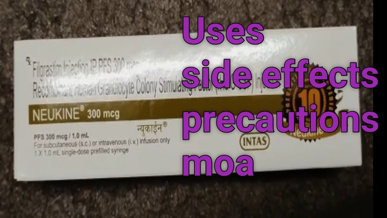
Neukine 300mcg injection/filgrastim (G-CSF)300mcg PFS/vials
47.7K
G-CSF (Granulocyte Colony-Stimulating Factor) is a growth factor that stimulates white blood cell production in bone marrow. It acts by binding to G-CSF receptors on hematopoietic stem cells, promoting differentiation into neutrophils—white blood cells constituting 50-70% of circulating WBCs that defend against bacterial and fungal infections. Clinical indications include neutropenia caused by chemotherapy, cancer treatment, or HIV/AIDS. Proper administration requires storing medication at room temperature for 15-30 minutes before use, inspecting vials for particulates, and avoiding administration within 24 hours before or after chemotherapy. Treatment duration depends on individual response and continues until neutrophil counts normalize (typically above 1,500 cells/μL).
Click to watch →
Interpretación de la BH. Clase 3
15.8K
Left shift indicates immature neutrophils (bands, metamyelocytes) in blood, suggesting active production. Right shift indicates hypersegmented neutrophils (>6 lobes), seen in megaloblastic anemia and myelodysplastic syndromes. Analysis should examine maturation progression from mature neutrophils through bands, metamyelocytes, myelocytes, to blasts. A 'stepped' left shift suggests acute infection; a non-stepped left shift may indicate leukemia. Neutrophil counts of 40,000-50,000/μL with left shift may indicate infection or early leukemia; counts above 50,000/μL with pronounced non-stepped left shift, basophilia, and eosinophilia suggest chronic leukemia.
Click to watch →

Снижение нейтрофилов и лейкоцитов на фоне химиотерапии (нейтропения). Как профилактировать?
869
Granulocyte-colony stimulating factor (G-CSF), primarily filgrastim, is the only evidence-based medication for preventing neutropenia. Treatment regimens are stratified into high, medium, and low risk categories. High-risk regimens require primary prophylaxis starting before treatment begins. Medium-risk regimens require individualized decisions based on age, comorbidities, and prior treatment history. G-CSF must be administered 24-72 hours after chemotherapy and no less than 48 hours before the next cycle to prevent paradoxical infection risk from excessive neutrophil stimulation. The drug prevents infection development, not merely improves laboratory values. If neutrophils are low on treatment day, chemotherapy should be postponed until counts normalize.
Click to watch →
Band Neutrophil
Toxic Granulation
Band Neutrophil
0:00- 1
Identifies a band neutrophil by its U-shape and unsegmented nucleus.
- 2
Distinguishes it from a hypo-segmented neutrophil, a younger cell type.
- 3
Notes differences in granule color and prominence, indicating toxic granulation.
Subjectivity and Quantitative Alternatives to Manual Toxic Granulation Grading
While manual assessment of toxic granulation is a traditional method for detecting systemic inflammation or infection, it is highly criticized in modern hematopathology for its subjectivity, lack of standardization, and susceptibility to pre-analytical artifacts (such as staining delays or pH variations). Many clinical pathologists argue that relying on manual visual grading is outdated. Instead, they advocate for objective, automated alternatives. These include quantitative neutrophil activation markers, flow cytometric measurement of CD64 expression, and automated hematology analyzer parameters (like cell population data or neutrophil granularity index). These modern technologies provide reproducible, standardized, and rapid assessments of neutrophil activation, reducing the inter-observer variability inherent in manual smear reviews.
Normal neutrophil morphology and life cycle, including their maturation stages in the bone marrow.
Fundamentals of the innate immune response, particularly the role of neutrophils in phagocytosis and acute inflammation.
Basic hematological staining techniques (such as Wright-Giemsa stain) used to visualize peripheral blood smears.
The concept of systemic inflammation, infection, and the physiological definition of sepsis.
Identification of other reactive neutrophil changes, such as Döhle bodies and cytoplasmic vacuolization.
Differential diagnosis between toxic granulation and genetic leukocyte disorders like Alder-Reilly anomaly or Chédiak-Higashi syndrome.
Distinguishing a leukemoid reaction (marked by toxic granulation) from Chronic Myelogenous Leukemia (CML).
Clinical application of neutrophil morphology in monitoring patients receiving granulocyte colony-stimulating factor (G-CSF) therapy.
13.3K views123likes2:04@JMGardnerMDOriginal Release: 2021-05-06
so now we're not looking at red cells we're looking at the leukocytes white cells all right so i see granules there um i can't tell if it's bilobed or like kind of a u-shape so that's probably more of a band a band so that would be that would be what a band would look like we talked about it earlier and i said imagine that the segments are gone so the segments are gone that's that's abandoned so a hypo segmented neutrophil basically no that's totally different is it really yeah oh yeah well i'm glad i asked them hypo segmented and i'll show you a picture in just a little bit means that they're literally less than the normal number of segments this is just a little bit younger and hasn't segmented oh okay i see oh and they mean totally different things okay uh so this is a man neutrophil but do you see how it's not quite that same sandy baby girl painting yeah i felt like the granules are a little different color and a little bit more prominent slightly bigger yeah not as fine right so this is called toxic granulation oh and this happens to be growth factor induced oh but it could look the same with sepsis so like maybe a growth factor maybe like someone's had a transplant a marrow transplant or something and they're giving them growth factor to regrow uh it means that the white count is low that can either be or they're getting an auto transplant and they are harvesting cd34 positive cells oh so they've given them so they've given them a stimulus for their their granulocytes um sometimes in mds you'll get given it sometimes after chemo they'll need a little boost with the especially if you end up with like a neutropenic sort of fever and you're trying to get your cranial sites back um but the the marrow is being stimulated to produce to produce more neutrophils basically right and that's so that's either endogenous your body's making it because of uh condition or you're getting a medication this is medication oh medication costs yeah this is the growth factor the medication okay sepsis can also look like this like you're you know your body is wrapped up fighting usually you know bacteria
Up Next
15:24
Red Blood Cell Morphology Abnormalities Explained
@MedLecturesMadeEasy
170.6K views•2015-10-16

48:47
Graphic Medicine: Comics for Collaborative Healthcare Communication
@nationalpatientadvocate
189 views•2023-12-04
9:29
Normal Blood Cell Morphology on Peripheral Smear | Neutrophils, Lymphocytes, Eosinophils & More | Hematopathology Education
@JMGardnerMD
21.6K views•2021-03-10

3:05
Stages of Labor and Vaginal Birth | Childbirth Animation
@nucleusmedicalmedia
52.1M views•2017-08-18
Related Study Plans & Knowledge Roadmaps
Structured learning paths in Medicine
medicine
Abnormal LFTs: Diagnostic Workup & Pathology
01Liver Anatomy and Basic Physiology
02Introduction to Liver Function Tests (LFTs)
03Hepatocellular vs. Cholestatic Patterns
+ 3 more learning modules
12h6 modules
Explore Roadmap
medicine
Analyzing Blood Smears: RBC, WBC & Platelets
01Introduction to Blood Components & Smear Preparation
02Normal Morphology of Blood Cells
03Erythrocyte Pathologies & Morphological Variants
+ 2 more learning modules
12h5 modules
Explore Roadmap
medicine
Mechanical Ventilation: Waveforms & Alarms
01Anatomy and Physiology of Breathing
02Introduction to Mechanical Ventilation & Core Modes
03Ventilator Settings and Waveform Interpretation
+ 2 more learning modules
12h5 modules
Explore Roadmap
medicine
Neuro Exam: Localization & Stroke Syndromes
01Foundations of Neuroanatomy and Vascular Supply
02The Neurological Examination: Step-by-Step
03Principles of Neurological Localization
+ 3 more learning modules
18h6 modules
Explore Roadmap