





























![SI NO SABES QUE ES UN HEMOGRAMA, RETIRATE DE LA CARRERA [LABORATORIO] S2 (Enfermedad hematológica)](https://i.ytimg.com/vi/PxadGgNqeT4/maxresdefault.jpg)
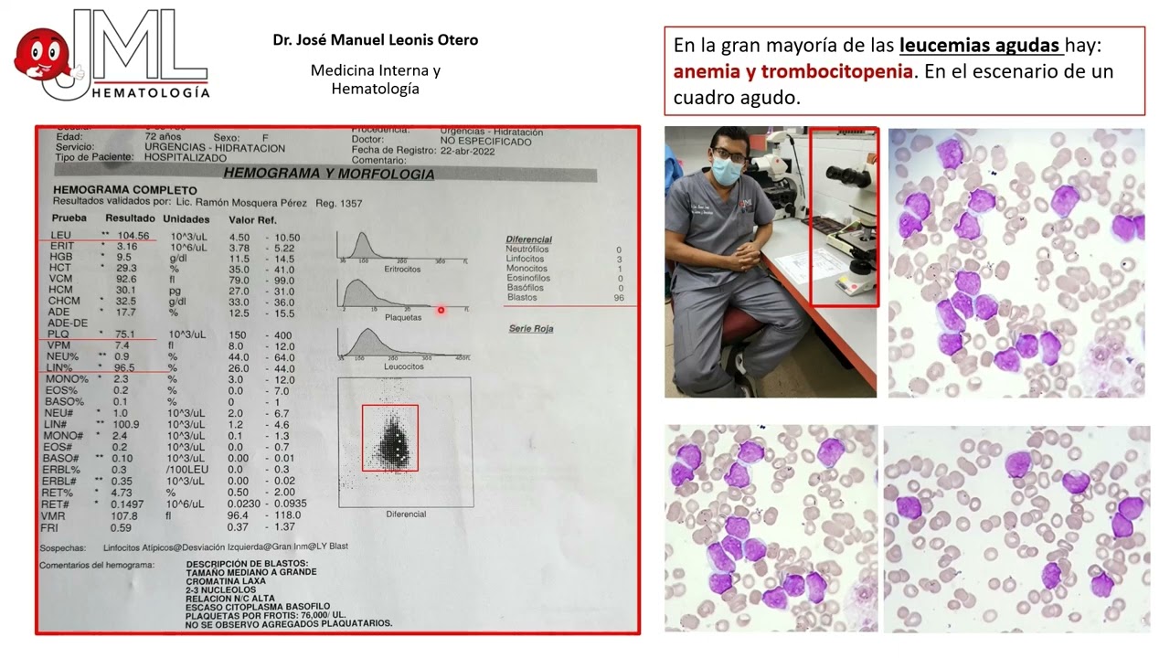
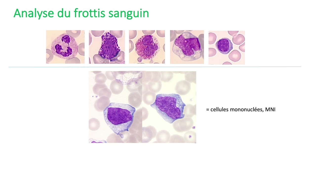
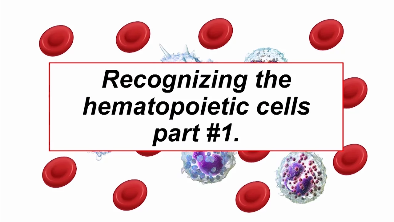
This video teaches the identification of normal peripheral blood cells: neutrophils (most common adult leukocytes with 3-5 nuclear segments and fine pink granules), lymphocytes (second most common in adults, nucleus similar to RBC size), monocytes (blue-gray cytoplasm with variable nucleus shape), eosinophils (easiest to identify with chunky hot pink granules), and basophils (least common with purple granules and bilobed nucleus); red blood cells are biconcave discs with central pallor occupying about one-third of the cell diameter, while platelets appear as small granular elements.
Normal Blood Cell Morphology on Peripheral Smear | Neutrophils, Lymphocytes, Eosinophils & More | Hematopathology Education
Added:Basic understanding of hematopoiesis, specifically the differentiation of myeloid and lymphoid cell lineages in the bone marrow.
The general physiological functions of the formed elements of blood, including erythrocytes, leukocytes, and thrombocytes (platelets).
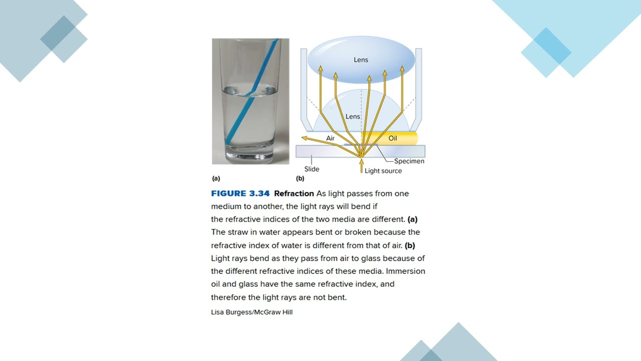
Fundamental principles of light microscopy, including magnification, oil immersion techniques, and the basis of Wright-Giemsa staining.
The clinical purpose of a Complete Blood Count (CBC) and the diagnostic indications for performing a manual peripheral blood smear.
Identification of abnormal white blood cell morphology, including reactive lymphocytes, toxic granulation, hypersegmented neutrophils, and immature blast cells.
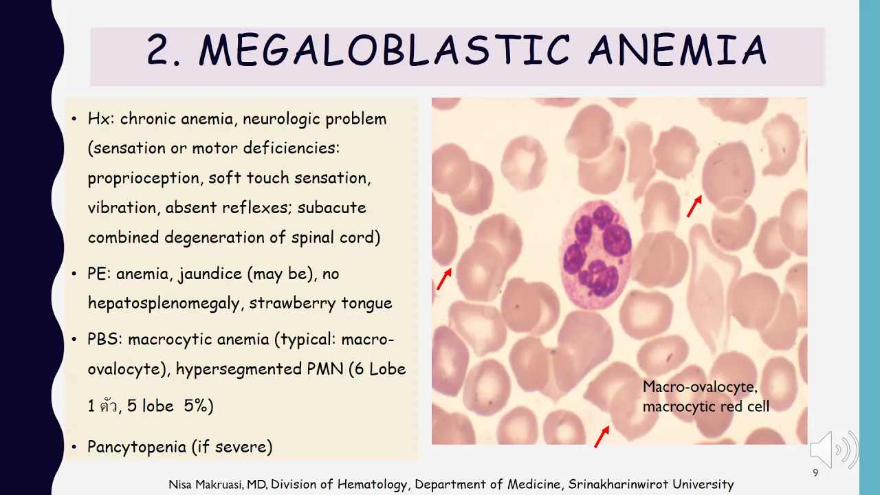
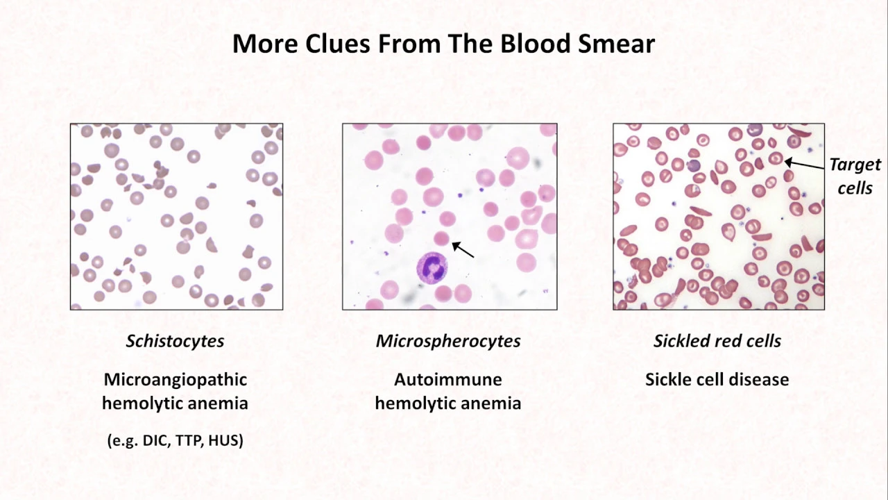
Evaluation of erythrocyte morphology variations, such as alterations in size (anisocytosis), shape (poikilocytosis like schistocytes, spherocytes, or sickle cells), and hemoglobin content.
Recognition of qualitative and quantitative platelet abnormalities, including giant platelets, platelet clumping, and manual estimation of platelet count.
Correlation of peripheral blood smear findings with specific pathological states such as leukemias, anemias, infections, and myelodysplastic syndromes.
Understanding the integration of manual differentials with laboratory automation, flow cytometry, and bone marrow aspirate evaluation.
21.6K views252likes9:29Original Release: 2021-03-10
so here is a higher power of our white blood cells uh or leukocytes so beautiful these are neutrophils in the adults they are the most common leukocyte uh they have what i call baby girl pink granules and they're more subtle usually than your eosinophils on your face so that's this background the this little fine granules here in the cytoplasm right yes and and what power are we on here like what magnification is this under oil this is under oil this is at a thousand a thousand x so that's why you can't like when i see these on um under my microscope i don't have an oil lens i look on a 40x and e to me white blood cells look a little pink but you definitely can't see those granules at least my eyes can't or and it's different in tissue h and e sections oh good point is on uh peripheral films um first of all h e sections are hematoxylin and eosin and versus right stain right stain okay so they look a little bit different they look similar but you'll notice if you go from bone marrow aspirates to your bone marrow biopsies looking at the right to the h and e you'll see some difference the color difference okay cool morphology and they generally have the neutrophils have three to five segments okay if you have neutrophils that have uh five segments and greater than five percent of your neutrophils or neutrophils that have more than five segments those are considered to be hyper segmented hypersegment neutrophils with less than three are considered to be hyposegmented okay but seeing five in just one cell doesn't necessarily mean there's some problem right no it's only if it's uh more than five percent you said of the total right and keep in mind if you look at cytospin slides that rule goes out the window because cytospin can artificially give you segmentation so this is a small mature lymphocyte the nucleus is about the size of a red blood cell you have a thin rim of blue cytoplasm and in adults it is the second most common leukocyte in newborn children is the most common nutrient leukocyte i didn't know that and it switches somewhere in the aging process this big guy is in monocyte it has more of a blue gray cytoplasm the nucleus can be variably shaped sometimes you'll have a nucleus that looks very much band shaped and your text will have a lot of problems with that so make sure you look at your cytoplasm it can be very very helpful in a band is that an immature or a left shifted neutrophil or no yes or no it's always more complicated than i think so the technical term for neutrophil that we see here is segmented neutrophil okay the technical term for band is band neutrophil okay so bands are technically also neutrophils so it is true that if you have increased bands so quote-unquote bandemia that that can be considered to be a left shift but if you have a few bands here and there that's fine totally fine when you get back a little bit more immature into the meta mylocytes i'm if i see even one meta i'm going to say either rare left grands or present or say like milo okay the point is a band band neutrophil can look a lot like a monocyte right that's right and so the main difference between a band neutrophil and a segmented neutrophil is that it basically is smooth all the way through there are no segments oh okay and then monocytes often will have vacuoles in the cytoplasm and that can be a real help especially if you have a reactive appearing lymphocyte which i'll show you later versus a monocyte so reactive lymphocytes can sometimes have vacuoles but most of the time the vacuoles are very reassuring that you know you're looking at your microscope but the chromatin pattern is how eventually you train your eyes to determine whether it's a reactive lymphocyte or a monocyte eosinophils are my favorite they're probably the easiest to identify they have very chunky hot pink granules um am i allowed to say that i call them barbie pink or is that trademarked you i think you could say i call them barbie pink because if i'm going to age myself now back when i played with barbies everything barbie owned was like this color pink her car her house her dresses and now barbie is is branching out a little bit more but i call this one baby girl pink that you know soft pink and this barbie pink so the color's a little different and the granules here are bigger and chunkier than here and they're even refractile if you're looking under like microscopy yeah on on h e i find that really helpful because i feel like the color of neutrophils and eos can be somewhat similar on h e at least from low power and i feel that oftentimes my residents will say oh i think they're eos and it's just neutrophils like in a derm path biopsy but i find that if i flip my condenser i can get that refractile looking you can really see the granules on the eo but again i can't on 40x cm on the neutrophil and i find that actually helpful in practice in tissue sections to tell them apart and then this guy here is a basophil now i find basophils to be the most morphologically variable of all the white blood cells circulating because if you look at a basophil cross-eyed it will degranulate which is kind of what this one has done most of this is degranulated and i'll show you one later uh that's not but you can still see that chunky uh purple granular these are the least common uh white blood cell and in fact is the only basophil on my entire peripheral which is why that is the example and not a better granulation you have low basophil count i think it must be something something serious i'm just joking the what you know what i did i've always wondered why don't we see basophils in tissue sections like i've never recognized the basophil in skin or anywhere else are they there and they just don't look like or do they only stay in the blood and never get out into the tissue is there any i don't know i've always wondered why is this thing that's in our blood never show up or at least i can never see it i well they're so rare even in the blood um i don't think you're really going to to pick them up they may not stain well on h and e i don't know because i've never actually really looked for them all unless they're not prominent in the bone marrow either okay um and then some people consider basophils and mass cells to be kind of cousins that's what i wondered if they are like mass cells but we see lots of mast cells in the skin way more than you would see in the blood so um some people don't consider them to be related it's it's sort of up in the air i they're not the same cell okay they are different cells they are different cells they look pretty similar they have that clumpy uh purple granules uh basophils should have this sort of bilobed appearance okay so you can see there's and mast cells don't have that and mass cells normal mast cells should have a centrally placed nucleus that you can barely see because it has so many grams okay that's cool that's good to know uh i the only time i really see bezos probably is in cml okay and this is a picture to highlight the red blood cells so the red blood cell in three dimensions is a biconcave disc which basically means that it comes together in the middle so it's fatter on the outside and thinner on the inside okay which makes it look like a donut yeah on the peripheral blood flow donuts so there you go you now have your first pathology food analogy for this session and it should be about a third so if you have an increased um you know pallor that would be sort of your likelihood should i look for iron deficiency okay or so the clear the clear part in the middle you're saying should be about a third of the space across the whole red cell right okay and that clear part is not really a hole right it's it's really connected it's just that the section we're seeing it looks like a right empty space okay it's not really an empty space so i mean the brushes it's a dip okay it's important to have that so your your red blood cell can fit through like all the capillaries yeah gold and stuff cool and there are platelets that you can actually see there is a little bitty platelet aggregate there and you know individual platelets here little bitty aggregates like this if they're few and far between i don't really mention because it's not really going to affect the platelet count but larger ones will and we'll talk about that a little bit later okay but now you can see them they're granular and that if you look at the megakaryocytes it looks like megakaryocyte granules okay and in the bigger megacare site i feel like those granules sort of look like the texture of a furry purple muppet and it's so your platelets should look a little bit furry okay
Up Next

15:57
How to Prepare & Stain a Peripheral Blood Smear | Hematology Lab Guide
@MedicalLabLadyGill
12.8K views•2020-09-15

48:47
Graphic Medicine: Comics for Collaborative Healthcare Communication
@nationalpatientadvocate
189 views•2023-12-04
2:04
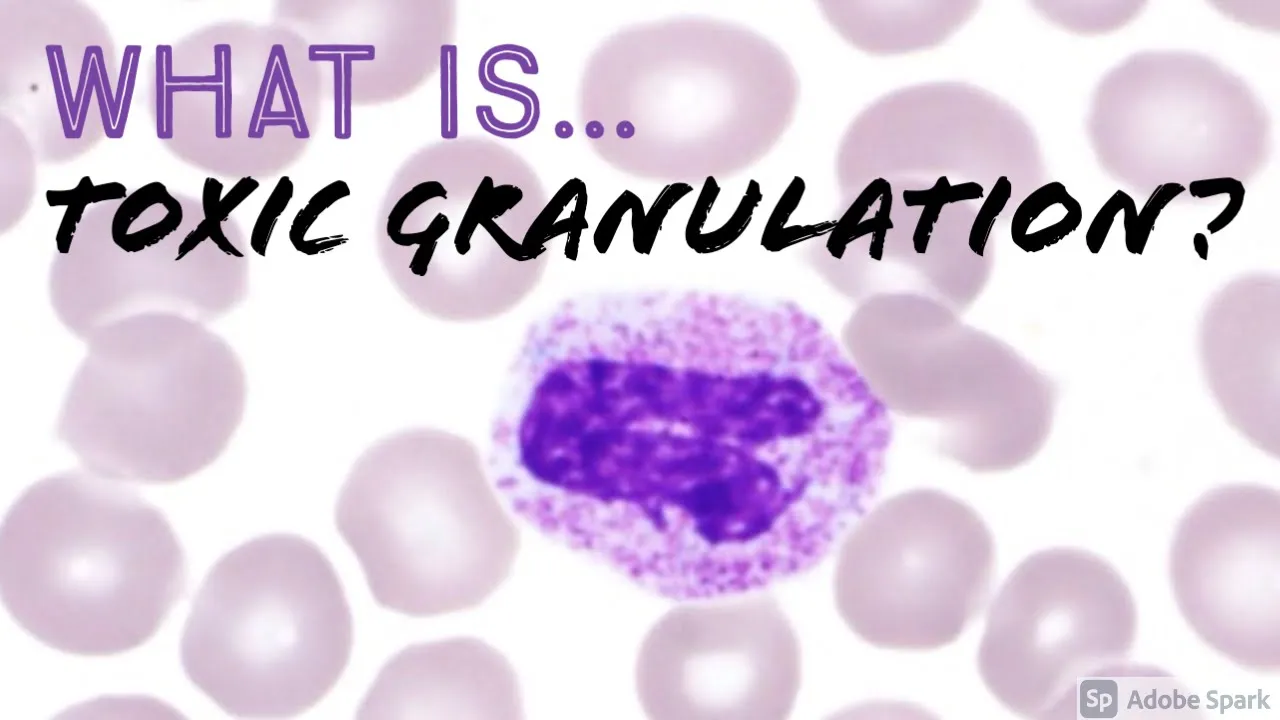
Toxic Granulation of Neutrophils: Causes and Hematopathology
@JMGardnerMD
13.3K views•2021-05-06

3:05
Stages of Labor and Vaginal Birth | Childbirth Animation
@nucleusmedicalmedia
52.1M views•2017-08-18
Related Study Plans & Knowledge Roadmaps
Structured learning paths in Medicine
medicine
Abnormal LFTs: Diagnostic Workup & Pathology
01Liver Anatomy and Basic Physiology
02Introduction to Liver Function Tests (LFTs)
03Hepatocellular vs. Cholestatic Patterns
+ 3 more learning modules
12h6 modules
Explore Roadmap
medicine
Analyzing Blood Smears: RBC, WBC & Platelets
01Introduction to Blood Components & Smear Preparation
02Normal Morphology of Blood Cells
03Erythrocyte Pathologies & Morphological Variants
+ 2 more learning modules
12h5 modules
Explore Roadmap
medicine
Mechanical Ventilation: Waveforms & Alarms
01Anatomy and Physiology of Breathing
02Introduction to Mechanical Ventilation & Core Modes
03Ventilator Settings and Waveform Interpretation
+ 2 more learning modules
12h5 modules
Explore Roadmap
medicine
Neuro Exam: Localization & Stroke Syndromes
01Foundations of Neuroanatomy and Vascular Supply
02The Neurological Examination: Step-by-Step
03Principles of Neurological Localization
+ 3 more learning modules
18h6 modules
Explore Roadmap