Blood is composed of plasma and three types of blood cells: red blood cells (erythrocytes) that carry oxygen from the lungs to body tissues using hemoglobin and return carbon dioxide for removal; white blood cells (leukocytes) that protect the body from infections by destroying harmful bacteria, viruses, and germs as part of the immune system; and platelets (thrombocytes) that are small cell fragments which gather at injury sites to form clots and stop bleeding, preventing excessive blood loss and aiding healing.
Blood Cell Functions Explained | Red & White Cells & Platelets Science
Added:Deep Dive
Basic cellular structure: Understanding what a cell is, including key components like the cell membrane, cytoplasm, and nucleus.

Cell membrane 1 || د.محمود الحسيني
2.2K
The cell is the basic unit of life, consisting of: (1) Cell membrane (outer boundary), (2) Nucleus (contains genetic material), (3) Cytoplasm (fluid-filled space), (4) Organelles (specialized structures like mitochondria, Golgi apparatus, endoplasmic reticulum), and (5) Vesicles (storage sacs). Organelles are classified as membrane-bound (mitochondria, Golgi, ER) or non-membrane-bound (ribosomes).
Click to watch →
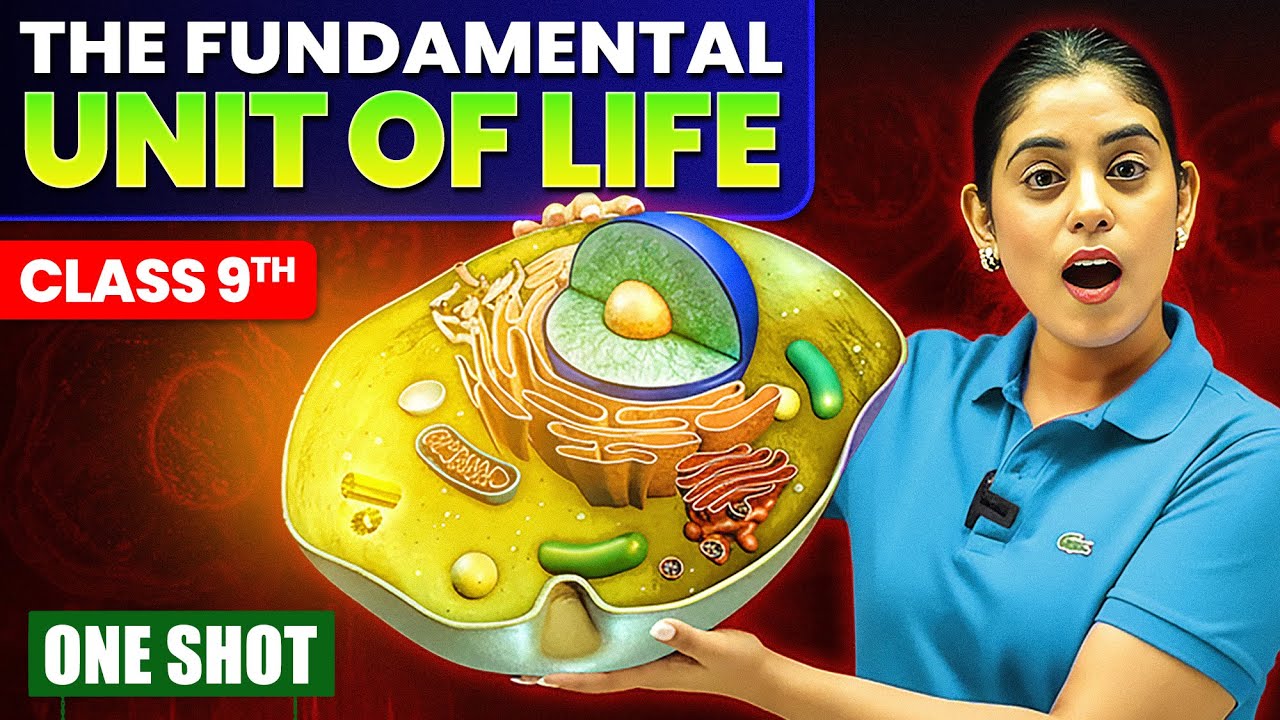
The Fundamental Unit of Life IN ONE SHOT ✅ | Class 9 Science Chapter 5 | NCERT + PYQs | Samridhi S.
2.7M
Cells have several basic structures: plasma membrane (cell membrane) as the outer boundary, cell wall (present in plants, bacteria, and fungi but not animals), nucleus (control center), cytoplasm (jelly-like substance), and cell organelles (specialized structures that perform specific functions). The plasma membrane controls what enters and exits the cell. The cell wall provides structural support and protection. The nucleus contains the cell's genetic material and controls all cellular activities. The cytoplasm provides a medium for cellular processes.
Click to watch →
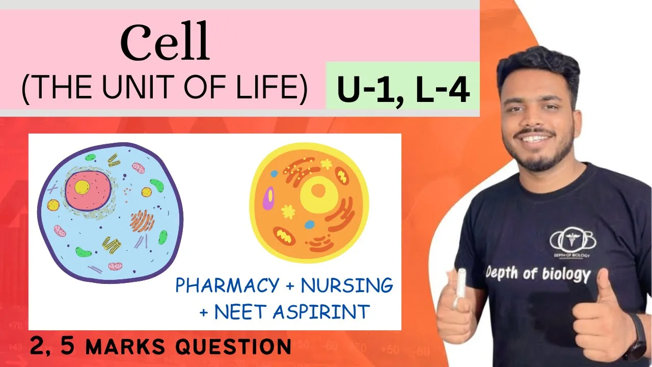
Cellular level of organization | Structure and functions of cell || transport across cell membrane
22K
Cells are the fundamental units of life, combining hierarchically to form tissues, organs, and organ systems. Robert Hooke first discovered cells. Cells require oxygen, water, and ATP for survival. The cell membrane is a thin, flexible barrier controlling substance movement. Plant cells have rigid cell walls made of cellulose for structural support, while animal cells lack this feature. The cytoplasm is jelly-like material containing organelles. Major organelles include the nucleus (genetic control), mitochondria (energy production), ribosomes (protein synthesis), endoplasmic reticulum (transport), Golgi apparatus (packaging), lysosomes (digestion), and chloroplasts (photosynthesis in plants).
Click to watch →

Estrutura celular básica
434
All living organisms are composed of cells, which share fundamental structures like the plasma membrane (a phospholipid bilayer with selective permeability) and cytoplasm, but differ significantly in genetic organization: eukaryotic cells have membrane-bound nuclei containing multiple linear chromosomes and specialized organelles, while prokaryotic cells have dispersed genetic material in the nucleoid region with a single circular chromosome and lack membrane-bound organelles.
Click to watch →
![[THE EPEX] 이펙스의 세포들 Ep.9 시골에서 펼쳐지는 이펙스의 자급자족 라이프 1 ⛰ (ENG SUB)](https://i.ytimg.com/vi/mGJf8DC_srs/maxresdefault.jpg)
[THE EPEX] 이펙스의 세포들 Ep.9 시골에서 펼쳐지는 이펙스의 자급자족 라이프 1 ⛰ (ENG SUB)
295.4K
Cells are the fundamental structural and functional units of all living organisms, serving as the basic building blocks that make up complex life forms.
Click to watch →
Deep Dive
The human circulatory system: A foundational understanding of how the heart, blood vessels, and blood transport materials throughout the body.

The Circulatory System
1.5M
The human circulatory system is a closed system where blood moves through blood vessels in a continuous loop, beginning at the heart which pumps oxygenated blood to the body via arteries and returns deoxygenated blood through veins; the heart has four chambers (two atria and two ventricles) that work in sequence: deoxygenated blood enters the right atrium, moves to the right ventricle, travels through the pulmonary artery to the lungs for oxygenation, returns to the left atrium, moves to the left ventricle, and exits through the aorta to deliver oxygen to body cells; blood consists of four main components—red blood cells (oxygen carriers), white blood cells (immune defense), platelets (clotting), and plasma (liquid matrix)—and the cardiac cycle involves coordinated contractions (systole) and relaxations (diastole) controlled by electrical signals from the sinoatrial node, with coronary arteries supplying oxygen to the heart muscle itself; a heart attack occurs when blockages in coronary arteries prevent oxygen delivery to heart tissue, causing cell death.
Click to watch →
مراجعه علوم الصف الخامس الابتدائي | علوم الصف الخامس الابتدائي الترم الاول
123.4K
The human circulatory system consists of the heart (four chambers: two atria and two ventricles), blood vessels (arteries carry blood away from the heart, veins return blood to the heart), and blood (carries oxygen and nutrients to cells). The heart pumps blood in one direction only. Arteries carry oxygen-rich blood to tissues, while veins return carbon dioxide and waste. Capillaries connect arteries and veins. This system delivers essential substances to all body cells and removes waste products.
Click to watch →

Human Circulatory System
7.3M
The human circulatory system consists of the heart, blood vessels, and blood working together to transport essential substances. The heart, no larger than a fist, beats approximately 100,000 times daily, pumping about 2,000 gallons of blood. Blood vessels include arteries (carrying blood away from the heart), veins (returning blood to the heart), and capillaries (connecting arteries and veins at tissue level). The heart has four chambers (right atrium, left atrium, right ventricle, left ventricle) and four valves (tricuspid, mitral, aortic, pulmonary) that regulate blood flow. Blood becomes oxygen-rich in the lungs and is pumped by the left side of the heart through the aorta to body organs, while deoxygenated blood returns via veins to the right side of the heart for re-oxygenation.
Click to watch →

Complete Biology in One Shot | Biology Maha Marathon 🏃🏻| Biology ICSE Class 10 | @sirtarunrupani
140K
The human circulatory system has double circulation: pulmonary (heart-lungs-heart) and systemic (heart-body-heart). The heart pumps oxygenated blood to body tissues and deoxygenated blood to lungs. Arteries have thick muscular walls and narrow lumens for high-pressure blood flow. Veins have thinner walls and wider lumens for low-pressure return flow. Capillaries enable gas and nutrient exchange. The universal donor is type O negative, while the universal recipient is type AB positive.
Click to watch →

مراجعه علوم الصف الخامس الابتدائي | مراجعه علوم الصف الخامس الابتدائي الترم الاول | ليلة الامتحان
135.1K
The human circulatory system (الجهاز الدوري) consists of three main components: the heart (pump), blood vessels (pipes), and blood (transport medium). The heart has four chambers: two atria (atria) and two ventricles (ventricles). Arteries carry blood away from the heart (oxygen-rich), while veins carry blood back to the heart (oxygen-poor). Capillaries connect arteries and veins.
Click to watch →
Deep Dive
The concept of diffusion: How gases like oxygen and carbon dioxide move across biological membranes.

Day 7 | Rainbow Flow by Akta Sehgal | Habuild Kids Summer Camp
3.5K
This segment introduces the concept of diffusion through the tea bag activity. The instructor demonstrates placing a tea bag in a cup and pouring hot water, showing how the color spreads throughout the water. The instructor explains that diffusion is the process where substances move from areas of high concentration to areas of low concentration. The instructor also demonstrates adding food coloring to water, showing how the color spreads automatically. The instructor emphasizes that observation and thinking about everyday phenomena is fun and important for learning science.
Click to watch →

What is Diffusion | Define Diffusion | meaning of Diffusion | define term Diffusion | Diffusion ?
302
Cells contain molecules that constantly move in and out through the cell membrane in gas or liquid forms. There are four methods of molecular movement: Diffusion, Facilitated Diffusion, Osmosis, and Filtration. Cells function as open systems where molecules continuously enter and exit. Diffusion is the movement of molecules from areas of higher concentration to areas of lower concentration along the concentration gradient without requiring external force. This is demonstrated through examples like dye spreading in water, air freshener scent spreading in a room, and cooking aroma spreading throughout a house. In biological systems, diffusion occurs across the cell membrane without cellular energy, enabling essential gas exchange where carbon dioxide moves out of cells and oxygen moves in, which is critical for cellular respiration.
Click to watch →
Class 9 Chemistry Chapter 1 🔥 Matter in Our Surroundings | One Shot Full Chapter | CBSE 2026
216
Diffusion is the intermixing phenomenon where particles of different matter mix with each other naturally. The teacher explains that when perfume is applied, its particles mix with air particles and spread throughout the environment. This demonstrates how particles of one matter intermingle with particles of another matter without external intervention. The concept establishes that diffusion is a fundamental property of matter particles.
Click to watch →

Modern Physics Lecture 30, foundations of the PN junction
72.5K
Diffusion is the spreading of particles from regions of high concentration to low concentration due to random collisions, driven by the second law of thermodynamics seeking uniform distribution. If there is a concentration gradient (dn/dx ≠ 0), particles will diffuse to eliminate this gradient. The flux (number of particles passing through unit area per unit time) is proportional to the negative of the concentration gradient: J = -D × (dn/dx), where D is the diffusion coefficient. The negative sign indicates that particles flow opposite to the gradient direction. This principle applies to uncharged particles like perfume molecules and charged particles alike.
Click to watch →

احياء السادس العلمي || الفصل الاول || المحاضرة الثامنة
885.6K
Diffusion (الانتشار) is defined as the movement of ions and molecules in a specific medium from regions of high concentration to regions of low concentration. This movement occurs because particles naturally tend to move from areas where they are more concentrated to areas where they are less concentrated, continuing until equilibrium is reached.
Click to watch →
Deep Dive
Introduction to pathogens: A basic familiarity with bacteria and viruses as external threats to human health.
![What are Pathogens with Full Information? – [Hindi] – Quick Support](https://i.ytimg.com/vi/BZ1wQOY3eLk/hqdefault.jpg)
What are Pathogens with Full Information? – [Hindi] – Quick Support
55.1K
Pathogens are harmful microorganisms that cause diseases in humans, animals, and plants. Unlike beneficial microbes that aid digestion and metabolism, pathogens cause harm and threaten lives. They enter the body through blood, air, water, food, and contaminated surfaces. Once inside, pathogens multiply rapidly and spread throughout the body, overwhelming the immune system and causing disease symptoms. The five major types of pathogens are bacteria, viruses, fungi, protozoa, and helminths. Each type has different characteristics and causes different diseases.
Click to watch →
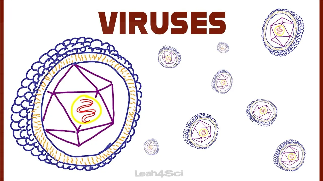
Viruses an Introduction
8K
Pathogens are infectious agents causing disease, including bacteria (prokaryotes), fungi (eukaryotes), parasites, and viruses. While some organisms in each category are pathogenic, many are beneficial—for example, intestinal bacteria produce vitamin K, and mold produces antibiotics like penicillin. Viruses are unique among pathogens as they range from 20-400 nanometers, making them 1,000 times smaller than bacteria. Critically, viruses are not considered living organisms because they lack cells, cannot obtain food, and cannot reproduce independently. Instead, they hijack living cells to replicate, functioning more like computer code that requires a host system to execute its instructions.
Click to watch →
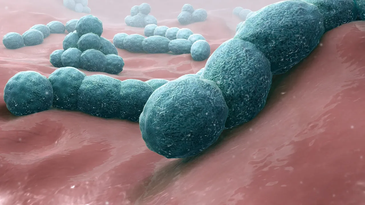
Introduction to the Microbial World
246.9K
Microorganisms are microscopic organisms that exist everywhere—in our environment, on our bodies, and even inside us. While most are harmless or beneficial, some are pathogens that cause diseases like colds, food poisoning, and chickenpox. The study of these tiny organisms began in the late 1600s when Anton van Leeuwenhoek discovered bacteria using his improved microscope, leading to the development of germ theory by Friedrich Henle and its confirmation by Louis Pasteur and Robert Koch. Microorganisms are classified into four main categories: viruses (the smallest, ranging from 18-300 nanometers), bacteria (prokaryotic unicellular organisms), fungi (eukaryotic organisms including yeast and mold), and parasites (ranging from unicellular to multicellular forms). Understanding these categories is essential for comprehending how pathogens interact with the human body to cause illness.
Click to watch →
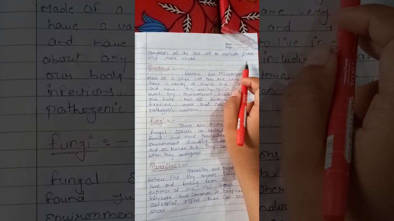
Pathogens and common disease
30.2K
Pathogens are microorganisms that cause disease. The human body naturally contains many microorganisms, but only certain ones cause problems. Pathogens can enter the body through various routes depending on their type. The four main types of pathogens are viruses, bacteria, fungi, and parasites. Each type has distinct characteristics and causes different kinds of diseases. Understanding these categories helps in identifying and treating infections appropriately.
Click to watch →

Vaccines|Immunity|Immunisation| Ch-2 | L-4 |Preventive healthcare | Social Pharmacy|D.Pharm Ist Year
41K
Immunity is the body's ability to defend itself against disease-causing organisms called pathogens. The immune system is the complete defense mechanism that fights diseases. Pathogens are organisms that can enter the body and potentially cause illness. The body constantly encounters pathogens through daily activities like eating, but only a few successfully cause disease because the immune system eliminates most of them. Antibodies are the 'soldiers' of the immune system that are released to fight against pathogens and protect the body from diseases.
Click to watch →
Prerequisite Knowledge
- Concept 01Basic cellular structure: Understanding what a cell is, including key components like the cell membrane, cytoplasm, and nucleus.
- Concept 02The human circulatory system: A foundational understanding of how the heart, blood vessels, and blood transport materials throughout the body.
- Concept 03The concept of diffusion: How gases like oxygen and carbon dioxide move across biological membranes.
- Concept 04Introduction to pathogens: A basic familiarity with bacteria and viruses as external threats to human health.
Subsequent Learning
- Step 01Hematopoiesis: The physiological process of blood cell production, regulation, and differentiation in the bone marrow.
- Step 02The coagulation cascade: The complex biochemical pathway involving clotting factors and platelets to prevent bleeding.
- Step 03Pathology of blood cells: Studying clinical disorders such as anemia, leukemia, hemophilia, and sickle cell disease.
- Step 04Adaptive immunity: Exploring how specific white blood cells (T-cells and B-cells) create antibodies and cellular memory.
- Step 05Blood typing and immunology: Understanding the ABO and Rh antigen systems and their critical role in blood transfusions.
Blood Basics
Blood Basics
0:00Playing Section
- 1
Blood transports oxygen, nutrients, and waste throughout the body.
- 2
Red cells carry oxygen, white cells fight infection, platelets clot.
- 3
Plasma and cells work together for protection and regulation.
Non-Canonical Cell Functions and Immunothrombosis
While traditional science education categorizes blood cells into rigid, distinct roles—red blood cells for oxygen transport, white blood cells for immunity, and platelets for clotting—modern hematology reveals these boundaries are highly blurred. Emerging research highlights 'non-canonical' functions and intense crosstalk between these cells. For instance, platelets are now recognized as key players in the innate immune response, directly interacting with pathogens and white blood cells in a process called 'immunothrombosis.' Similarly, red blood cells actively participate in immune signaling and vascular regulation, rather than acting as passive oxygen carriers. Introducing students to these overlapping, multifunctional roles challenges the traditional compartmentalized view of blood physiology.
Deep Dive
Hematopoiesis: The physiological process of blood cell production, regulation, and differentiation in the bone marrow.

An introduction to Haematopoesis
537.5K
Hematopoiesis is the process by which hematopoietic stem cells in the bone marrow differentiate into all blood cell types, including red blood cells (erythrocytes), white blood cells (lymphocytes, granulocytes, monocytes, dendritic cells, mast cells), and platelets; this process follows two main lineages—the lymphoid lineage producing immune cells like B cells, T cells, and natural killer cells, and the myeloid lineage producing granulocytes, monocytes, erythrocytes, and platelets—under hormonal regulation by erythropoietin and thrombopoietin, with hematopoiesis occurring primarily in the bone marrow in adults but also in other organs like the liver, spleen, and lymph nodes during certain conditions.
Click to watch →

Hematopoiesis | Formation of Bood | Formation of Hemoglobin | Part 2 Unit 3 | Anatomy and Physiology
139.3K
Hematopoiesis is the process of blood cell formation occurring primarily in the bone marrow, where hematopoietic stem cells differentiate into myeloid and lymphoid progenitors that produce red blood cells (erythropoiesis), white blood cells (leukopoiesis), and platelets (thrombopoiesis); hemoglobin, the oxygen-carrying protein in RBCs, consists of four heme units (each containing iron) attached to globin polypeptide chains (two alpha and two beta chains), and is synthesized through a complex pathway involving succinyl-CoA, glycine, and iron to form the oxygen-binding molecule essential for cellular respiration.
Click to watch →

Hematopoyesis
63K
Blood consists of plasma and cellular components: erythrocytes (oxygen transport), granulocytes (pathogen defense), monocytes/macrophages (phagocytosis), dendritic cells (immune sentinels), thrombocytes (hemostasis), and lymphocytes (adaptive immunity). All blood cells originate from hematopoietic stem cells with self-renewal and differentiation capabilities. Intrauterine hematopoiesis occurs in yolk sac, AGM region, and placenta, producing primitive erythrocytes with nuclei. Hemoglobin structure involves alpha and beta globin gene clusters, with different types (Gower 1, Gower 2, Portland, Fetal, Adult) appearing sequentially. The liver becomes the major hematopoietic organ intrauterine, shifting to bone marrow postnatally. Stem cells are classified by potency: totipotent (complete organisms), pluripotent (all three germ layers), multipotent (specific tissue lineages). Hematopoiesis produces lymphoid (B, T, NK cells) and myeloid (erythrocytes, megakaryocytes, granulocytes, monocytes) lineages. Hematopoietic stem cells are extremely rare (0.01% of bone marrow cells) and identified by surface markers (CD34+, CD90+, CD117+, CD133+). Differentiation follows a recipe-like process requiring specific factors (thrombopoietin, erythropoietin, interleukins). Clinical applications include using these factors to stimulate blood cell production in deficient patients.
Click to watch →

Blood Physiology By Dr Sree Teja
125.9K
Hematopoiesis begins in the yolk sac around day 19 of embryonic development. By 3-4 months, it shifts to liver, spleen, and lymph nodes. At birth, it becomes exclusive to bone marrow. During puberty, hematopoiesis becomes limited to membranous bones (vertebrae, sternum, ribs) rather than long bones. This spatial and temporal organization ensures continuous blood cell production throughout life.
Click to watch →
Railway Vacancy 2025 | रेल Foundation 2025 | Blood #2 | Biology Demo Class 02 | Bhawna Mam
53.7K
Hematopoiesis is the process of blood formation with three stages. The yolk sac stage is the first site of blood formation, where the mesoderm layer produces blood cells. The hepatic stage involves blood formation in the liver, typically occurring around 24-28 weeks of fetal development. During the fetal stage, blood is formed in both the liver and spleen. The marrow stage involves blood formation in the bone marrow, which is a spongy material inside bones. This is the primary and final site of blood cell production in adults, and blood formation continues in the bone marrow throughout life. Red blood cells are called erythrocytes.
Click to watch →
Deep Dive
The coagulation cascade: The complex biochemical pathway involving clotting factors and platelets to prevent bleeding.

Coagulation cascade | Human anatomy and physiology | Health & Medicine | Khan Academy
1.3M
The coagulation cascade is a complex series of enzymatic reactions that stops bleeding after blood vessel injury, involving two main pathways—the intrinsic pathway (activated by damaged endothelium) and the extrinsic pathway (activated by tissue factor)—which converge to activate thrombin, which then converts fibrinogen to fibrin strands that form a mesh holding the platelet plug together; this cascade includes negative feedback loops to prevent excessive clotting and deficiencies in specific clotting factors (such as VIII, IX, or XI) can lead to hemophilia.
Click to watch →
![High Cholesterol is Healthy! [with David Diamond, PhD]](https://i.ytimg.com/vi/XfJNUbJE_V0/maxresdefault.jpg)
High Cholesterol is Healthy! [with David Diamond, PhD]
1.1M
The coagulation cascade is a complex system controlling blood clotting, with each step having both pro-coagulant accelerators and anti-coagulant brakes. When this system malfunctions, inappropriate clots form more easily. Stress, hypertension, and high blood sugar activate this pathway because the body interprets these conditions as hemorrhage threats, preparing to form protective clots. This evolutionary response was adaptive for survival in ancestral environments but becomes pathological in modern society with constant stress and carbohydrate consumption. Understanding this mechanism explains why controlling blood sugar, blood pressure, and stress is more important than simply lowering cholesterol for heart disease prevention.
Click to watch →

The Coagulation Cascade - Intrinsic & Extrinsic- Most COMPREHENSIVE Explanation - Hematology
353.1K
The coagulation cascade is a series of enzymatic reactions that convert fibrinogen into fibrin to form a stable blood clot, involving two main pathways: the intrinsic pathway (activated by subendothelial collagen exposure, involving Factors XII, XI, IX, and VIII) and the extrinsic pathway (activated by tissue factor released from damaged tissue, involving Factor VII); both pathways converge at Factor X to activate prothrombin into thrombin, which then converts fibrinogen into fibrin fibers that trap red blood cells, with Factor XIII providing final stabilization of the fibrin meshwork.
Click to watch →
Coagulation Cascade SIMPLEST EXPLANATION !! The Extrinsic and Intrinsic Pathway of HEMOSTASIS
977.6K
The coagulation cascade is a sequential biochemical process that stops bleeding through clot formation, involving two main pathways—the extrinsic pathway triggered by tissue trauma releasing tissue factor that activates Factor VII to convert Factor X, and the intrinsic pathway triggered by blood cell damage or collagen exposure that activates Factor XII to sequentially convert Factors XI and IX; both pathways converge to form prothrombin activator (activated Factor X), which converts prothrombin to thrombin, which then converts fibrinogen to fibrin monomers, with Factor XIII stabilizing these monomers into cross-linked fibrin polymers that attract platelets and phospholipids to form a stable blood clot and achieve hemostasis.
Click to watch →

360 Degree Series 🔥| 360 Degree Most Imp MCQs #1479 | NORCET & All Nursing Exam| Siddharth Sandu Sir
18.3K
The coagulation cascade is a series of reactions that leads to blood clot formation, involving 13 clotting factors (proteins except factor IV which is calcium). The process begins with platelet plug formation, followed by the extrinsic pathway (initiated by tissue thromboplastin) and intrinsic pathway (initiated by collagen exposure), both converging to form prothrombin activator. This converts prothrombin to thrombin, which then converts fibrinogen to fibrin, forming a stable clot. Factor XIII stabilizes the clot. Vitamin K is essential for synthesizing factors II, VII, IX, and X.
Click to watch →
Deep Dive
Pathology of blood cells: Studying clinical disorders such as anemia, leukemia, hemophilia, and sickle cell disease.
Hematological test for the diagnosis of blood disorders #patho by "Yash Sharma" Bsc nursing 3rd sem
2.6K
Pathology is the study of diseases - how they occur and develop. Blood is a connective tissue in liquid form that carries oxygen. Blood consists of plasma (55%) and formed elements (45%). Plasma contains water, proteins, and salts. Formed elements include red blood cells (erythrocytes, biconcave discs without nucleus, 7.5 micrometers, 120-day lifespan, 4.5-5.5 million/cmm), white blood cells (nucleated, 4,000-11,000/cmm, classified as agranulocytes and granulocytes), and platelets (small irregular fragments without nucleus, 2-4 micrometers, 7-14 days lifespan, 2-4 lakh/cmm).
Click to watch →

Red Blood Cell Pathology | Ch-5 | Red Blood Cell Pathology Pathoma | Blood Pathology Lectures
348
Red blood cell pathology encompasses various conditions affecting RBC structure, function, and lifespan, classified morphologically by MCV (microcytic <80 fL, normocytic 80-100 fL, macrocytic >100 fL) and etiologically by decreased production (iron deficiency, B12/folate deficiency, thalassemia), increased destruction (hemolytic anemias like sickle cell, spherocytosis, PNH), or blood loss. Iron deficiency anemia, the most common worldwide, presents with microcytic hypochromic cells and pencil cells. Thalassemia involves globin chain synthesis defects (alpha on chromosome 16, beta on chromosome 11), with major forms requiring transfusions. Sickle cell anemia results from HbS polymerization under low oxygen, causing vaso-occlusive crises. PNH involves GPI-anchored protein deficiency leading to complement-mediated hemolysis.
Click to watch →

MORPHOLOGIE EN HEMATOLOGIE EPISODE 10
1K
This segment covers two important morphological abnormalities in blood cells. Stomatocytes are red blood cells with an oval central pallor appearing as an elongated slit, concave on one side only, observed in alcoholic cirrhosis, obstructive jaundice, and May-Hegglin anomaly. Döhle bodies are blue-staining cytoplasmic inclusions in neutrophils, representing rough endoplasmic reticulum remnants from immature granulocytes. They are 1-2 micrometers, well-defined, and appear in May-Hegglin anomaly, certain anemias, and infectious states like scarlet fever. Any variation in normal blood cell morphology indicates cellular pathology.
Click to watch →
![Лекция 1. Фармфак. Вводная и лейкоцитозы. [поправлен звук]](https://i.ytimg.com/vi/vTW-Jkz2aBQ/hqdefault.jpg?sqp=-oaymwEmCOADEOgC8quKqQMa8AEB-AH-BIAC6AKKAgwIABABGFIgSyhlMA8=&rs=AOn4CLD2SgS6e514LBT8qymF7Lu3CUHVgQ)
Лекция 1. Фармфак. Вводная и лейкоцитозы. [поправлен звук]
528
General pathology consists of three parts: pathological anatomy (morphological changes), pathophysiology (functions of sick organism), and clinical pathology (clinical manifestations). Pathophysiology studies causes, mechanisms, and laws of disease occurrence, development, and termination. Causes include external factors (physical: radiation, temperature; chemical: acids, alkalis, medications; biological: viruses, bacteria) and internal factors (hereditary, genetic defects). Conditions include environmental, lifestyle, and constitutional factors. The etiotropic principle targets specific causes for optimal treatment. Disease mechanisms include damage mechanisms and protective/compensatory mechanisms. The ratio between protection and damage determines prognosis. Peripheral blood contains erythrocytes, leukocytes (4-8 × 10^9/L), and thrombocytes. Leukocytes are divided into granulocytes (basophils 0-1%, eosinophils 2-4%, neutrophils 55-65%) and agranulocytes (lymphocytes 25-35%, monocytes). Bone marrow is located in flat bones and epiphyses of long bones. All blood cells originate from hematopoietic stem cells differentiating into lymphoid and myeloid lineages. Leukocytosis is physiological (after eating, exertion, pregnancy) or pathological (concentration from fluid loss, production from specific cell increases). Basophils contain heparin and histamine, indicating tissue necrosis or allergic reactions. Eosinophils contain histaminase, indicating allergic reactions, helminth infections, or infections with allergic reactions.
Click to watch →

Tout savoir sur les globules blancs et rouges_leurs rôles dans le sang...
353
This comprehensive lesson covers blood cells and their clinical importance. White blood cells (leukocytes) are immune cells named from Greek 'leukos' (white) and 'kytos' (cells), circulating in blood, lymph, and lymphoid organs. Classified into granulocytes (40-80%), lymphocytes (20-40%), and monocytes (2-10%), they participate in innate immunity (immediate, non-specific response) and adaptive immunity (specific, memory-based response). Pathologies include autoimmune diseases, allergies, HIV/AIDS, and cancers. Diagnostic tests include hemogram, myelogram, and urine examination. Red blood cells (erythrocytes) transport oxygen via hemoglobin, are nucleus-free cells living 120 days with biconcave disc shape. Normal count is 4.5-5.5 million per mm³ (men) or 3.8-5.3 million per mm³ (women). Analysis evaluates hemoglobin, MCV, MCH, MCHC, hematocrit, and morphology. Abnormal shapes indicate diseases like sickle cell disease, thalassemia, or spherocytosis. Results outside normal ranges require medical interpretation.
Click to watch →
Deep Dive
Adaptive immunity: Exploring how specific white blood cells (T-cells and B-cells) create antibodies and cellular memory.

Вячеслав Дубынин "Иммунитет — вечный бой с инфекцией"
117.5K
Adaptive immunity allows the immune system to learn and adapt to specific pathogens. B-lymphocytes produce antibodies with unique binding sites for specific antigens. T-lymphocytes coordinate immune responses and destroy infected cells. Memory cells persist for years or decades, enabling rapid responses upon re-exposure. This system requires initial exposure to develop, taking days to weeks, but provides long-lasting protection. The principle of vaccination exploits this memory, introducing antigens to train the immune system without causing full disease.
Click to watch →
Immunology | Adaptive Immunity
1M
Adaptive immunity consists of two main branches: humoral immunity mediated by B cells and cell-mediated immunity mediated by cytotoxic T cells. In humoral immunity, B cells recognize free extracellular antigens through their B cell receptors (BCRs), undergo receptor-mediated endocytosis, and present processed antigens via MHC class II molecules to activate T helper cells. Activated T helper cells secrete cytokines like IL-4 and IL-5 that stimulate B cell proliferation and differentiation into plasma cells (which secrete antibodies) and memory B cells. In cell-mediated immunity, cytotoxic T cells recognize infected or cancerous cells displaying foreign peptides on MHC class I molecules; upon recognition, they release perforin and granzymes that induce apoptosis in target cells. This dual system provides comprehensive protection against both extracellular pathogens (humoral) and intracellular pathogens/cellular damage (cell-mediated).
Click to watch →
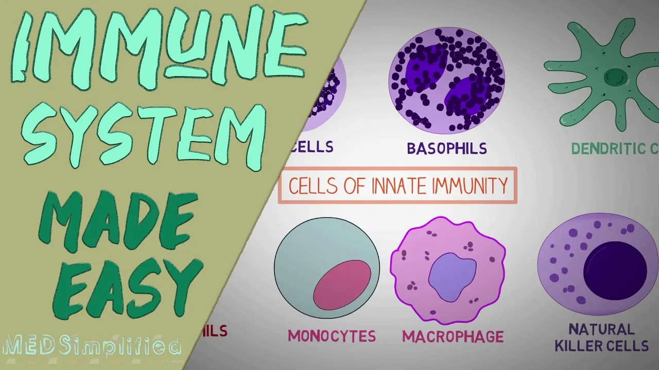
IMMUNE SYSTEM MADE EASY- IMMUNOLOGY INNATE AND ADAPTIVE IMMUNITY SIMPLE ANIMATION
2.5M
Adaptive immunity (acquired/specific immunity) develops after birth in response to pathogen exposure. It consists of T lymphocytes and B lymphocytes (20-40% of WBCs, total mass comparable to brain/liver). T cells include helper T cells (CD4+) that coordinate immune responses by secreting growth factors and cytokines essential for other cell maturation, and cytotoxic T cells (CD8+) that directly kill infected cells. AIDS destroys CD4+ T helper cells, crippling the immune system. B cells produce antibodies (Y-shaped proteins) that recognize and capture pathogen antigens. Upon first exposure, adaptive response is minimal but stores immunological memory. Subsequent exposures trigger amplified, specific responses. Humoral immunity (B cells/antibodies) and cellular immunity (T cells) work together to eliminate pathogens.
Click to watch →

مع د. مجدي فودة، هنتعلم كل تفاصيل باب المناعة بطريقة بسيطة وسهلة. استعدوا لشرح كامل !
469.3K
Adaptive immunity is acquired after birth through exposure to specific pathogens. When an organism encounters a particular pathogen, its immune system develops specific defenses against it. This type of immunity is specific to particular pathogens and provides long-lasting protection. If the same pathogen attacks again, the immune system can quickly recognize and eliminate it.
Click to watch →

فرض الفصل الثالث في العلوم الطبيعية السنة الرابعة متوسط 2025
123.3K
Adaptive immunity (third line of defense) is characterized by three key properties: (1) Acquired immunity - not innate but develops through exposure or vaccination, (2) Transferable immunity - can be transferred between individuals via serum containing antibodies, (3) Specificity - each pathogen requires its own specific antibody response. The mechanism involves B lymphocytes recognizing pathogens, proliferating, and differentiating into plasma cells that produce antibodies. These antibodies bind to specific antigens, forming immune complexes that neutralize pathogens and prevent their spread.
Click to watch →
Deep Dive
Blood typing and immunology: Understanding the ABO and Rh antigen systems and their critical role in blood transfusions.

Blood Type (ABO and Rh) Made Simple!
428.4K
This comprehensive section covers the fundamental immunological principles underlying blood typing. It begins with defining antigens as molecular markers on cell surfaces that identify cell type, and antibodies as Y-shaped immunoglobulins produced by the immune system that specifically bind to particular antigens. The body normally eliminates self-reactive antibodies through tolerance mechanisms, but failure leads to autoimmune reactions. The lecture then transitions to the ABO blood group system, explaining how Type A expresses A antigens, Type B expresses B antigens, Type AB expresses both, and Type O expresses neither. The Rh system is introduced as determining D antigen presence (Rh positive) versus absence (Rh negative). Laboratory testing methods using anti-A and anti-B antibodies are explained, demonstrating how positive/negative reactions determine blood type through antigen-antibody binding.
Click to watch →

ABO Blood Types Made Easy!
429.2K
Blood typing is based on antigens (sugar molecules) on red blood cell surfaces that determine identity. The ABO system classifies blood into four types: A (only A antigen), B (only B antigen), AB (both A and B antigens), and O (no antigens). Each blood type produces specific antibodies against foreign antigens: Type A produces anti-B antibodies, Type B produces anti-A antibodies, Type AB produces neither, and Type O produces both. This antigen-antibody relationship explains why incompatible blood transfusions trigger immune attacks, making proper blood typing essential for safe transfusions.
Click to watch →
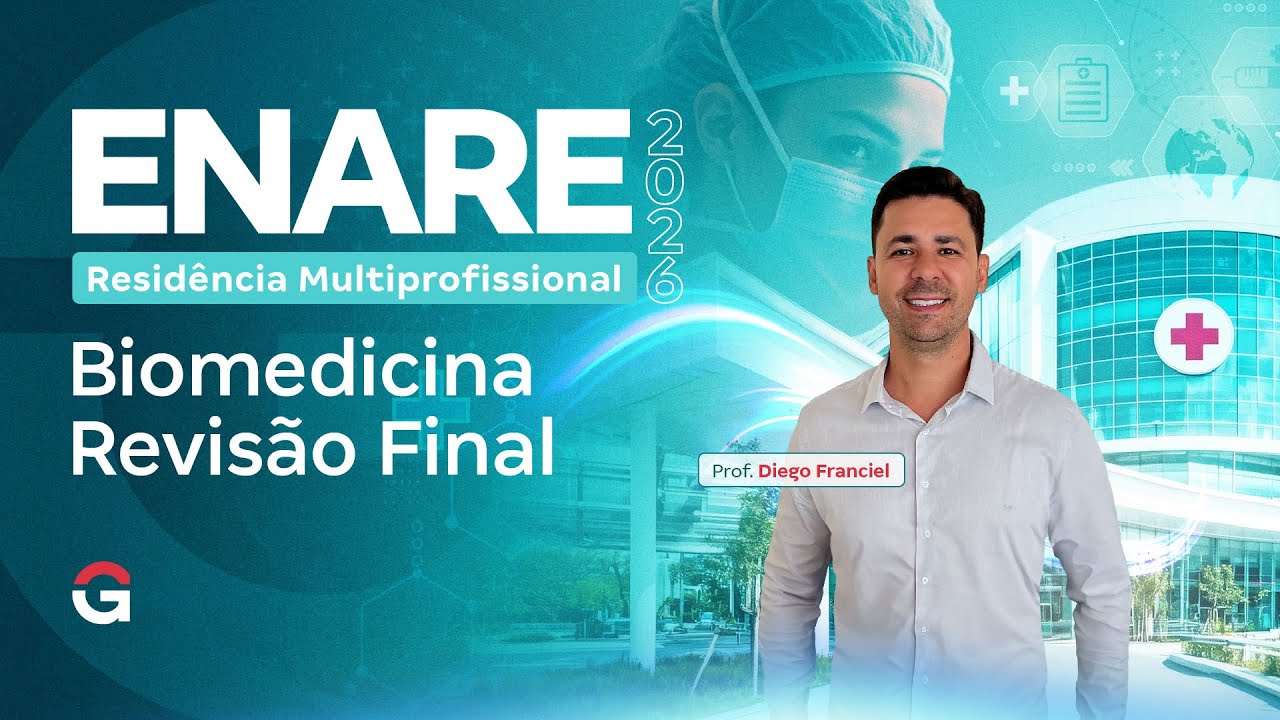
Residência ENARE Biomedicina | Revisão Final
698
This segment covers essential hematology and immunology topics for FGV exams. ABO blood typing is defined by antigens on RBCs and natural antibodies in serum. Direct Coombs test detects antibodies or complement bound to RBC surface by adding patient RBCs with commercial antisera and antiglobulin reagent. Indirect Coombs test detects free antibodies in serum by adding patient serum with known antigen-positive RBCs, then adding antiglobulin reagent. Key distinction: direct test detects antibodies on RBC membrane, indirect test detects antibodies in serum. The antiglobulin reagent acts as an anti-antibody that bridges antibody Fc regions, making agglutination visible. Essential topics include RBC morphology alterations (drepanocytosis, sickle cell, target cells), hemoglobinopathies (thalassemias, sickle cell disease), antigen-antibody reactions, autoimmunity, and immunological techniques (ELISA, immunofluorescence, flow cytometry). Rapid test principles (chromatography) are also covered.
Click to watch →
Repaso Sistema Sanguineo - Respiratorio | Anato y fisio humana | UPCH
287
Blood types are determined by antigens on red blood cells: Type A (A antigen), Type B (B antigen), Type AB (both antigens), Type O (neither). Rh factor adds positive/negative classification. Blood typing uses agglutination: mixing blood with anti-A, anti-B, and anti-Rh antibodies. Agglutination indicates antigen presence. Immunity includes innate (non-specific, present at birth: skin, mucous membranes, tears, inflammatory response) and adaptive (specific, requires prior exposure, provides memory). Lymphocytes coordinate adaptive immunity: T cells direct responses, B cells produce antibodies (immunoglobulins: IgA, IgE, IgG, IgM, IgD).
Click to watch →

Физиология. Глава 4. Кровь. Группы крови
42.2K
Blood groups are determined by specific antigens on red blood cell surfaces. The ABO system classifies blood into four groups based on A and B antigens: Group I (O) has no antigens, Group II (A) has A antigen, Group III (B) has B antigen, and Group IV (AB) has both. The Rh system adds the D antigen classification (positive/negative). Antigens are molecular structures that can bind to antibodies; agglutinogens refer specifically to red blood cell antigens, while agglutinins are the corresponding antibodies. Each blood group produces antibodies against antigens it lacks: Group I produces anti-A and anti-B, Group II produces anti-B, Group III produces anti-A, and Group IV produces none. When incompatible blood is transfused, recipient antibodies attack donor antigens, causing dangerous agglutination reactions.
Click to watch →
Blood Basics
Blood Basics
0:00- 1
Blood transports oxygen, nutrients, and waste throughout the body.
- 2
Red cells carry oxygen, white cells fight infection, platelets clot.
- 3
Plasma and cells work together for protection and regulation.
Non-Canonical Cell Functions and Immunothrombosis
While traditional science education categorizes blood cells into rigid, distinct roles—red blood cells for oxygen transport, white blood cells for immunity, and platelets for clotting—modern hematology reveals these boundaries are highly blurred. Emerging research highlights 'non-canonical' functions and intense crosstalk between these cells. For instance, platelets are now recognized as key players in the innate immune response, directly interacting with pathogens and white blood cells in a process called 'immunothrombosis.' Similarly, red blood cells actively participate in immune signaling and vascular regulation, rather than acting as passive oxygen carriers. Introducing students to these overlapping, multifunctional roles challenges the traditional compartmentalized view of blood physiology.
Basic cellular structure: Understanding what a cell is, including key components like the cell membrane, cytoplasm, and nucleus.
The human circulatory system: A foundational understanding of how the heart, blood vessels, and blood transport materials throughout the body.
The concept of diffusion: How gases like oxygen and carbon dioxide move across biological membranes.
Introduction to pathogens: A basic familiarity with bacteria and viruses as external threats to human health.
Hematopoiesis: The physiological process of blood cell production, regulation, and differentiation in the bone marrow.
The coagulation cascade: The complex biochemical pathway involving clotting factors and platelets to prevent bleeding.
Pathology of blood cells: Studying clinical disorders such as anemia, leukemia, hemophilia, and sickle cell disease.
Adaptive immunity: Exploring how specific white blood cells (T-cells and B-cells) create antibodies and cellular memory.
Blood typing and immunology: Understanding the ABO and Rh antigen systems and their critical role in blood transfusions.
783 views63likes1:36@priyakumar.OOriginal Release: 2026-01-19
Have you ever wondered what is flowing inside your body every second to keep you alive?
Blood.
Blood is an important fluid that circulates throughout the human body and helps in transport, protection, and regulation. It is made up of plasma and blood cells. There are three main types of blood cells. Red blood cells, white blood cells, and platelets. each having a specific function.
Red blood cells, also called iththraittes, carry oxygen from the lungs to all parts of the body. They contain a protein called hemoglobin, which binds with oxygen and gives blood its red color. Red blood cells also carry carbon dioxide from the body back to the lungs for removal.
White blood cells, also known as luccoytes, play a vital role in protecting the body from diseases. They help fight infections by destroying harmful bacteria, viruses, and other germs. White blood cells are an important part of the immune system and help the body stay healthy.
Platelets or thrombocytes are very small cell fragments that help in blood clotting. When an injury occurs, platelets gather at the sight of the wound and form a clot to stop bleeding.
This helps in healing and prevents excessive blood loss.
For more fun learning videos, please like, share, and subscribe.
Up Next

71:26
Reactive vs Neoplastic Lymphocytes: Morphology Guide
@gillianrozenbergmicroscopi6968
13.8K views•2021-12-28

50:12
Bacterial Communication: Quorum Sensing and Biofilm Formation Explained
@JHUAAP
12.6K views•2011-06-28

5:35
Enteric Nervous System Explained: The Gut's Brain | Neurobiology Lecture
@alumniu6029
438 views•2018-09-12

7:08
Bacteriophages: Earth's Deadliest Killers and Future Antibiotics
@kurzgesagt
34.6M views•2018-05-13
Related Study Plans & Knowledge Roadmaps
Structured learning paths in Biology
biology
RNAi: Biogenesis, RISC & Gene Silencing
01Molecular Biology Foundations: DNA to Protein
02Introduction to RNA Interference (RNAi)
03Molecular Biogenesis of miRNA and siRNA
+ 2 more learning modules
10h5 modules
Explore Roadmap
biology
Mycorrhizae: Signaling, Biophysics & Ecology
01Foundations of Mycorrhizal Symbiosis
02Molecular Signaling and Root Colonization
03Biophysics and Bioenergetics of Nutrient Exchange
+ 1 more learning modules
11h4 modules
Explore Roadmap
biology
Circadian Clocks: TTFL, SCN & Periphery
01Foundations of Molecular Biology & Gene Expression
02Introduction to Chronobiology and Actograms
03The Core TTFL: Molecular Architecture of the Clock
+ 3 more learning modules
14h6 modules
Explore Roadmap
biology
Population Genetics: Math, Drift & Selection
01Genetic & Probability Foundations
02The Hardy-Weinberg Principle and Baseline Models
03Mathematical Modeling of Natural Selection
+ 3 more learning modules
18h6 modules
Explore Roadmap