
















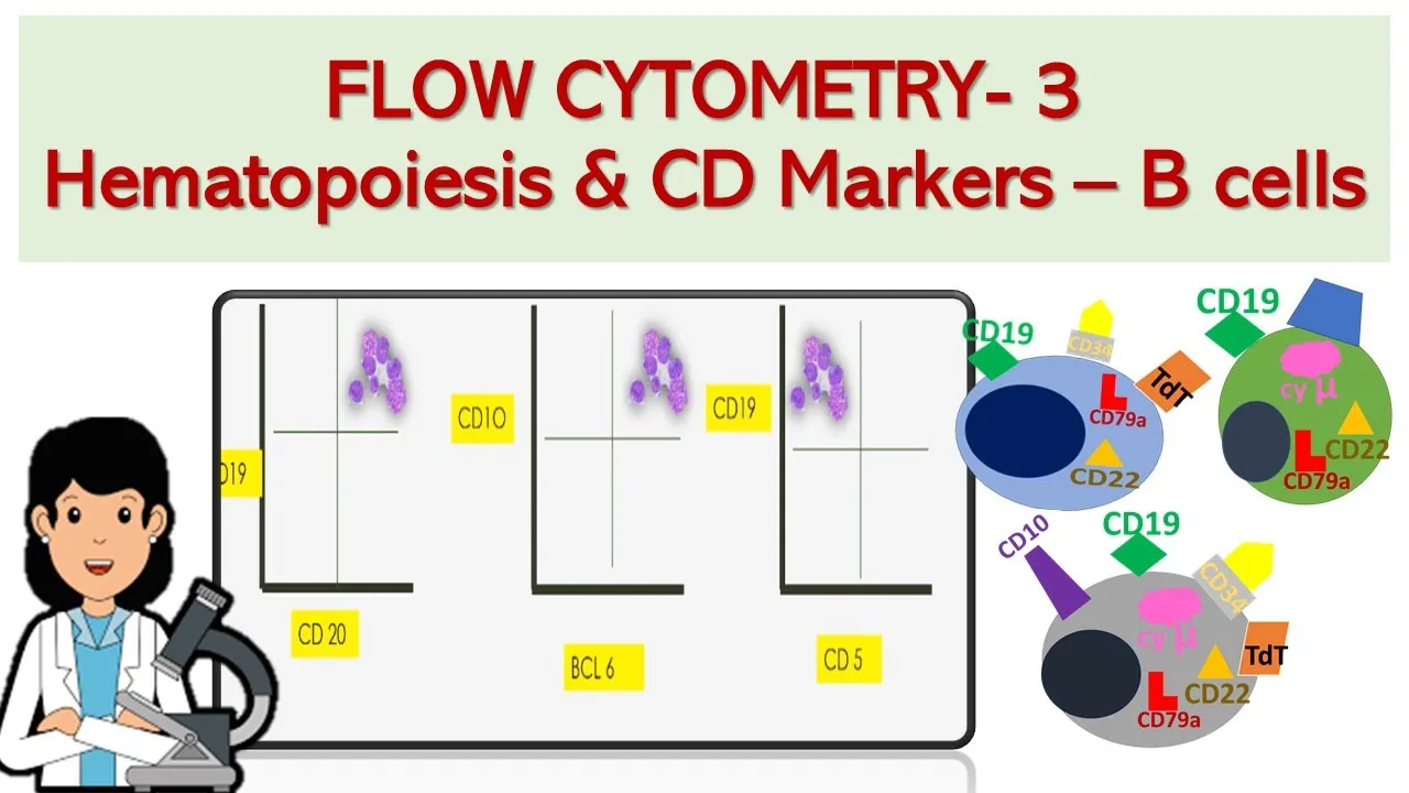










Reactive lymphocytes are activated immune cells responding to infections or inflammation, characterized by increased size, flowing basophilic cytoplasm, enlarged nuclei with fine chromatin, and prominent nucleoli; they commonly appear in viral infections like infectious mononucleosis, CMV, and dengue. In contrast, neoplastic lymphocytes represent clonal proliferative disorders with distinct morphological features including non-flowing cytoplasm, less basophilic staining, coarser chromatin, and more prominent nucleoli, forming monomorphic populations associated with cytopenia, splenomegaly, and lymphadenopathy. Key diagnostic markers help differentiate these conditions: B-cell neoplasms show CD19/CD20 positivity, while T-cell neoplasms show CD3 positivity, and specific immunophenotyping aids in identifying subtypes such as CLL/SLL, hairy cell leukemia, or Burkitt lymphoma.
Reactive vs Neoplastic Lymphocytes: Morphology Guide
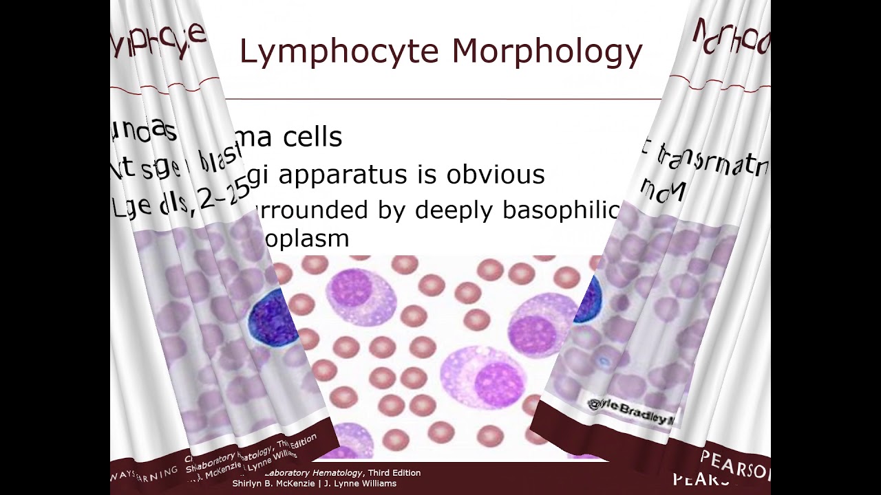
Added:so our next topic today is lymphocytes reactive versus neoplastic so lymphocytes are the cells of the body that respond to foreign antigens small lymphocytes range in size from 9 to 12 microns while large lymphocytes range from 12 to 16 microns small lymphocytes have a round nucleus occupying about 90 percent of the cells the chromatin is dense and the cytoplasm is basophilic and here's a nice example of a small lymphocyte you can see the nucleus is approximately seven microns in diameter so large lymphocytes have more cytoplasm and usually contain five to fifteen small asiophilic granules they are a sub-population making up five percent of the lymphocytes seen in normal blood so i'm afraid i must apologize because this is not a very good image of a large lymphocyte they don't usually have these cytoplasm flowing like this but once you've made a made this workshop set in concrete it's a lot of work to get rid of it and place it with a good one but that's a large lymphocyte we don't see only 100 5 to 15 will be on a normal blood film so lymphocytes consist of distinct subsets that are quite different in their function and protein products even though they appear morphologically similar there are three classes of lymphocytes the b lymphocytes these are derived from the bone marrow and are the only cells capable of producing antibodies the antigen receptors of b lymphocytes are membrane-bound forms of antibodies interaction of antigens with these membrane antibody molecules initiates a sequence of b cell activation culminating in the development of plasma cells that actively secrete antibody molecules t lymphocytes these arise from the bone marrow and then migrate and migrate to and mature in the thymus t lymphocytes are further divided into functionally distinct populations the helper t cells are the cd4 cells and cytotoxic t cells are cd8 positive t cells do not produce antibodies their principal function is to regulate all immune responses to protein antigens and to serve as effector cells for the elimination of intracellular bacteria so large granule lymphocytes or the natural killer cells these lymphocytes do not express markers of either t or b cells and were initially called null cells natural killer cells are capable of lysing virus infected cells as well as tumor cells without overt antigenic stimulation so now we're going to move on to reactive lymphocytes i might say at this point in time that sometimes people use the incorrect term they call them atypical and atypical should never be used ever according to the icsh atypical is finished so always think of these lymphocytes as reactive so in reaction to viral infections when there is antigen recognition the cd8 cytotoxic lymphocytes are attracted towards the antigen target and commenced to produce cytokines activated cd8 cytotoxic lymphocytes are increased in size and have flowing basophilic cytoplasm the nucleus is enlarged and the chromatin fine with one or more nucleoli and i've listed here for you some of them more probably the majority of disorders where you see reactive t cells so infectious mononucleosis cytomite cytomegalovirus toxoplasmosis viral hepatitis dengue fever measles chickenpox bordetella pertussis and lymphocytosis of acute infection and bordetella pertussis is in italics because it is actually not a viral infection it is a bacterial infection but most of the textbooks will refer to the lymphocytes in bordetella pertussis as being reactive so just concentrate on the lymphocytes here the red cells are not very good in this particular image but these are the reactive lymphocytes of infectious mononucleosis so you can see that the nucleus is quite pleomorphic the cytoplasm is increased it's very basophilic and often when you look carefully the actual cytoplasm border is quite dark blue with the romanovsky stain of course all these films are stained with the romanovsky stain this is an example of a cytomegalovirus very similar to that of infectious mononucleosis again this is another example of three reactive lymphocytes in somebody who's got the cytomegalovirus infection this is an example these are examples of turk cells this is a case of a patient with viral hepatitis and the turk cells are quite large they have very round nuclei the cytoplasm is very basophilic but it doesn't flow like that it does in the infectious mononucleosis for example they're quite large and this is a patient who had viral hepatitis and this is a patient again who's got a turk cell um and that's the turk cell and i've photographed a small lymphocyte nucleus and put it next to it so you can see the difference they stand out on the blood film very clearly they're quite basophilic cytoplasm an eccentrically placed nucleus and a large cell so you can call these turk cells or you can actually call them reactive lymphocytes i might say reactive lymphocytes and then in brackets turk cells because they're a classical feature of dengue fever and this is just another single just to impress on you this is a terxil or a reactive lymphocyte seen in dengue fever also seen in malaria infections as well often if somebody has got a significant number of malarial parasites infecting the red cells these cells react it's basically like a cd8 positive t cell reacting to an infection and then in malaria it's reacting to the presence of malarial parasites infecting the red cells this is a case of varicella infection chickenpox and here's the bordetella pertussis which is a bacterial infection and the classic feature in bordetella pertussis is that the lymphocyte nuclei are cleaved and not all of them will be cleaved so it should be a significant feature if you want to ask me the question what percentage i can't tell you it's probably most classically found in a young child who will have a lymphocytosis for no known reason except that when you get that lymphocytosis in a young child i would look very carefully from field to field to see how many of the lymphocytes have got a cleaved nucleus and when you see that i would report it definitely report it and where i work i make sure that the scientists will ring the consultant uh about this child and let them know that we've seen lymphocytes significant or commonly found in bordetella pertussis it's highly infectious so it's important that you do notify somebody on the medical team looking after the child and this is the last case this is lymphocytosis of acute infection in a child these were cd4 positive t cells and this is probably the only case i have seen you can see the t cells often do have cleaved nuclei and this was a little child who was three years of age who had a very high nuclear lymphocyte count and had a very severely infected finger and i watched this patient from progress and after about three weeks these all disappeared but this is actually a recognized disorder which is reported in some of the pediatric textbooks and my only case that i've ever seen so let's now go on to neoplastic lymphocytes so neoplastic lymphocytes have many of the features of reactive lymphocytes but they're part of a lymphoproliferative clone they're almost always b cells and they show b cell markers depending on their degree of immaturity of course you do get t-cell disorders but they're t-cell neoplasms but they're much less common so their cytoplasm the b-cells they're cytoplasm is contained and it does not flow to the same extent as it does in the reactive lymphocytes the cytoplasm is less basophilic and the chromatin pattern of the nucleus is more coarse and the nucleoli are more prominent so being lymphocytes form a monomorphic population there is a progressive increase in the number of neoplastic cells its associated with they are associated with a concomitant cytopenia and or a leukoerythroplastic blood picture they show normal or hypogamoglobin anemia on serum epg and they may show a paraprotein they're associated with non-tender splenomegaly and lymphadenopathy so now i have listed for you all the b-cell lymphoid neoplasms and these nomenclature will follow the latest who classification so b lymphoblastic leukemia lymphoma not otherwise specified often shortened as all chronic lymphocytic leukemia small and pacific lymphoma which is shortened as cll sll then there's pro lymphocytic leukemia splenic marginal zone lymphoma hairy cell leukemia lymphoplasmacetic lymphoma and this is a more recent term for waldenstrom macroglobulin anemia plasma cell myeloma as i said this is the more recent who class classification so we don't call it multiple more like multiple myeloma is not used now it's plasma cell myeloma and then plasmacytoma non-hodgkin lymphoma and burkett cell lymphoma leukemia so the t cell neoplasms t cell lymphoblastic leukemia lymphoma large granular lymphocytic leukemia aggressive natural kelly cell leukemia adult t cell leukemia lymphoma pro-lymphocytic leukemia cutaneous t cell lymphoma so we'll go through that second group at the end of this workshop we're now going to go through the b cell lymphoblastic neoplasms or precursor neoplasms so we're going to talk now about be lymphoblastic leukemia slash lymphoma not otherwise specified so the blasts range from those with a high nc ratio fine to clumped chromatin pattern with scanty cytoplasm and inconspicuous nucleoli now just to say to you at this point in time some of you that may remember the fab classification the french american and british classification of these disorders this first group here these has been was actually known as the all l1 and the second group i'm just going to describe now which is to those that are heterogeneous in size and shape with fine to coarse chromatin pattern often cleaved indented and folded nuclei nuclear are nearly always present with variability in size and number and also a variable amount of cytoplasm so if you're in the generation that used to know the fab classification this is the l2 group here and what the who have done they've linked the two together just as a general description of the lymphoblasts that you see in b cell lymphoblastic leukemia lymphoma and here's a really nice slide this is showing you a case of b lymphoblastic leukemia lymphoma and these would be the l1 type of lymphoblasts with a very high nc ratio um reasonably find a clumpy chromatin pattern basophilic cytoplasm and in this case inconspicuous nucleoli another one here inconspicuous nucleoli this is another these two about the same so they would be the ill ones these are a little bit different this is happened to be patient with a mixture so perhaps possibly that's why the who have combined the two together in these two here these are classically lymphoblasts uh with a high nc ratio slightly more fine chromatin pattern with a clear nucleolus this is a nucleolus here and a nucleolus here there's possibly one here but they're usually described as being inconspicuous in this particular type of being infoblastic leukemia lymphoma not otherwise specified this is the bone marrow of the same patient and you can see it's an overwhelming number and this is very common in children um this is the classical bone marrow which is filled with a very high lymph blast cell count uh very high in c ratio inconspicuous nucleoli and something to note from experience that often when you've got a bead in for plastic leukemia or even a tea you sometimes see vaculated um vacuoles appearing even superimposing the nucleus and it's a common feature that has no specific significance now i'm taking you through some trafines because i think it's very interesting to look at it you're fine scientists may not look at the true fines but you should make it your job to if you see a case and you're looking at the marrow just see if you can find that refine and have a look at it so basically that refine gives you the architecture of what's going on in the marrow and they basically core down after they've done the aspirate they cool down and take a cylinder of bone and that refine is sent to anatomical pathology where it is decalcified and stained and this is a hema this is an h stain hematoxylin and eosin stain and this is part of the bone which has been decalcified so when they've done that they're actually embedded in wax and they cut it on the micro term and stain it so you can see here that we've got really a monomorphic population of cells which are just replacing the entire bone marrow this is a slightly higher power so this is that first one was times 200 magnification this is times 400 and just to give you an idea that you've just got the marrow being replaced by a clonal population of lymphoblasts so just to make it clear for you because we call this disorder lymphoblastic leukemia slash lymphoma so the term benefiblastic leukemia applies when there is involvement of the bone marrow and peripheral blood and the term lymphoblastic lymphoma when there is significant nodal or extranodal involvement and the distinction between the two names is arbitrary so now i'm as i explained to you in some of the other workshops these are morphology workshops and i'm going to concentrate on the morphology features but i obviously have to include immunophenotyping of the cells and also the cytogenetics so i'll just go through some of the markers and point out to you that these are b cells so the b cell markers here the cd10 cd19 22 and i've already told you about cd 34 which is a progenitor marker so okay so there are actually four different categories in the immunophenotyping and they're actually grouped in so this is called an early ball this is a common all and they're grouped according to the actual maturation of the cells the most common that we get especially in children is the precursor b all and um you can see with the precursor b allele the cd34 in this case in that particular group is negative and then there's just a straight out b cell all and that's all put into those categories according to the immunophenotype cytogenetics we're not going to go into cytogenetics that much because it's not really um because if you really want to if we're a registrar studying these workshops you may want to know this but these are just the most common cytogenetics but i'm not going to go into those and explain those to you so now we're moving to the next group which is the mature b cell neoplasm and we're going to look at chronic lymphocytic leukemia small lymphocytic lymphoma and classified shortened often on the request form as cll sll and it occurs in older males with a median age of 65 years there's a clonal lymphocytosis with more than ten by ten to the ninth per liter you get small lymphocytes with round nuclei clumped chromatin pattern and basophilic cytoplasm sometimes you'll see smudge cells on a film with someone with this cll sll and just to make sure you understand the smudge shell is these cells in this particular neoplasm are quite um delicate and when you spread them even with the when you make a manual blood film but especially when the blood goes through the analyzer you can produce a lot of smudge cells it's just because they are fragile and the smudge cell is really just a broken lymphocyte and you all what you're looking at is the nucleus which has been a little bit distorted uh less than two percent pro lymphocytes are present in cll so uh it's not really even worthwhile reporting you have to have more than 55 percent of pro-lymphocytes you can put it into a pro-lymphocytic leukemia so the occasional pro-lymphocytic cell is not really that significant in a cll and here's a classical picture of cll chronic lymphocytic leukemia all of those lymphocytes look okay they're nice they're normal i think when i probably started my workshops doing the very first workshops i emphasized on the fact that you look at the age of the patient and also look at the sex of the patient remember that cll is a classic feature of older males so you put it all together it's all important all these features the clinical picture it's all important um to give you an understanding of what you're looking at no point just reporting it and not knowing what you're looking at that's not the idea of morphology morphology is very exciting and when you put it all together you can make a diagnosis yourself so the cd markers are important in that when we go through all of these b-cell neoplasms cll sll is the only one that is cd5 positive and cd5 is a t cell marker but it's positive when we look at some of the others which we're going to look at in this group it might look a bit like cll lymphocytes they if they're only going to be cll if they're cd5 positive okay so that's something to remember cytogenetics is a whole lot of cytogenetics i'm not going to go into those so now moving to prolimb acidic leukemia this occurs again in older males with a median age of 65 to 69 they have a clonal lymphocytosis it's usually a very high count of more than a hundred by ten to the ninth per liter the pro lymphocytes must exceed 55 of the lymphoid cells in the peripheral blood to put it into this category as i just said to you pro-lymphocytic leukemia pro-lymphocytes larger than lymphocytes are larger than the lymphocytes of cll because the cellular lymphocytes are mature pro-lymphocytes are always just a little bit less mature and larger in size they have abundant cytoplasm around nucleus with moderately condensed chromatin pattern but what is standout feature of a protein beside is they have a single and large nucleolus it's almost like an eye looking out at you and this is the blood film of a patient with pro lymphocytic leukemia and i'll just point out to you the most classical of this film they're all classical to me but for you if you're still studying morphology that is just classical of a pro-lymphocyte it's got a single nucleolus the chromatin pattern is is you know slightly clumpy slightly immature it is not a blast because the nc ratio is much too low the nc ratio in a blast is very high as you saw so that's a classical pro lymphocyte this is another one here there's a bit of a nucleolus here which is not so obvious but here the really clear nucleolus here here and here i'll show you another image of this patient uh as i said they have a very high white cell count usually more than a hundred by ten to the ninth per liter and here he has got a very high count so he's gradually getting worse this particular patient i remember very well because he was in his terminal stage of his disease and his wife's account kept going up and so the hematologist looking after him um lucoferist him took off some of the lymphocytes to bring down the count and a few weeks later sadly the count went back up again so that's a classical feature of pro-lymphocytic leukemia he's a single nucleolus here here here here and here just like an eye looking out at you and here's a smudge sill actually these two are smudge cells okay i would only report smudge cells in someone who's got chronic cll um but you do sometimes notice smudge cells cll is just a classic classic disorder so not to mention smudge sales but you can see the nucleus is being released from the cytoplasm you can see the nucleolus there right on the edge and these are two smudge cells so that's what a smudge cell looks like just a broken still with a distorted nucleus there's your immunophenotyping [Music] still looking at the b cells but see look this is a see the cd5 here with the pro lymphocytic leukemia cd5 negative slash positive whereas in cll it's just straight cd5 positive 19 20 22 23 they're all b cell markers and we're just going to disregard the cytogenetics so now we're moving to the next disorder which is splenic b cell marginal zone lymphoma and it occurs predominantly in males with a median age of 60 to 70 years they have a splenomegaly or an enlarged spleen and in some cases there is a lymphoid there's a lymphadenopathy and a middle hepatomegaly so a large a large um liver as well and lots of lymph nodes so the white muscle count is usually raised but it rarely exceeds 25 by 10 to the ninth per liter so the lymphoma cells have a round to oval shaped nucleus variable amounts of basophilic cytoplasm with thin short villi unevenly distributed and often concentrated to one pole of the cell that's the differentiating feature morphologically so what i've done here i've shown you an image of a splenic marginal zone lymphoma cell you can see that the nucleus is just slightly towards one periphery of that cell and see the villi here towards one pole and i'm just compared a normal small lymphocyte so you can see the difference and again looking at the immunophenotyping cd5 negative okay so now moving to hairy cell leukemia here is leukemia is an indolent so it it's short you know it responds reasonably very well to treatment these days it's not such a frightful thing to have put it that way so it's an indolent neoplasm it occurs more often in males and females there's a ratio of five to one with a median age of about 50 years there are two forms the classical form presents with a pancytopenia so all three cell lines reduced including a neutropenia and a monocytopenia and then the variant form presents with a mean white cell count of 88 by 10 to the ninth per liter and it's not neutropenic or monocytopenic i just want to talk about this 88 the latest books the latest two who books go from the two latest books talk about um 30 to 35 by 10 to the ninth per liter some of these workshops have been going for a number of years and this was um one that was an earlier version of the wh opened if you look up the who now i think the second last one sets 35 um here he sells by 10 to the ninth per liter and the most recent one says 30.
so just look at the difference in the actual classif actual morphology of these two types so looking at the classical form the cells vary in size from 10 to 20 microns in diameter the cytoplasm is basophilic with many fine hairlike projections around the entire circumference and the nucleus is eccentric and round to over in shape and the variant form the cells are smaller with a diameter of 10 to 15 microns nucleus is generally centrally placed rather than eccentrically and in both forms the marrow is usually filled when you diagnose the patient with these hairy cells but you can often see hairy cells in the peripheral blood as well so the bone marrow shows patchy or diffuse infiltrate and it may result in a dry tap and i have explained that to you before and i'll just re-revise that dry tap the dry tap is when you can't aspirate because the marrow is just so full of cells that you just can't aspirate so you get a dry tap the drafine which you would take it refined if you had a dry tap shows an infiltrative lymphocyte nuclei which is surrounded by halos of pale staining cytoplasm and i'm going to show you a case of that later on not in this workshop i'm showing it to you in the case studies which are going to follow and i've put it in for the basically for the registrars because often they may be given an image of a classical hairy cell trophine which is not that common and it's very classically diagnosed so i'll point that out to you when we do our case studies so here we are this is a hairy cell this is a variant with a much higher white cell count okay but these are you can see that these are quite different where the villi around the entire circumference of the cell not like the splenic marginal zone inferno when it's cl when it's to one pole of the cell now the classical and the variant can be undifferentiated as well by the immunophenotyping you can see if you look at the cd103 in the classical it's positive and in the variant it's positive negative and in cd25 in the classical it's positive and cd25 in the variant is negative i'm skipping the cytogenetics but anyone who wants them it's all there all around say all the most specific findings are there so now let's look at the um lymphoplasmasitic lymphoma or i still remember it as being wardens from macroglobulin anemia and it's a subset modern strong macroglobulinem is a subset of the lymphoplasmacetic lymphoma recently changed the nomenclature and when we look at the morphology you all understand why they changed it to lymphoplasmacytic lymphoma it occurs mainly in males with an e median age of 60 years they have a normochromic normocytic anemia they also have a monoclonal igm causing hyper viscosities so they often have a very high esr user site sedimentation rate and they may have cryoglobulins present so there's an increase in the number of lymphoplasmacytoid lymphocytes in the marrow and sometimes you'll see those on the peripheral blood the lymphocytes are small with a round sometimes eccentrically placed nucleus and basophilic cytoplasm often this is a bit hard to recognize so you just i guess how i can tell you how to do this is to just keep going over these workshops over and over um and looking at age of the patient the sex of the patient any history that you can get in this particular disorder of the lymphoplasmacytic you'll get a raised esr so this is at times a thousand magnification and normally we would not i would not take a photo where the red cells were sort of forming in mind a bit of rouleaux rulo shows up very strongly on times 10 magnification but this makes me think when i see this sort of picture with the red cells and i see that these um these are um slightly lymphoplasma xytoid you know a bit looking a bit like a plasma cell but they're not and that's why it's been called lymphoplasma city and lymphoma this is a better one this is a more on the same film this was quite an advanced case of lymphoplasmacetic lymphoma see how they're all slightly eccentrically placed in the cytoplasm so much more easily recognized in this particular blood film again i'm just emphasizing this this is cd5 negative and then these are the cytogenetics so now let's look at plasma cell myeloma so proliferation of plasma cells within the bone marrow secreting abnormal amounts of monoclonal immunoglobulin m-protein or paraprotein in the serum and in the urine and in the urine it's called a benz jones protein so it can be an iga an igg really it could be an igd or an ige it occurs most commonly in males between 60 and 70 years of age and one of the classical features that they get is bone pain sometimes they present with pathological fractures so when i see an elderly male with perhaps a low hemoglobin and the history saying bone pain i'm going to look very carefully for rouleaux because that's the classical feature that i see on the blood film the pathological fracture is because these cells do penetrate into the bones and so the bones become weakened and a pathological fracture is somebody that actually doesn't put much weight on perhaps their arm or their leg perhaps and fractures their femur let's just say their feet forget the arm but just pathological fracture without any effort they fracture the bone that's a classical feature of this disorder so they have a normochromic normocytic anemia never raised tears are very important to raise deer so depending on where they aspirate they can have between 10 and 100 plasma cells in the bone marrow so it's a very patchy disease plasma cell leukemia is usually the terminal stage of plasma cell myeloma and to have plasma cell leukemia you have to have more than two by ten to the ninth plasma cells in the peripheral blood so these plasma cells have round eccentrically placed nuclei intensely basophilic cytoplasm and in perinucleate halo representing the golgi zone and this is a classical plasma cell in plasma cell myeloma so it's an eccentrically placed nucleus with a very basophilic cytoplasm and that's the peri-nuclear halo or the golgi zone this is another plasma cell here and i put this cell here just to show you this is a small lymphocyte it possibly could be even a large lymphocyte because it's got a few granules in the cytoplasm but just the difference between the plasma cell and the lymphocyte this is a plasma cell leukemia so this is a terminal disease terminal stage of the disease these are not so classical but i want to point out to you that nothing's black and white in hematology so you put everything together all the information you can find and work out what's going on um if somebody had plasma cell leukemia i'm sure you'd have in the clinical notes that the patient started off with plasma cell myeloma see how they're eccentrically placed all of them eccentrically placed the perinucleate halo are not sowing so significantly on this particular blood film this is the immunophenotyping and there are some markers which i think stand out which i try to remember so cd39 cd3938 sorry cd38 is a classical marker for plasma cells as is cd79a and these are both positive so there are certain markers that i remember because i actually not a flow cytometrist i'm obviously a morphologist lots of cytogenetics possibly so now this is interesting this is a solitary plasmacytoma of the bone now you remember in the myeloid cells when we talked about a myeloid sarcoma this is the same sort of thing happening in the b cells a plasmacytoma so it's a solitary tumor of plasma cells identical to those that occur in plasma cell myeloma they show up on x-ray in areas such as the vertebrae the ribs and the skull and the immunophenotyping and the cytogenetics are identical to those of plasma cell myeloma now occasionally i'll put some interesting slides in for you not to confuse you but this was a csf on a patient who had a plasmacytoma it was a very well collected csf because there's only one red cell there okay and looking at these without knowing anything about the patient uh you'd be a bit confused as to what was going on but if you do it and follow the way i teach you you look at the age of the patient you look at the clinical notes which hopefully you will have been put someone has put into your patient notes if they had myeloma or plasma cell myeloma then you can tell that this is a really severe terminal stage of this could either be plasma cell myeloma or a plasmocytoma because as i said they mark identically now we're moving to the lymphomas so this is non-hodgkin lymphoma these are malignant cells found only occasionally in the peripheral blood the lymphoid cells may be large with a high nc ratio a non-cleaved nucleus and prominent nucleoli or smaller with more basophilic cytoplasm and a cleaved nucleus so these are classical lymphoma cells in a patient who's got a mantle cell lymphoma there's lots of different types of lymphomas and if you're a real expert you can tell the difference on the proof of blood but you don't necessarily have to be what we have to be is to may be able to recognize these as lymphoma cells because they do have a high nc ratio you can see the cytoplasm there it's a bit of cytoplasm here all right but you need to be re able to recognize that the chromosome pattern is mature it's not immature like a blast cell uh and the nucleoli in this case are not obvious so these are mature cells with a high nc ratio and they should be counted as lymphoma cells this is another case of mental cell lymphoma morphologically a little different this looks like a very angry case to me um but this is also in the bone marrow as well with the high nc ratio but you wouldn't want to call this a blast cell because it's so mature it's so mature this one is a touch immature it's got a nucleolus there but you need to move from field to field and get a general feeling of the cells and this is another patient a poor patient this is um the acidic fluid of somebody who had mental cell lymphoma and this is a terminal state of the disorder just to give you an idea we look at a lot of blood fluids where i work um sometimes it can get a bit overpowering so many blood and so many fluids but this is a patient who's got acidic or psyches and terminal stage of mantle cell inferno this is a follicular lymphoma follicular movements tend to be just slightly convoluted slightly [Music] but we don't need to be able to recognize the difference if you bought lymphoma cells with a qap and don't i'm talking about their well in australia the royal college of pathologists quality assurance program i'll just start again if you said inferno score before because i was a referee for the qap for long things that i'm not so sure about the way the qap in australia mark or scores the lymphoma cases here's another lymphoma this is a diffuse large b-cell lymphoma okay there's a very angry lymphoma these are lymphoma cells so you can see the nucleolus there um this one's got a nucleolus here probably a couple of feel like this one's a tiny little bit squashed but this is the fuselage be still inferno in okay so do that first i blood okay so now we've got um these are the markers so this is actually a mold lymphoma i'm just going to go back to another slide yeah okay so we've just skipped to malt lymphoma i haven't got an image of a mold lymphoma but these are the immunophenotyping here you can see again cd5 negative i'm just going over the immunophenotyping here's the funicular lymphoma as well these are the markers the immunophenotyping the mantle cell lymphoma and the diffuse large b cell lymphoma none of them have got cd5 positive so often when the doctors deal organ diagnose these cases they just call them um b cell lymphoid disorders it's not until you see the actual markers that you can actually put it into the right category we'll skip the cytogenetics so now burkitt's lymphoma that's a highly aggressive b-cell malignancy occurring in both children and adults the cells are large heterogeneous blasts they have round nuclei with finely stippled nuclear chromatin pattern and one or more nucleoli they have intensely basophilic cytoplasm and these cells stand out because they have prominent vacculation in this intensely basophilic cytoplasm lymphoblasts are confined to the nodal or extranodal sites if that's the case it's diagnosed as a burkitt lymphoma so if the lymphoblasts invade the bone marrow and spill over into the proof of blood then it's called a burkitt cell leukemia the latest who classification is burkitt leukemia variant they keep changing changing nomenclature i would just call this a burkett cell leukemia and the image i'm going to show you is a burkitt leukemia they now call it a burkitt variant you can see here it's a bergen variant this is a classical berkel leukemia in the peripheral blood you can see the nuclear layer quite often not that obvious there's such a lot going on there's nuclear there's vacuoles in the cytoplasm and actually vacuoles can overly the nucleus as well and just for fun i once gave a case of this into anatomical pathology and asked them to do an oil red oat stain for me on the case because i wanted to know what was in the vacuoles and i'll just tell you it's lipid with it stained up positive for oil red oh so again this is the immunophenotyping markers here cd5 negative and cytogenetics so we've gone through all the b cells and the b cells are the most common that we see t cells are much more rare and i think having a t-cell neoplasm slightly less a lesser prognosis not so good so let's look at t lymphoblastic leukemia lymphoma not otherwise specified so the blasts are small to medium in size with a high nc ratio they have moderately condensed chromatin pattern and inconspicuous nucleoli the nuclei are often folded and cleaved so t cell acute lymphoblastic leukemia occurs most commonly in adolescents rather than in young children so basically the features are much the same as the b cell but you do get these cleaved folded nuclei and i guess it's a bit sad that it's more the older adolescent children and it's a hospital where i work at prince here in sydney australia they have a separate oncologist a separate pediatric oncologist who looks after the adolescents so looking at the proof of blood you can see if you can think in your mind back to the b-cell peripheral blood i think we only had about six blast cells on that picture but the total lymphocyte count or lymphoblast count on the t cells is very high they have a very high blood cell count um you can see i've tried to show you some of the nuclei are cleaved so there's a cleft there in this cell here there's a cleft here in this blast so these are all blasts with a high nc ratio this cell here is probably cleaved as well so that's a classical feature there's another bit of a cleft here i think as well and as i said you can sometimes see vacuoles so you can see that in the t cells as well not so many in this particular case this is the marrow but there are some more vacuoles in this one in the bone marrow and these are classical this is a classical teething for plastic leukemia slash lymphoma um we see some cleaves that looks a bit cleaved here i would never actually admit to a doctor even if i thought i had the right diagnosis that i thought this was a a t cell because um you need to make sure you never report you just report these as being blast you never report that these could be t cells if the parent of a child ever got to read a report that said um suggestive of a t cell all if they look that up on google they'd know that had a very poor prognosis so that would be something that you would never do and just reiterating again that the term tedium for blastic leukemia applies when there's involvement of the peripheral blood and bone marrow and t lymphoblastic lymphoma when there is significant nodal or extranodal involvement in the terms the distinction between the two terms or names is arbitrary now as i told you when i was talking about some of the other myeloid disorders that the w.h.o book still talks about cytochemistry so i have shown you some cytochemistry in this particular t-cell leukemia something that we don't do anymore um but the actual according to the who the t cell lymphoblasts show focal positivity with acid phosphatase stain and you can pick that on the peripheral blood and bone marrow and these are the t cell markers so they're easy to remember um two three four five six and seven and eight these are the markers on this particular case lots of cytogenetic possibilities and i have shown you an acid phosphatase it's quite an old slide but this is what we used to do before we had the immunophenotyping so this is the acid phosphatase showing is a focal positivity so this is a bone marrow slide and we've treated it with acid phosphatase and so this is the way we used to pick this is a t cell leukemia now we've got much more precise markers i put this in for interest again this is a t lymphoblastic lymphoma in the csf this is actually a beautiful i shouldn't say this but i morphologists do talk about terrible disorders being beautiful to look at so these are this is a beautifully collected csf again with no red cells these were t lymphoblasts with a high nc ratio the nucleoli are really very prominent there's a prominent one there and there and there they're very obvious you can see them everywhere and the rt so you do see some sort of um you can see cleaves and you get an idea that there's some that are a bit cleaved not quite as good as i thought it might be but a big but a classical csf so cns relapse probably in this particular case now um little boys can relapse so i'm talking about the t still and in the adolescence as well they can relapse in the testicle and so this is a case of the testicular relapse in a little boy and this is um a biopsy which has been taken and stained with the hematoxylin essay in ease and stain in anatomical pathology so i'm just going to go on to a higher power to show you this is under 100 oil immersion and you can see clearly with asiany staying that these are blast cells you can see the nucleoli standing out very prominently and these are blasts so this was a boy he was an adolescent who had t cell all and sadly relapsed now if it was again a case of unable to get a trafine to make a diagnosis or you wanted a quick diagnosis from that biopsy taken initially if you did an imprint of that biopsy onto a glass slide so this is a tick this is the actual testicle tissue which has been just touch prep onto a glass slide and you give it to the ana to hematology department they'll do romanovsky stain for you and prove to you uh in a short period of time that these cells are lymphoblasts you can see this one's cleaved starting to be shown cleaved and it's starting to show a bit of a cleft here as well so now moving to the t cell pro lymphocytic leukemias again just like the bees commonly seen in elderly males again the white cell count very high more than a hundred by ten to the ninth per liter the lymphocytes are small to medium-sized with non-granular basophilic cytoplasm round to oval-shaped nuclei sometimes clover sheep clothed for clover leaf shaped with a single nucleolus and again i'm just taking it from the who saying positively with acid phosphatase so this is quite rare and these are showing you the cloverleaf shape of the nucleus um there's a nucleolus there there's possibly nucleolus here and one here and even one here but this is a t cell and again this is another case beautiful nucleolus there with the cleaves nuclei here cleave a couple of clips here the nucleolus here nucleolus here t cell markers and cytogenetics so now some of the really really rare ones i don't have examples of all of these so this is a t-cell large granular lymphocytic leukemia commonly shortened as an lgl it's a clonal proliferation of cd3 positive large granular lymphocytes afflicting older males and females with equal frequency they have a lymphocytosis between 2 and 20 by 10 to the 9th per liter they're severely neutropenic with less than 5.5 by 10 to the ninth neutrophils and often they have a positive direct antiglobulin test so they have a hemolytic anemia so not all cases of t-cell lgl have increased absolute lymphocyte counts so careful observation of the blood film is necessary for a diagnosis of this neoplasm they have a very indolent clinical course they can go on for a long time and 30 of cases will have rheumatoid arthritis the lymphocytes are large with abundant basophilic cytoplasm containing fine to coarse atrophilic granules the nucleus is round to oval in shape and smudge cells interestingly are not a feature of this neoplasm and here i do have a really nice example to show you these large granular lymphocytes you can see the granules in most disease here's the immunophenotyping and cytogenetics no specific specific cytogenetic abnormality so now looking at the aggressive um natural killer cell leukemia it occurs in teenagers and young adults both male and female they have large granular lymphocytes with basophilic cytoplasm containing fine to coarse azeophilic granules it's a very aggressive disorder and the patient can die within days or weeks unfortunately i have not an image i have not seen a case so it's very rare thank goodness it's very rare here's the immunophenotyping markers and often not many cytogenetic markers if it's present then this is the most common disorder so now moving on to the next one this is adult t-cell leukemia lymphoma so less than five percent of cases of cll slash sll are atl diffuse marrow infiltration cutaneous tumor involvement of characterize this atl is caused by the human t-cell leukemia virus type 1 htov1 and they have an absolute lymphocyte count which is higher than that in cll sll in most cases the lymphocytes are morphologically indistinguishable from those of cll sll however in some case some cases show characteristic convolutions of the t-cells i think you have to use your imagination but that's a bit possibly this is what you're going to see slightly indented this is the immunophenotyping and the cytogenetics so now moving to cutaneous t cell lymphoma and of course cutaneous means referring to the skin so this occurs mostly in males over 50 years of age some t-cell lymphomas are associated with the htlv-1 retrovirus cesare syndrome and mycosis fungoides are both t-cell lymphomas and have a predisposition for the skin so you get skin lesions with severe itching the lesions develop into plaques which in time develop into cutaneous tumors and you get generalized exfoliative erythroderma so cesari cells may be either large or small with a high nc ratio they have fine sorry that they're round two they have round to overall nucleus with condensed chromatin pattern with a characteristic hyper convoluted or cerebral form appearance that's quite difficult to photograph cesari cells this is the best that i could do and if you look at this cell look at it carefully move from field to field when you're trying to work out what a cell really looks like move from field to field and look at the fringe that it's keeping but you can tell that there's something not normal about that nucleus with a bit of imagination you can see that there are convolutions going through that chromatin pattern and here's another example slightly different angle you can see that there are convolutions going through so this was a patient that was classically had cesari syndrome and i tried to do the best to get really nice looking cesari cells for you now again because i often have registrars doing my face-to-face workshops this was just a slide for the registrar something a bit informative that to differentiate um between cesari syndrome and microsis fungoides so in cesare syndrome uh you get one by ten to the ninth per liter cesarean cells in the peripheral blood and the cd4 to cd8 ratio is more than ten to one and in my coses fungoides less than one by ten to the nines per liter cesare cells in the peripheral blood and the cd4 to cd8 ratio is less than ten to one and sometimes we're asked to look for sid cesari cells on a blood film they might be queering mycosis fungoides and that's what you've got to look for so to differentiate between the two cesarean syndrome and microces best done with immunophenotyping you can see that when you come to um cd7 cd7 in the cesare syndrome is positive negative where it's negative in [Music] my coses fungoides and yeah these are both negative cda to both negative so you really need to look at your cd7 to differentiate a hundred percent these are the science genetics but no specific cytogenetic finding has been identified in cesarean syndrome and again the same in microsoft you're basically looking at your immunophenotyping and so that ends the session on lymphocytes
Up Next
Organs-on-Chip: Electromechanical Design & MEMS Integration
@cnr-ieiit740
180 views•2024-04-19

Bacterial Communication: Quorum Sensing and Biofilm Formation Explained
@JHUAAP
12.6K views•2011-06-28

Enteric Nervous System Explained: The Gut's Brain | Neurobiology Lecture
@alumniu6029
438 views•2018-09-12

Bacteriophages: Earth's Deadliest Killers and Future Antibiotics
@kurzgesagt
34.6M views•2018-05-13
Related Study Plans & Knowledge Roadmaps
Structured learning paths in Biology