





















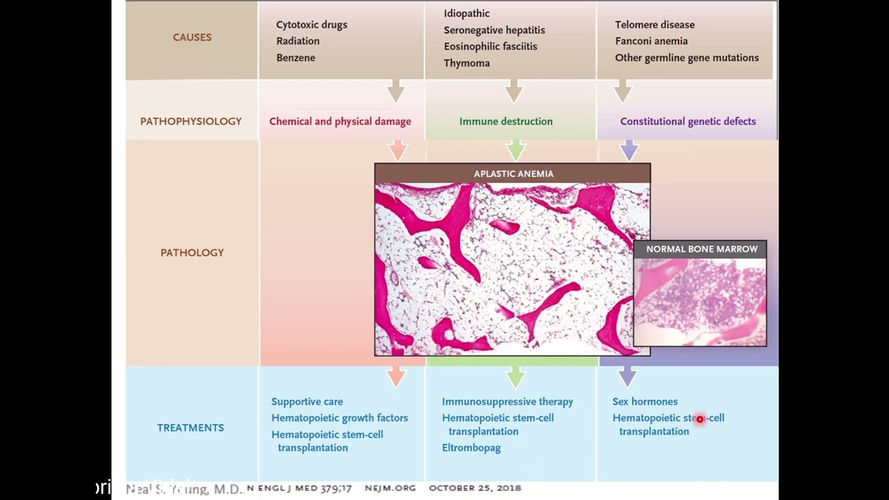





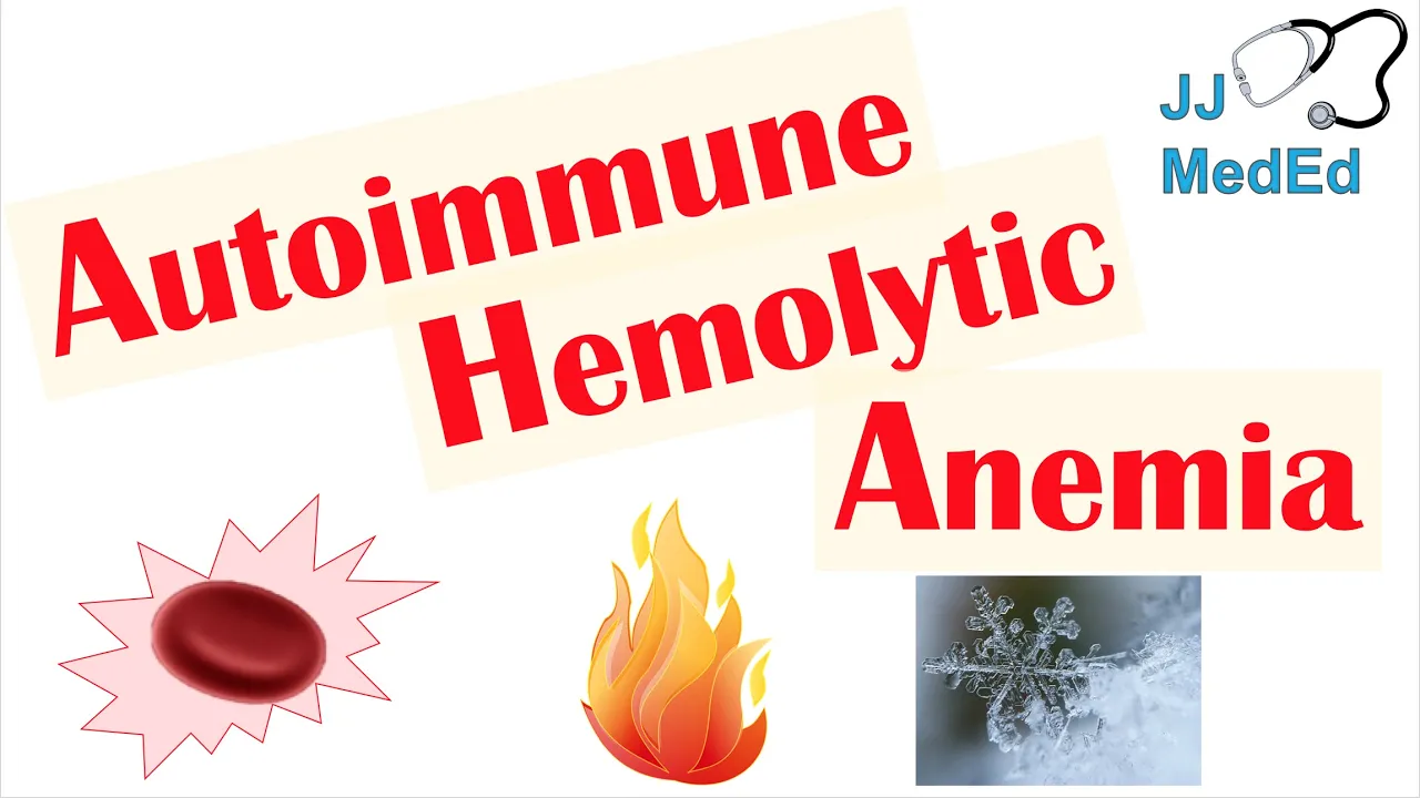

A systematic approach to diagnosing anemia begins with calculating the Reticulocyte Production Index (RPI) to determine whether the anemia results from decreased red blood cell production (RPI < 2%) or increased destruction/loss (RPI > 2%). For decreased production, categorize by MCV: microcytic anemia (MCV < 80) suggests iron deficiency or anemia of chronic disease, while macrocytic anemia (MCV > 100) suggests B12/folate deficiency or alcohol-related causes. For increased destruction, order LDH and haptoglobin; elevated LDH with low haptoglobin indicates hemolytic anemia, requiring DAT testing to differentiate between warm antibody (IgG-mediated) and cold antibody (IgM-mediated) types. Always consider underlying conditions like thalassemia, myelodysplastic syndromes, or paroxysmal nocturnal hemoglobinuria when initial tests are inconclusive.
Anemia Diagnosis & Workup: A Systematic Approach | Medical Lecture
Added:this I'm Brian moof I'm one of the attendings at the VA hospital for the medicine department and I'm just going to talk to you about the general approach for anemia the purpose of this lecture is a very focused kind of lecture on how you should approach the problem of anemia both for your board examinations and in your clinical practice so it's going to be very practically focused and then we're going to move into questions at the end and just discuss that so there's actually going to be very few slides I'm not going to spend a lot of your time on pathophysiology and processes like this this is about how to actually look at a patient with anemia both on your boards and in practice so keep that in mind so there's three main mechanisms by which people will become anemic there's either Hemorrhage or loss of blood cells there's increased destruction of red blood cells or there's a decreased production of red blood cells if you keep that in mind you can kind of understand the testing later on so out of those categories we could add Hemorrhage generally Hemorrhage is going to be obvious so a patient who comes in who's anemic with bright red blood rectum or hemo timus or melenic stool we don't really need to know this Approach at all so please don't feel like you need to order these tests if someone had active ongoing very obvious bleeding then you would go down and work up for whatever their particular bleeding disorder would be what we're talking about here is someone who's anemic with Hemorrhage without an obvious source with an occult source of blood loss so what are we talking about generally it's going to be a patient who's on blood thinners the majority of people don't get spontaneous bleeds that are not clinically obvious without trauma or GI source so if someone who's on blood thinners though they can have bleeding in the retrop parent Neal space and what you look for there we always memorize this for hemorragic pancreatitis but Fox colon and gray Turner signs are all signs of retr bleeding Fox's sign is where it goes down the inguinal ligament gry Turner is on the flank and colon sign is around the umbilicus so look for Hemorrhage in those locations in a patient if it appears a cult and hemorragic um thighs and humoral areas can bleed I have had patients admitted before and the residents don't know why either anemic and look like they bled and I go in the room and the guy's thigh is like three times its normal size with osis so make sure that you pay attention to other organ sites where you can bleed but generally you can only really bleed massively in your retrop parental space your abdominal cavity your thigh or your humoral area otherwise compartments and other aspects tampon out the bleeding and prevent massive loss so please don't ever arite that you think someone with plural space or brain is really the source of their anemia if it's not clear when you look at a patient you you want to look for those retrop parent Neal sources and that would be considering a CAT scan of the abdomen pelvis or if you suspect an Ault GI that perhaps hasn't reach the rectum yet or the patients of Po historian then you might consider GI Scopes the other option is that the patient has decreased production of red cells so when we say someone has a decreased production of red cells that means that there's generally either a nutritional or endocrine disorder causing it there's low arthro production or a lack of available iron or the patient has a pulmonary disorder that's actually decreasing the immature cells that then can come out and produce red cells so under the category of nutritional indicine deficiencies we have iron deficiency anemia which is the classic one we have D12 or folate deficiency we have hypothyroidism and rarely adrenal insufficiency in the rectin state the most common cause is anov iron is anoc chronic disease in which iron is sequestered within the red blood within the uh the bed cell as well as in the liver and other organs and they also have a drop in the production of ropin so it's actually a two hit mechanism in aneman chronic disease CIT blastic anemas are abnormal iron storage again within the cells that make the iron not available and then there's anemia dialysis where patients kidneys cannot produce the arthlin that's usually required and pure Red Cell plasia which is an antibod E8 disease where antibody binds and uh prevents the proper function of aop and in the production of immature cells bone marrow disorders include toxins that poison the bone marrow most commonly alcohol that's what we see most frequently m is plastic syndrome which is very common in the elderly multiple Myoma which you should Su in someone with kidney dysfunction and anemia who's elderly lucap I mean leukemia or lymphomas a plastic anemia which generally will be a pan cytopenia so I'm not going to worry too much about it in this what we're talking about today CU we're Focus on more anemia and then pure Red Cell plasia which is just a red cell decrease and it is in both categories because it's actually just a catchall of causes some of the causes are antibod mediated and some of the causes are t- cell mediated so T cell mediated B toxicity is different then we can have increased disruption our increased disruption of cells in patients generally comes from either an inherited inherited hemolytic syndrome which is going to be either an enzyme deficiency such is pro at kise which is really only in kids so I'm not going to worry about it or a G6PD which can affect up to 10% of the US population intrinsic membrane defects such as spherocytosis and elliptocytosis and hemoglobinopathies such as CLE cell and phalos we can also have acquired hemolytic processes most commonly mechanical through my angiopathic hemolytic anemia such as GIC TTP hus malignant hypertension Health syndrome the can or valves or per valvular leaks that destroy those cells in the periphery we can also have autoimmune hemog hemolytic anemia which is warm and cold and proyal nocturnal hemoglobin Nuria I tend to prefer this approach of just thinking about the hemolytic processes as opposed to all that stuff intravascular extravascular which doesn't really help you pathophysiologically to know what's happening with a patient so we talked about that we either have destruction or loss of blood cells or you have a decreased production so how can we tell that the cell type that represents the production of new cells is immature blood cells and the most common one we say we could see nucleated rbcs but those are pretty rare most of the time what we're seeing in patients is going to be a reticulocytosis so reticulocytes do affect that response if our particular site count is low we have decreased production and if it's high it represents destruction or loss unfortunately it's a relative term the more anemic I am the more reticul sites I can produ should produce so let's say maybe the last marks 3% reticular sites as high that's not really high if my hemoglobin is three or four my count should actually be much much higher than that so when we look at that we have to account for that we do that with What's called the reticulite production index the reticulite production index is the reticulite count times the patient's hemat divided by normal hemat which generally is around 45 and then we have to divide that by a correction factor from the table below so whenever you get a lb value take a look at it do it now for your board examination you're obviously not going to be spending a lot of time you're not going to have this table and they're not going to give it to you on your boards though fortunately for testing purposes they're going to make it really really really obvious so a guy's going to have a particular site count that's very high like eight or nine or it's going to have a articul site count that's like one or less than one so they'll make the values for you on your test very clear so don't stress about it on boards but in the real world things may be variable so make sure that you actually do this when you see a patient in the real world so now I really only have a couple slides on actually how to approach anemia once you memorize this you will get 100% of the questions right on your board examinations because in the boards everything is perfect the first step you need to do when you look at a patient is look at what we just talked about the reticulite production index ifite production index is less than 2% there's decreased production if it's more than 2% there's going to be increased destru uction it's really not real reliable until it's more than 3% kind of in between a little bit of a gray Zone in the boards your articul side counts will be perfect in the real world there's not a reason why I could not have two problems for instance let's say that I have a chronic ulcer in my stomach I have chronic blood loss and I get iron deficiency anemia I therefore have a decreased production problem then I get an acute Hemorrhage from it and my hemoglobin drops precipitously what is my reticulite count going to be it's still going to be inadequate because of the iron deficiency that I've accumulated you before I can also have multiple health problems I could develop a hemolytic process and have a cancer associated with that and that gives me an anemia chronic disease and therefore I don't produce the reticul size on the boards it will be perfect in the real world this is only valuable about 40% of the time what's really important is that you recognize that a high reticulite production index is very very very helpful but a low reticulite production index does not guarantee that you don't have a hemic or a hemorrhagic process right so once I know which category I'm in based off the RPI the next step is if it is decreased and shows a decreased production is that determine what category you are in and the easiest way to do that is to look at the MCV of the patient we can divide our patients into microtic normocytic or macrocytic patients why do we do this we do it because it determines what lab test we really need if I'm microtic it is highly unlikely that I have a problem with my thyroid B12 folate so I don't really need to send those tests if I have a patient who's very macro cdic it's very unlikely that I need have a problem with iron deficiency and generally I would stick with my B12 Folly TSH type testing if I get a value in between micro ciic and macro ciic which is going to be 80 to 100 then I'm normic and at that point it's a little hard to know what's really happening with the patient so in that case I need to do both I need to do both sets of testing so whenever you get a patient find which category they are in and then order the appropriate test iron studies consist of iron fadin and tibc tibc represents indirectly transfer we also want to pay very close attention when we get into our mosic group about the patients um peripheral smear findings their RDW and their RBC count okay when you get ready for your board exam and you get a question you're going to look at the MCV and the patient's microtic the next thing you want to look at is what's the RDW the RDW can be high in multiple diseases but the majority of patients with a low MCV in the real world as well are going to be microtic anemia from iron deficiency or anemia cryic disease and the RDW is generally normal anemia chronic disease what that means is that the cells are all about the same size when I get iron defici efficiency I have variable intake variable access variable cell production I get anisocytosis which means variability in the size of the cells and that causes my RDW to be high that does unfortunately happen with the other two ideologies up here as well so look at your RDW on your boards if the RDW is normal it's a very big clue that the patient has an inoc chronic disease and that does work out somewhat in the in the real world as well so once I look at the RDW the next thing I want to do is look at my RBC count why I look at my RBC count because my RBC count should be decreased in most of these disorders however in phemia which are more a cell size disorder more than a red cell count disorder generally my RBC count will not be marketly reduced once I noticed that a patient RBC count is not marketly reduced I can consider doing what's called a ner index the idea of this is actually pretty simple what I do is I'm taking the patient MCV and I'm dividing it by their RBC count everyone understand the idea of what's going on here the cells are are going to be extraordinarily small in a phalia generally the MCB is going to be less than 70 generally you won't see that any other causes so really low MCD is a big Thalia clue because my MCD is disproportionately low while my RBC is not particularly low that allows me to calculate this index and if that number is less than 13 that suggests that I actually have very large cells but not much of a decrease in the number and therefore I have a ium if I had an iron deficiency for example I would have a low cell count and a low MCV if my MCV gets down to 69 for minor deficiency I'm probably going to have a very profound anemia as a result of that severe of the disease and therefore my Count's going to be very low soone understand that idea on it okay so look for that sort of red flag on your boards in the real world it'd be rare to have an adult who hasn't had that diagnosed before I've had one but it does happen all right if I get my look at it and I don't think that they have a phalia or a Cito blastic anemia which would be recognized by basilic stippling or papenheim bodies which are C blastic iron inclusion bodies within the red cells then I would consider that my patient has iron deficiency or anemia chronic disease what do I do at that point I look at my iron studies now when I say iron studies people frequently don't really understand the best way to interpret that when I look at iron studies any I do not look at the iron the tibc and the fadin I actually look at what's called the iron stat percent and The Fad the iron sa percent is a patient serum iron divided by their tibc as this value gets lower and lower it becomes more and more likely iron deficiency at values below 20 it starts increasing below 15 it's much more specific as it gets lower and lower it becomes more reliable of an indicator this is important because transference values can fluctuate serum iron levels can fluctuate but the ratio between those will tend to still show you what's going on with the patient this is fairly sensitive it's fairly specific but it's not as good as the fadin the fadin is the most specific test in patients with iron deficiency an fadin values for your boards just try to remember less than 50 the lower the fadin gets the more reliable it is and they'll probably make it pretty obvious a fadin of 14 or lower is almost 100% specific for iron deficiency anemia they did a study where they looked at veterans and people with multiple health problems and what they found though is that Farin values when you look at F marob biopsies as a gold standard are very variable depending upon what your disease state is so if I have a chronic disease state where I'm hospitalized with an acute infection my fadin values are not reliable this is why most hematologists do not recommend checking labs for iron deficiency anemia on people with admitted to the hospital if the guy comes in for anemia and has nothing else wrong he fine but if the guy comes in and he has pneumonia and you notice he's anemia your labs are very likely to be distorted by the acute inflammatory process fadin will go up in that case so in this paper what they found was anyone who has an elevated CRP a value of less than 100 actually was pretty reliable with about an 86 to 88% specificity so people who have and they broke it down by individual disease state but that's kind of the ballp part figure was around 100 each one had a little different th give you a pretty good specificity so we have a normal type person who's just an emate without a chronic inflammatory disease such as rheumatoid arthritis renal failure or anything else that patient should be less than 50 I mean less than 50 suggest iron deficiency in a patient who has a chronic inflammatory state which could be reflected by checking a serum CRP level a value of less than 100 would suggest iron deficiency and if I'm on dialysis patients who have a value of less than 200 may have iron deficiency now there's been some newer stuff AR even 400 on some but I think 200 is probably a more reasonable value so memorize these kind of categories for the real for your board they make it real obvious with a really low VAR type if I think the patient RW was normal anoc chronic disease what I'm mainly looking for is that my iron stat is higher than 20% and my fadin is more than 100% that will confirm the diagnosis for me if I don't find either either of those type of values on my testing and I don't have the clear answer with a low MCV then I start looking at my thas and my C blastic anas um if I saw one of there's Flags like the Red Cell town in admin index or some of the smear findings that I'm already there but if I don't find any of that and it doesn't match then I come and look at this review the history Thalia are usually hereditary I want to make sure I a good history the one patient I had no one had ever asked him what his heredity was his mother immigrated from Greece so people do have kind of a history if you look there if you want to look forward and confirm the diagnosis you can do an electr electroforesis for beta Thalia minor because remember we're talking about Minor here major would be clinically obvious or you have to do Gene studies for Alpha if you have a CIT blastic anemia you suspect because of one of these risk factors over here um generally you're going to have to do a bone marrow biopsy to really confirm that so one of the things that associated with C blastic anemia is when you look at your board's question with the history of the patient you're seeing alcohol is a very common one actually underdiagnosed in a paper where they looked at that mile plastic syndrome with citro blast is one of the most common that you'll see and then we have drugs that cause puod oxine deficiency such as IID interestingly laid which is a newer drug that you free frequently chlorinol and cycloserine both of which are not used very often anymore lead poisoning is a classic one for your board with basilic stippling and papenheim body but unfortunately Bic stippling also happens in palac and the numerous other diseases so it's not path aonic so make sure you pay attention to the whole question um copper deficiency which happen in gastric bypass or severe Mal absorptive disorders can happen it's super rare if you don't have that um because because coppers and everything you eat same thing with zinc zinc poisoning from Bad Venture cream could cause copper deficiency which then causes the problem and then you can have patients with congenital C blastic anemas but generally we won't see that in adult anemia that's why I didn't list that there's a whole host of uh conal defects like that any questions on that part of it okay if I don't have a micro ciic the other category I want to talk about with you is a macro ciic macro ciic doesn't have as quite as nice of a lab breakdown it's a little more sense than just a few Labs so in that case we want our B12 our folate and our TSH and that's going to account for the majority of our cases hypothyroidism B12 or fate deficiency um or Al alcohol abuse alcohol changes both the nucleic acid production in cells as well as the RBC membrane and causes macrocytosis if I don't have one of those causes on my initial testing and my history and social review then I would consider that the patient may have a bone Mar disorder myis plastic syndrome with CIT blast may be here but the most common types of MDS are actually macro ciic or normic anemia so if I get a macrocytosis in a 70y old and all this first part is blank most likely I have either multiple myom or MDS if a patient has renal failure I want to consider multiple Myoma and would do spep and upep testing which is positive in over 80% of those cases um acute leukemias are more rare especially neutr neutrophilic leukemias and then we have a whole host of drugs that affect nucleic acid production Baum cyclophos chemotherapies like 5fu remember the HIV drug zyoin and steine hydroxyurea which again is a poison in the B and then we have drugs that affect the uh the uh utilization of folate directly such as methotraxate isine and six murine interestingly it's a common side effect of Metformin which we see almost everyone on so you may see quite a few patients who don't have anything explained and maybe on that for is possible it is related to that sulfasalazine and inflammatory bowel disease seizure drugs are a big contributor particularly fenin and valproic acid and valacyclovir lastly we can have RBC membrane defects this is one of those common reasons you see it in people with chronic liver disease it doesn't have to be from an alcohol toxicity you can get macrocytosis from the RBC membrane processing that happens in the liver being affected by sosis um and those patients on a smear will have additional findings of a caneyes which are a sign of either uremia or a liver disease and again they frequently have target cells and then splenomegaly which may be affiliated with liver disease as well can also lead to membrane product uh processing problems I used to list triglycerides but actually that was disproved a few years back so hyper triglyceridemia does not actually cause um a high MCB lastly I can have a normic anemia when I have a normic anemia it's most commonly due to iron problems b1248 or TSH or some combination thereof if I have early iron or early client disease I may not have the microcytosis I expect or I could have a combined set of disorders I could have MDS and then have an iron deficiency on top of it so when I look at a patient here I check those labs and see if I can find that they match either iron deficiency NE chronic disease or hypothyroidism B12 something like that if I don't find it there's some other diseases that are more unique to it um I could really have a hemolytic process with bone marrow suppression I might consider going down the hemolytic pathway or I could have a mix disease um of which uh inoc chronic disease is the number one MDS remember again in elderly patients is one of the most common causes of anemia that's either normic or maic and then the two I want to point out for your board exams and the real world that you need to keep an eye out for is a plastic anemia is inure Red Cell aasia now logically if I have a plastic anemia I'm going to have other cell lines affected I'm really going to be doing an approach for p cytopenia however pure red cell aplasia is just of red cells what is the recognition fact for both of these as well as chemotherapy or other direct toxins a patient may have been exposed to is a very very low reticula site count normally when I have a problem with my Bal marrow I still have reticula sites unless I get a massive cancer replacement I get a chemotherapy agent or some sort of poisoning I'm not going to have a drop and then I have my autoimmune disorder which is the pure Red Cell of plagia so if you get a question with an extraordinarily low reticul site count keep that differential in your mind look through the history that was provided and consider testing for Pure Red Cell plasia which would involve checking Reen values and B A biops any questions on this slide yes do you ever order a soluble transpar Interceptor going back to the MCV like iron between iron deficiency and Ne chronic disease so that's getting into like if you don't kind of trust your results it's kind of like an RBC faway over here you could do an RBC fway I'm not sure the cost and availability are worth it most of the time some people Advocate different settings and different values and if you're on the borderline kind of ordering more specific tests um those kind of things are much more expensive tbcs are dir cheap and wildly available um I haven't really ever seen a situation where you really need that stuff very much for instance if you if there's some Publications some people suggest if you have a B12 value that's not real specific meaning it's between 200 and there's a new paper maybe 500 you would consider doing a methanic acid and a homocysteine but you still use to B12 because if it's really low you know they have it and you don't need to do that um and for folate it's between two to four is a little bit of an indeterminate area that's where an RBC folate or again a homocysteine or methanic acid would be useful on a board exam if they give you a situation with that sort of stuff they'll give you the methon and that end with it I don't know that they would ask you to what would be the next best test and you'd have to choose that usually they want you to interpret those results and remember a methanic acid becomes uh elevated in both in a sorry uh B12 deficiency but not folate deficiency but homosysteine goes up in both particularly very high homosysteine and folate so it's the folate case you would check it and then if they gave you a board's question MMA and folate and homoy were both High you would know that actually the patient had B12 deficiency that was underdiagnosed by the B12 serum level that kind of answer your question about it it's just those all there's there's several different factors and ways of looking at iron deficiency that are very expensive I can't remember the Nam one really newer one that they they had in one of the papers but um those aren't really available and they're probably not going to be on your boarding yet yes when you order the here come immature how do you interpret it uh I called over here last year when I did this and actually it's still just Rous I to you still have to do the RPI cuz some people were under the impression that is the RPI but that's not the RPI yeah so or or sometimes they can give you an absolute count um there is a separate formula I didn't include it here because this is the easiest one um but if they give you and the board's don't use if they give you an absolute count there is another formula for accounting for that that's like the RPI are they giving you an absolute number of cells are they G absolute imature factor and percent yeah so you can use either test either way you want to out of that the the r count is the the percentage they're giving you it is not a Rous production index they're not doing the mathematical adjustment when they do that all right second algorithm is if my RPI is high my RPI is high I have either destruction or loss of cells so I have a hemolytic process so what is the next best test for my boards and the real world you need to order whole bunch of stuff and LDH and a haptoglobin will be back within a couple hours so an LDH and haptoglobin are the next test the LDH and haptoglobin are not elevated or abnormal and warning on half to globin half globin is produced squ does anyone know produced in your liver so an instake cerotic patient always will have a low haobin level that doesn't mean they have a moyses um so be aware of that when you order it but if both with with that with that exception and the forge will always give you a great easy case if the patient has uh normal values that means that it is blood loss and now I need to find the blood loss and that's why I was saying go back re exam the Pati make sure you miss anything with the arms and legs look for any of those refal bleed findings if you don't find it it'd be r to miss the GI track do another rectal maybe considering in tube nothing showing up GI you might want to consider a scan for a retr paral bleed usually that's going to be a patient on warfront and it's a SE muscle a very small tear that then bled in the ref parum because of the warfront and we get several admissions of those each year at the VA where a lot of people have atrial fibrillation and blood thinners um if on the other hand I get a patient who has high values I now know that they probably have a hemolytic process a high LDH and a low half Lo once I know that a person has that process it behooves me to figure out what that process is um there's several Publications on ways to do this I'm giving you a way that I think works very well for the boards and as long as you keep it in mind it works pretty well for the real world so generally when we did our diagram we have kind of two general processes of RBC destruction we have kind of our mechanical causes and we have sort of immune mediating causes and we have our cell membrane causes okay some people Advocate doing a peripheral smear first looking at the findings and then doing a direct antibody test which is also a kums direct test we don't really care about the indirect here so adap or direct antibody test so I prefer looking at it from the D standpoint first the caveat is 10% of people walking the street right now are test positive and don't have hemolytic anemia we get transient antibodies throughout our life and throughout year to year and our body wipes them out they go down they go away we don't know what trigger them that's true for all these antibod test so you always have to take it with a grain of salt and you still should look at the peripheral smear so in the real world you need to do is smear and look at the DAT test and then think about the whole picture and what you find but for a boards in a thought process function 90% of the time the D test is going to be helpful to you so if the D test is done on a patient and it's negative then I have a nonimmune be hemolytic anemia if my dad is positive I now think the patient has a hemic process and on the boards I can pretty much guarantee you they're not going to try to trick you if they give you a d value and it's positive the patient has a hemolytic an autoimmune hemolytic anemia once I know you have a hemic anemia now for my board's exam in the real world I need to figure out which one you have because warm antibod hemolytic anemas are treated with immunosuppressant and cold are generally managed with supportive care because they're usually not really severe now there's obviously caveats on both those ends and you have to read more about it but you need to figure out what category the patient's in so how do I do that you have to look at the antibody pattern warm antibod hemolytic anemia is mediated by IGG antibodies with compliment deposition cold antibody is deposited through IGM and compliment so you might think on my boards test I need to order next an IGM or an IGG well they're not going ask to order it whenever you get a DAT they usually will run these values for you they run the complement which is a deposition kind of thing you're looking for here it should be positive in the case confirming they have a htic anemia the next thing you want to look at is imunoglobulin a few research labs and I think Louisville actually is a place that does this runs the IGM automatically most places and on your board exams I do not believe they will run an IGM so they will only give you the IGG don't get confused if I have IG positive compliment positive I have a warm antibod anemia from a viral infection transfusion type reaction Hodgkins non Hodgkins and F CL of which 11% of people with cl actually get this disorder multiple Myoma Thoma or I have a connective tissue disorder of some kind such as lupus arthritis or I have a drug induced hemolytic anemia classically from sephos sorin penicillin inid or quinine really remember those penicilin type antibiotics or I can have a Dan longstein antibody which is IGG positive but only causes hysis in sort of a cold set so these are my possibilities if I get positive if my IGG is negative and I have complement on the surface of the cell it got there somehow and it got there through IGM so an IGG negative is the same as saying IGM positive do anyone understand that idea so don't look for the IG and go there no IGM I don't know what's going on in this question IGG negative complement positive is a antibod and that is most commonly through mitlas canity through lymphomas and leukemias evv can cause it cancers occasionally can cause it cold of glutenin disease in elderly patients can sometimes occur and then syphilis it's a common factor there okay any questions on that part so just make sure you look at that IGG mainly if I have a patient who's that negative now I need to look for obvious causes in the patient's history does my patient have a history of a hemoglobin opathy such as CLE cell Etc they have G6PD deficiency or they at risk for it they get an antibi antibiotic recently such is vrum and they we don't know that they have G6PD so they have a history of liver disease or splenomegaly that can be consuming and destroying the cells there throughout a history of spider or snake fight that's really rare but it could happen rattlesnake poisoning um do I have a history of clotting why I look for clotting because proyal nocturnal hemoglobin Uria not onlya cuses homolysis it causes clotting disorders as well classically of the mesenteric and portal veins do I have any trauma such as counting my feet you can get Marathon human that'd be really rare again the patient came in and said they did that burn patients but we're probably not seeing many of those but they have fever a patient who has fever in a hemolytic process makes you think infectious and then you think of things like malaria bosis Etc um drug review you want to make sure they're not any of the drugs that are associated with it such as dapson ibig which causes an autoimmune hemolytic anemia G really um or do they have other cell lines that are off which might suggest a BAL marrow Source or cancer um that's associated with it if you get the patient and I don't have an obvious history here I really want to play really close attention to my peripheral smear I want to go look at and I want to look for certain cell types so I look for my RBC membrane disorders so this is what spherocytosis looks like see the rounded shape of the cells I can have elliptocytosis which you can see here with the elliptical shape of the cells those are both hereditary disorders but I suppose they could be missed sickle cell disease sickling of the cells again this would usually be someone who already has a diagnosis andless you see a younger patient in phemia we look for target cells unfortunately again target cells are not specific they happen in a lot of hemolytic processes but uh more than five of these per high powered field would be suggestive of disorder that's generally true for all the cell out more than five for high fire field is a lot U it suggests a real disorder as opposed to slide prep microangiopathic htic anemia is critical because he killed the patients such as the things we talk about hypertension help syndrome di TTP these patients you can see helmet cells and you can see just a sites so if you see more than five of those per High P field that'd be a sign of a problem if the patient has liver disease we could see e caneyes blister cells are a classic finding in G6PD and if you see it before its processed they have hindes bodies which are small inclusion bodies within the cell um we could possibly have malaria bosis or Bartell which are this is a bartella which would be inclusion bodies why is that not removing and then we could have teardrop or nucleated cells so one of the most important things medically for you to recognize is a patient with a process of anemia that appears to have what's called a lucco orro blastic picture so whenever you look at a smear if you see a patient who has a nucleated red cell like this and also has a lot of immature myoides metaloides prom myoides or white cells that's called a luthro blastic tiure and that means they have an underlying leukemia in this case what it would mean probably is they have a leukemia and the anemia may not really be from that the LDH may be a false result from the leukemia or other process that's going on um you want to look for RBC ghost which are a sign of toxicity and sepsis which could cause this usually cherial and then basophilic stippling from lead disease yes I listed lead disease under microscopic decreased production um but it is technically a hula process just like the phas phemia also really tend to have a decreased production of immature cells and don't really end up on this part of the algorithm but you could see it on the smear if things are are misleading so now that we've kind of gone through that I want to go through cases we're going to try to go through them quickly I'm going to ask you guys to give a respones everyone shout it out I'm not going to single anyone out maybe if no one says anything I may single people out okay we're going to go through them so that we kind of submit this algorithmic approach to looking at the patient we can recognize it quickly so in our board exam we can do the question very fast get it correct and move on okay so we have a 56y old man with a history of feeling weak for three weeks he has no changes to his stool or urination and no hemimysis or a mesia his CBC shows a normal white cell and platelet count and he has a hemoglobin of 8.5 what is the next best test in this patient it's what no I we look at the RPI right always order TI side this is the number one say no one ever words were tick had a patient admitted one time for severe anemia PCP on iron ordered all these tests never the RPI I came in RPI was through the roof you had a hemolytic anemia so always make sure you look at that first it's just good medicine again it's only helpful to tie though RPI was 0.9 that means what's the patient's problem the decreased production so what's the next best test now we look at MCV mCP is 78 that means the patient is microtic what's the next best test but I say it's a shortcut on the boards and in the real world we look for RDW RDW is high suggest iron deficiency one their causes RDW is normal suest chronic disease RDW is 24 which is high that me he has anisocytosis if you look at a smear what's the next test now now we want to look at our iron studies the patient has a transparent sat which is the same as an iron sat should have change the names on you guys sorry transparent sat or iron s of 133% and a fertin of 13 and a low RBC count I should have put RBC count SE so he has a low RBC count in these values so what do you think of these tests what does this suggest the patient has right and what is test here is the most specific for that the fair the fadin of 13 clenches it the iron stats are a lot less reliable on specificity and just so you know the fadin DI the specificity varies it's only 86% specific it's 50 in that paper I mentioned for normal kind of people it becomes closer and closer to 100% the closer you get to 14 okay all right so the next step then is to look for the cause of it that would be looking for EGD and ccope probably cope first EGD you look for bowel sources of bleeding which are the most common cause consider doing in UA if he has chronic blood loss talking about M seeing blood in his school C he cough up blood so look for a source of the blood loss when you talk to the patient case number two we have a 66y old black man with a history of rheumatoid arthritis he comes to the clinic with some fatigue his hemoglobin is 10.2 his white count is within normal limits and his platelet count is slightly low at 129 he has FY syndrome but what what's the next best test what's the next best test in this patient it's always the same that's why I want you guys to get it so we look at the RPI RPI is 1.1 what does that mean he has decrease production so what's the next best step we look at the MCV MCV is 74 which is low you micro ciic plus the next step does look at his RDW his RDW is 17 which is normal now in our board's question we're already thinking what anoc chronic disease in the real world too we get iron studies anyway to make sure we're not missing something or have a compound anemia chronic disease with that remember we could have a chronic disease and have iron efficiency usually the RDW be high RDW is pretty good for picking up iron efficiency actually so we look at the iron studies the transferin SAT is 42% and the fan is 210 and the RBC count is low what does that suggest the patient's diagnosis is and fr disease from premature arthritis okay patient his 56y old man with a history of iron deficiency anemia for chart he sees you in the clinic taking iron tablets but his hemoglobin is 12 what is the next best step decreased so what's the next best test MCD is 57 what does that tell you when you see a 57 this question this patient probably has a Thalia right could be other causes it be very rare be below 60 even in the high 60s is very rare for enoc chronic disease or iron deficiency you very profound anemic so without even doing a calculation what did we notice there's a dissoci between the MCB and and the hemoglobin right so that would suggest that he will have what calculation yeah but we look at the rdws the next step to make sure the RDW is normal in this case so maybe it's even chronic disease then we get our iron studies and we look at our RBC count the RBC count is normal the iron studies are kind of non-specific the f is actually a little high taking iron tablets and whatever else is wrong RBC count in this case is normal so again we look at that we compare that to our MCV and we calculate what Miner's Index right and it was 11 what number were we looking for 1 less than 13 again for your boards just be aware of looking for that fact that the hemoglobin or the RBC Camp doesn't match the degree of MCV decrease and MCV decrease is really really low in I is going to be a very profound anemia um so this would suggest the patient has Almia so go get a a family history and youd want to consider electri forist since beta th Miner is one of the more common ones let sensitivity on insert on what on Min I actually saw that but I don't remember the number I think it's 80 something yeah it's not that's what I remembered I look at there's a couple other indexes you can do too yeah there's Miner and there's uh it starts with a w it's like w wall seeing or something there's another index you can calculate as well it's the same idea though it's still uses the MCV I think that one MCV time RDW / the patient RBC count they do they add the the RDW into it um so you can do either one I just think that one's easiest because it doesn't have three factors and it only has two um if the patient is a 60-year-old veteran with severe alcohol abuse and HIV is admitted found down drunk by police and his ER hemoglobin was 9.3 rest of his counts are miraculously normal so what's the best test it's what RPI RPI is low less than 2% what's our next test MCV MCV 72 what's our next best step RDW is high what's our next best test now we're thinking iron deficiency versus one of the other causes so what's next Iron studies iron studies show a a transfer s of 47 should be% and a fairing of 112 So the next test it it doesn't quite fit with it looks like iron deficiency but it doesn't have the labs of iron deficiency so now we're thinking Thalia C blastic anemia his MCV wasn't particularly low oh I didn't put the count on here his RBC count actually was low so in this case it doesn't look like the miner index fits what would be the next thing in this patient look at the smear and this is what a smear looks like does anyone know what that is so that's basophilic stippling so this patient has probably has CIT blastic anemia probably from the alcohol and the next step would be to either just watching him off booze or to do a balar biopsy I think the majority of people in this case would just tell him to stop drinking all right next case we have an 85-year-old man with a history of partial gastrectomy 25 years ago what does that make us think of on our board stuff right lost of inity factor he was amused with increasing Falls and dementia and was unable to cooperate with the exam but in the ER he had a hemoglobin of 11 what's the next best test RPI RPI is 1.1 less than 2% so it's low what's the next best test MCB is 112 that shows that he is I in fact he's close to megal Black megal blast is 115 or higher if it's that high it either has to be chemotherapy agents that affect folate or B12 or fate deficiency what's the next best test that's right because we're in that category we don't need our iron studies B12 was 210 fate 612 and TSH is 8.4 so little high but it wouldn't give you a severe anemia that's not really true disease folate was above our cut off of before we talked about earlier what is the B12 it's what yeah so that's indeterminant so what do we want to order next on our board step okay methanic acid is high and homocysteine or high that's consistent with B12 so it confirmed our diagnosis bew that New England thing recently showed they used to say 200 to 300 I don't know what your boards will have but they found that 12% of cases of B12 were actually miss by using a cut off of 300 that's why I'm saying if it's anywhere if it's over 500 it's still good at ruling it out if it's under 200 it's good for saying they have it you don't need anything else but in between that you probably want to do a meth monic acid in homos next case the 43-year old black female aded the hospital with severe fatigue and weakness following a recent viral illness her hemoglobin was 4.6 in the ER this is pretty bad what's the next best test okay her KCK count is 0.1 what's everyone think of that with ti count so that's a red flag for for what a plastic anemia B you know pure red cop plasia chemotherapy something else we don't have a history giving any of that okay next thing we look at our MCV MCV is 86 which is noros cdic so we go ahead and do our B12 col8 TSH iron studies all of them are unremarkable so in this case with that very low count what do you think the next best test would be yeah so be a b biopsy and left off an levels so you want to check that and see what's going on with the patient that's a very low value look for that in the real world and the boards we have a 58-year-old with a history of mechanical malro valve he's on warring therapy he's admitted for Syncopy and he's kind to have a hemoglobin of 5.4 so the question itself if you read the board is already making me think either retrop parent Neil bleed or hemolytic process from the valve right okay what's the next best test it's always the same RPI RPI is 6.8 that's more than 2% more than 3% so that suggests the patient has he has a loss or destruction so what's the next best step LH and half the glob it LGH is normal half the glob it is normal what does that make you think you bled right okay so what would be the next best test in this case I think I wrote this on the side you to go back and do your exam right if your exam doesn't show anything you want to consider getting a CAT scan to look through a retrop bleed from the S muscle all right next case 71y old with a history of mechanical a Val seen clinic for hog of 10.1 that has had a steady decline over the past several months he has slight jaundice on examinations what's the next best test or what's some test we're going to do RPI RPI RPI is 4.5% what's the next best test LH is high half the globin is low what's the next best test KS test I recommend thinking of that way you really order a kums and a periphal smear at the same time that was negative therefore we think it is not it's not autoimmune right that's why I really like doing this you know I have that 10% risk of getting a false result with the da but if it's negative I can trust it and then I can move on so the D's negative so what's the next best steps now I know it's not so what's next peripheral smear well look at the history he a mechanical valve and you want to look at a smear and what does the smear look like all right so we have helmet cells see look like a helmet we have some shis sites around we have more than five per high power field you can have a few just from slide prep but more than five for high field so this patient it's probably from mechanical micro valve unfortunately there's not a lot they can do for that you image it make sure it doesn't have a per Val leak or anything else on it that's the worsening it but um next case is 73y old with cl who's seen in your clinic has a hemoglobin of 12 with slightly eeric scera his Labs show that his Billy ruin is slightly elevated 2.6 and it's hemoglobin is 9.6 what's the next best test all right let's shout it RPI RPI is high that suggest the patient hasis or loss what's the next best step lhn half to glob LDH is high half the globin is low that suggests the patient has hemis either through destruction or autoimmune so what's the next best test his dad is positive what do we want to do next we look and see what the IGG what the imog globulin type is does anyone want to bet what it will be in this case it would be warm because he has CL so it's going to be IGG positive right okay so the patient is IGG positive complement positive but he would probably end up on steroids next case is 32-year-old history of C for one week productive of scan green speedum and fever to 101 and the ER is hemoglobin is 13.6 and it's Billy Ruben is 3.4 all of it in you already have a big clue what's going on right so what's the next best step RPI still stick with it RPI is high what's the next best step LH is high have to GL blow what's the next best test we do our bad and our d is positive what do we do next we look at the IG and the compliment values what do you think's wrong with this patient micoplasma occurs in 25% of cases microplasma pneumonia so if I get the test result I get back these IGG negative and complement positive so that's consistent with what I thought was wrong with the patient I'm kind of done if I had a patient with these symptoms and none of this stuff and end up cold still can occasionally be when F linium I want to go back examine it more closely think about other testing or see what happens have to do I'm sorry what you don't need to doig well I was saying a lot of labs don't do it uh you can if you want to I saying U ofl I think actually does IGM automatically on all positive dat test um so I think I think here you get that information most places it's just if it's IGG negative you assume it's IGM positive if you were doubtful or you weren't sure you could send a confirmatory IGM test so yes you probably have to send that out most places so maybe the U all right next case 62y old with no history of illness or substance abuse presents with severely low hemoglobin or sorry plet count hemoglobin of 7.2 and he has renal failure his total ability is elevated and it all appears to be indirect what's the next best test RPI is high at 7.2 what's the next best test I didn't make these I could have thrown it it was not high but I mean if you find else and go through and you don't have an answer and something clinically is jumping out to you please feel free to ignore that because remember I warned you that RPI I should have done that all these question RPI is not always reliable on the board's always so okay so LDH is high half to globin is low what's the next best test that that is negative that means he is non autoimmune so what's the next best test want look at his history is anything jumping out on History does this history sound suspicious right I'm sorry what it looks suspicious for H or or TTP right okay hus generally happens in children PTT generally in adults so the next test I would want to do in this patient is the uh smear I look through he doesn't have any drugs that would cause it anything else there's no history that's jumping out to me except for the renal failure and all and I see this slide what is this a slide of which means the patient has the whole reason we do the smear and we do it as quickly as we can after we get in that category is to look for this so this means the patient has a microangiopathic hinia without hypertension he's not pregnant anything like that now we think the patient you know could have TTP hus just to take you even further down the road let's say the guy had some diarrhea as well what test would you consider if you had diarrhea we can check for shot toxin shigal toxin that actually would be diagnostic of hus it's fairly good test not perfect and if we thought it was TTP what test is actually proving to be pretty good for that atam ts13 right so that's kind of how you decide if you get a category where a case looks similar and it could be either one that's what you kind of choose on it 22-year- old man with a hip prior office with no prior office visits with a hand of 10.2 and a normos ciic pattern what would be your next best test okay look at his RPI RPI is 3.4 so what's the next best test LH in half the globin LDH is high half the globin is low what's the next best test I know it's repetitive but I'm trying to get you guys to remember it so when you see people it's easy that is negative what's the next best step do a smear and history the history has no red flags when we look at our obvious causes first and then do our look for our smear for those findings but there's no red flags here we look at his case and what is this a picture of yeah see how there's no central power so this patient he's younger he's 20 he's never seen anyone before he has spherocytosis I think that's the only clinical scenario where you you would probably pick that up 34-year-old recent travel to Africa has current fevers and night sweats and my allerg since returning so what's the red flag on this question so far well Africa but fever right so fever with the cell Adali friends noticed her eyes were were yellowish um at the office Vis she has joh us and her hemoglobin is 8.2 what's the next best test RPI RPI is 99.2 what's the next best test LBH is high half the globin is low where's the next best test that is negative what's the next best test clinical history and what was the red PL in fever and travel to Africa so we think it's something infectious in this case um I wrote peripheral smear you might want to do a thick blood smear which can be used for some of these inclusion things as well um and the smear on this patient looks like this and what is that a picture of f so 50-year-old socialite presents after a recent visit to the New England area she complains of feeling fatigued and having intermittent rigors and fever with diarrhea her exam is not very remarkable her hemoglobin is 6.8 what's the next best test RPI RPI is high what's the next best test L and ha globin high and low so it istic what's the next best test that is oops that that off that was negative what's the next best test hisory okay history out of History what's the red flag on your board's question fevers right and New England so what do you think it is before you look at it fever johnice in New England and anemia is what is that at the Mal cross which means she has theosis so the board's likes giving questions with smear pictures sometimes on it so that's why I'm putting these up so you can regiz almost done 73y old black man presents with the history feeling fatigued in disn he had no major history but his Labs two weeks ago were normal he received backr for a UTI at that time exam had a white count exam shows minor ter clle he 7.8 Philly was 6.2 and almost all of it's indirect what's the next best test on your board's question always look at the RPI RPI is a little bit High what's the next best test LDH in half the globin high and low what's the next best test that is negative so we know it's not our immune what's the next best test and history remember history and smear history what on here is the red flag you got back from right so you had a recent issue all right so we look at his smear and we see this what is this which is a sign of G6PD right perfect world perfect board exam none of it's resolved okay last case 42-year-old white man with a history of Prior DBT he's on warer for 3 months at that time but had no therapy in 2 years he presents with abdominal pain his Imaging reveals portal vein trombosis but no sping on megal therosis or liver masses his PCP has previously tested his patient for for an e of 10.2 and he was iron deficient on his Labs there was no that should be P there was no reticul side production index done at that time what is this question a red flag for okay why does this patient have iron deficiency yeah because proxim hemoglobin when I go to sleep I heme I release iron it goes into my urine I pee off the iron and I become iron deficient so a combination of iron deficiency plus thrombosis should make you concerned about p& that's one of the red flags in a patient's history only with clots and iron deficiency or plots in hemolytic process a lot of times you don't pick up the hemolysis when you see them okay his RPI is 4.0 he had IR deficiency it could have been falsely low what's the next best step and again I don't think the board are tricky LDH is high half the globin is low next test is that that is negative next best test is history and smear his smear is unremarkable his history doesn't reveal much except what we talked about above with the clots and all a little bit of a red flag what's the next best step so oh I I think I may have gloss through that on our slides when you get there if you don't have an answer on a patient you can consider an ultrasound to look for liver disease or splen megal that could be destroying the cells if you don't find an answer on that you can consider that the patient may have p& which is causing their problems and what is the next test for p& does anybody know we can do a urine hemo remember we said iron's being released in the urine at night right so what happens is is the tubular cells become stained with iron and then if I check your urine I can see that those cells come out and I can see hemosin deposits in the urine so you're in hemosin I'm going to warn you it doesn't mean you have p& you're in hemosin means you have a hemolytic anemia so if I find a patient looks like a hemolytic anemia but all my evaluation everything is negative you're in hemosin would confirm it once I know he human you're hemo more I'm sorry it represents a chronic hemolytic anemia so there's a lot of those up there that could be that so we got to rule out everything else first I don't have any other causes nothing else is going on and I have a chronic hemolytic anemia now I think p& so the next process test I would do at that point is I would do the cell marker testing for p h uh for the surface markers and all that on the cells and I would consider testing all right that's it any questions
Up Next
Red Blood Cell Morphology Abnormalities Explained
@MedLecturesMadeEasy
170.6K views•2015-10-16

Graphic Medicine: Comics for Collaborative Healthcare Communication
@nationalpatientadvocate
189 views•2023-12-04

Antimicrobial Stewardship: Principles, Benefits & Resources
@UofLIM
9.9K views•2023-07-21

Stages of Labor and Vaginal Birth | Childbirth Animation
@nucleusmedicalmedia
52.1M views•2017-08-18
Related Study Plans & Knowledge Roadmaps
Structured learning paths in Medicine