








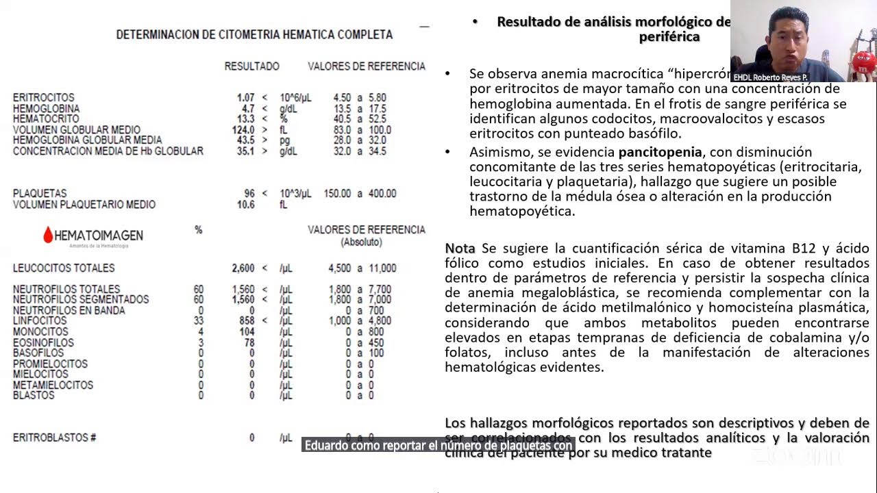





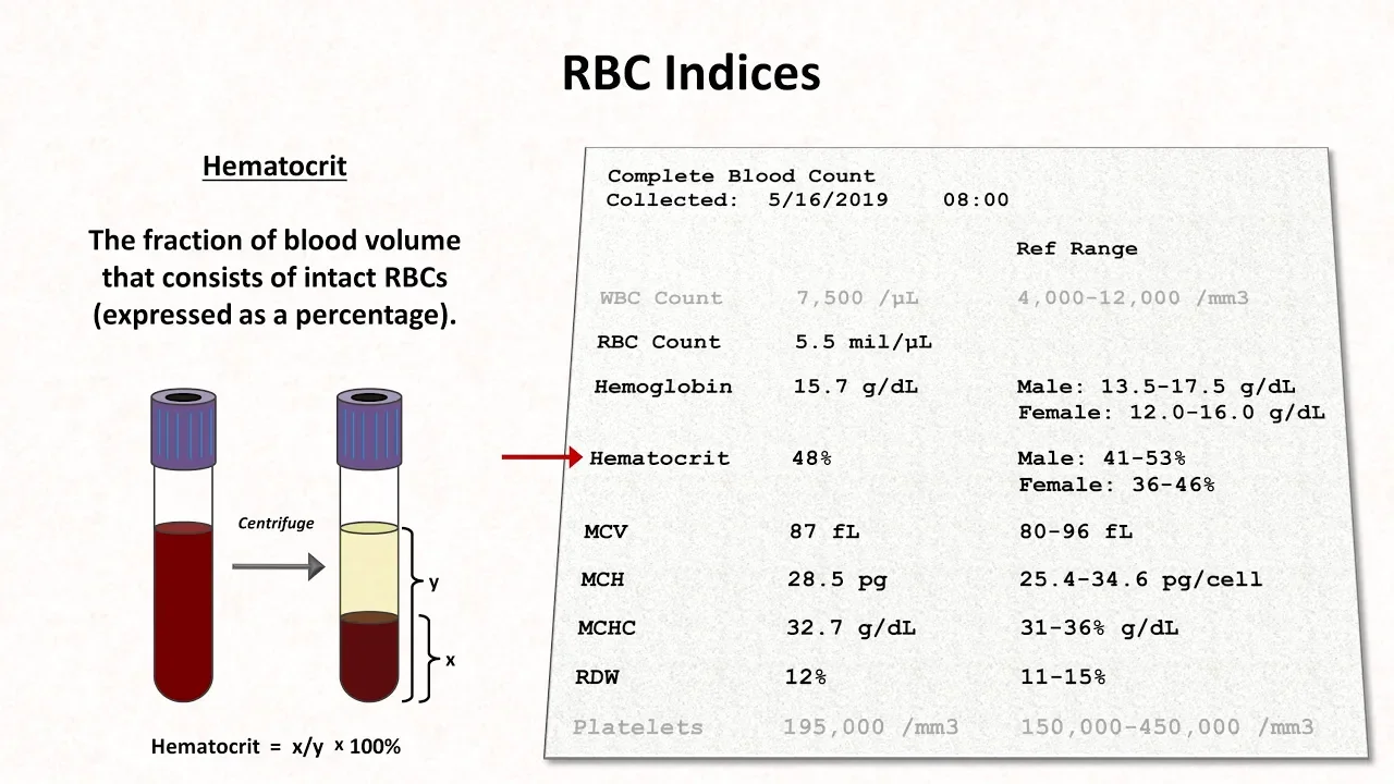

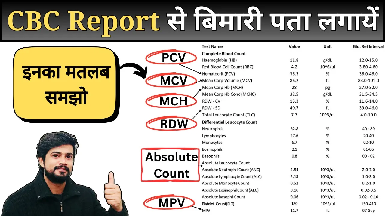
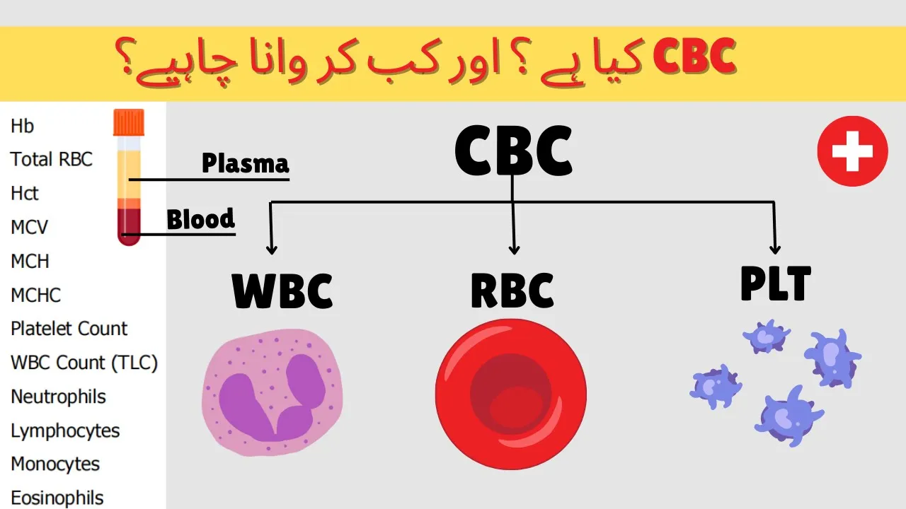










Morphology examination of peripheral blood smears remains the central pillar of hematology diagnosis, serving as a fundamental complement to automated cell counters by providing essential information on red blood cell size, shape, and inclusions (such as distinguishing iron deficiency anemia from thalassemia trait through RDW and RBC count analysis), white blood cell morphology (identifying toxic changes, blasts, and dysplasia for leukemia and myelodysplastic syndromes), and platelet characteristics (detecting pseudothrombocytopenia, giant platelets, and clumping), while also revealing critical findings like schistocytes indicating microangiopathic hemolytic anemia, spherocytes suggesting hereditary or autoimmune hemolytic anemia, and parasites such as malaria or microfilaria.
Morphology: The Central Pillar of Hematology | Peripheral Blood Film Decoded
Added:i think nadim i have some problem in the desktop because even in the last lecture by professor neelam i was not able to connect it in the desktop [Laughter] so all set we have one minute to go we are live on youtube sir so should we start yes i'm ready yes all right yeah i'm ready a good evening everybody welcome to this evening episode of pursue this is pursue 14c we are with hematology general and fundamental and we have two extraordinary hematologists here to want to moderate another one to present and the topic of the day is morphology the central pillar of hematology decoding the peripheral blood film which is so very important i take pleasure in introducing the moderator and doctor and moderator will introduce the presenter the moderator is none other than dr connell deepak mishra who's an mbbs md from fmc pune is a post-doctoral fellowship in hematology from ames new delhi senior consultant in laboratory hematology and molecular pathology and director department of laboratory science tata medical center kolkata he's an mbbs honors with gold medalist had educated the best medical graduate in 1983 from his college he did with md pathology from fmc pune his major interest is flow cytometry molecular genetics and hematology including mrd and acute and chronic leukemia including mds mpns and multiple myeloma he's also developed interest in solid molecular pathology including liquid biopsies trained in hematology at multiple international centers and he's also the examiner for mbbs md dm dnb in hematology clinical pathology hematology and transfusion medicine in various centers he is a visiting professor at iit kharagpur nit role killer for its ms phd program in biomedical science has published more than 190 international and in international and national journals is a fellow of the indian college of pathologist and indian society of hematology member of the national academy of medical science chair of the icmr task force subcommittee on mds and national convener of the indian mds registry initiative current president of molecular pathology association of india and former president of iapm ishbt former secretary of ipm and former editor of the indian journal of hematology and blood transfusion currently a board member in the board of directors of international society of laboratory hematology and the chair of the american society of clinical pathology for certification program for india member of the eha sir i would uh request you to introduce the speaker and then we'll start from there so you please take over dr mishra please very very good evening to everybody and at the outset i must thank dr nadeeb to have put this in place the way he was introducing me look like it he will take over the attack today is a very important talk in the fundamental series of hematology and as you know that morphology is the central pillar to any diagnosis and peripheral blood smear examination in hematology is one of the most important basic fundamental parameters for any hematological diagnosis and even into non-hematological problems so today we have a distinguished speaker with us who is none other than professor devdak basu who is the professor and head of pathology at the prestigious gypmar purucheri i have been to his department at the fantastic department and currently he is presiding over it and as he's a hematopathologist i'm sure the hematopathology has got a preeminence in that center i think uh that department had the dm program and due to some reason probably the dm program has gone a little behind because of uh teachers of issues dr devdata is very well published more than 200 publications he did his senior residency uh from the uh prestigious malnourished medical college and since then he is a faculty in pondicherry i have been to his department number of times for empty pathology exams and i have seen him working very diligent astute morphologist and he has a great interest in morphology in hematopathology he had a great interest in automation in lymphomas and into various other hematological disorders so with that uh brief introduction i will not come in between devdata and you i would request professor basu to start his talk okay thank you dr deepak and uh very good evening to all of you and uh a special thanks to dr nadeem for inviting me and getting me on board in this pursuit so without further ado i will start the presentation give me a minute yes so we can see your screw all right so let's pursue the peripheral blood this evening any building or any monument needs pillars okay whether it is the uh ancient uh parthenon at the greece or the thousand pillar corridor at rameshwaram temple or whether it be a modern absolutely a modern architecture or for the people in kolkata the food court of the south city mall all buildings all edifices all institutions need pillars and if the pillars have inscriptions this is a pillar in the ancient greece in the in the pyramids where the inscription of the pictograph of hieroglyphics was decoded by trophy or shampoo so that makes uh the event even better so this evening thanks to dr nadeem he coined the title for this talk this session as morphology the central pillar decoding the peripheral blood film so in the next possibly an hour we will uh find out clues in the peripheral blood films that help us in diagnosis you know the basics of hematology is a smear the counter and the microscope and let us see how the peripheral blood and looking at a peripheral smear helps us in solving problems i always put this slide up whenever i talk of anything in hematology i mean this is something that happened in 1845 200 years ago probably electricity was also not so developed so can you imagine someone sitting on a microscope and looking at a drop of blood and arriving at a diagnosis john hughes bennett did this morphologic diagnosis of leukemia in that seminal case report of his where he called the patient at splenomegaly possibly due to separation of blood this this panel here shows a drop of blood unstained under the microscope and after adding adding acetic acid you know acetic acid is a component of your turks fluid uh where you do the wbc used to do wbc counts uh the rbc's get uh dissolved and all that is left is a is are these sheets of myeloid cells neutrophils 200 years ago this was the first kind of morphologic diagnosis of leukemia and the disease was diagnosed by just a drop of blood under the microscope so what might a blood film help us to do decoding the morphology may help us in diagnose a disease prognosticate a disease we'll come to all of these in a bit monitor the disease give information on complications and side effects detect pre-analytical or analytical errors in the output of an automated instrument over the past few years as you know automation or the cell counters have taken over hematology and the need for doing a peripheral smear has really come down but peripheral smear acts as a complement to a automated cell counter it confirms or explains the flags that are given by the instrument and at times tells us something that no automated instrument can yet tell us it in other words opens the window to the world of clinical diagnosis so what are the indications of looking at a peripheral smear i mean as i said before with automation once you get the counts probably the indications of peripheral smear over the years has decreased but still when a physician initiates a request based on the clinical indications or clinical features or abnormalities in a past blood count so that that's one of the indication where we need to do a peripheral blood when the indication is based on the laboratory requests either as an abnormality in the count that is detected on the cell counter or the flags that are given by the cell counter or as a policy like some institutes have a policy of making smears for all the cases that come or you you base it on certain guidelines uh if that depends on lab to lab so the the requests are initiated both at the clinical level as well as at the laboratory level or as a part of curiosity so whatever it be there are certain consensus criteria and if you look at this article that individual follow labs follow their own criteria depending on the patient population the type of analyzer use etc but generally if your mean corpuscular volume is less than 70 or more than 105 rdw more than 22 platelets low platelets increased a first sample in a neonate no wbc differential or incomplete differential count available on the counter all these are some of the consensus laboratory each department follows their own different criteria for looking at a smear however this is something that i always tell my post graduates or trainees that if you have to master the art of peripheral smear examination look at each and every case possible whether normal or abnormal in the beginning of your career the first month or the first two months maybe the first six months look at all the smears that come so that by the end of that period you are so familiar with the normal that you would be able to pick up what is abnormal in a jiffy so that should be your aim look at smears till you know that what i am looking at is abnormal this finding is not what is normal that is where you will start your learning and as always correlation with the clinical history histograms automated parameters always resolves most of the issues and these are four articles those those who are into training i have always shared these four important landmark articles which help you in looking at a smear and getting guided by these these articles so let's start at the very beginning i mean i hope you all recognize this snapshot or from the film sound of music where before you had to sing you had to learn your doremi or before you knew your alphabets it was abc so let's start at the very beginning and the very beginning of looking at a smear comes looking at the gross slide itself a good look at the slide with a naked eye will tell you a lot of things about the case the quality of preparation and staining these are all four well-made prototypic blood smears as we say but it tells you gives you a lot of diagnosis if you can look at it the one on the absolute left one in the second third and the fourth each of them tell a story the one that is on the extreme left is obviously a polycythemic smear the next one is a normal smear the third one is an anemic smear and the fourth one is there is some kind of protein in the blood and that is what is causing the gammopathy and the bluish discoloration many times samples sent by the clinician from the central line which is heparinized also has this blue color okay so this looking at a gross appearance of the slide is good enough we all must be aware by now as to what we look at call the optimal assessment area or the zone of morphology it is something something over here not at the head end of the smear neither at the extreme feathered tail edge but it is always somewhere in between and how do we know that somewhere in between is correct if you look at it this is the head end this is the very tail end and this is where we have to look so what do we look for we look for rbcs that are kind of separated from each other not something that is flat not something that is crushed not something that is close to each other looking like a ruler formation so look for an area where rbc's are just sticking to each other there the morphology is the best so you do all your looking at the differential counts everything only in this this area of the smear that's why we insist that the smear has to be made well i will not go into the details of how to make a smear and all that because that would not be a part of this talk so what are the features of a well-stained smear macroscopically the color should be pink to purple now you must know your colors i hope all of you are aware of what is pink and purple and blue and violet that's how we look at a slide microscopically the rbcs should be the red cell should be pale orange to salmon pink what is the color of an rbc anyone knows what is this color of this rbc what is this color neither brown nor orange not pink the color is called puff and that's the color of the rbc how it should be a wbc the nucleus should be purple to blue that's of a neutrophil mainly the cytoplasm pink to tan the granules are lilac to violet geosynophils are important they have this orange colored granules and the basophils have dark blue to blackish granules which often obscures the nucleus now this is all basic i'm sure all of you are familiar with this now this is a normal smear how it looks it's also very important that the ph of the stain that you use is well maintained and if you get a too basic a smear you will get your eosinophils gray granules and the red cells are gray in color so again this is not a well stained smear that there is a problem and you might land into problems looking at a smear like that so train yourself and train your technicians to make good quality smears that's the first trick of the trade now let's look at one particular case on a routine checkup the cbc came as this hemoglobin 2.4 tlc 500 platelet count 1200 all right so there's obviously marked cytopenia that the cell counter has given you so now what do you do what we are seeing is nothing but fibrin strands and platelets that have been caught up in it so this particular sample had a little bit of a clot which was missed while the cell counting was going on the blood went and the cells were not there so the cell counter gave it as a severe pancytopenic because the entire cell population was caught up in the clot one problem that this plotted samples could do is it could clog your tubes of the cell counter so that was a problem but at very low magnification itself at 10 x this is what we do we look at the distribution of cells you select out your zone of morphology look up 10 good low power fields you can pick up ruler formation you can pick up agglutinated rbcs you can pick up clumps of platelets and you can pick up even big parasites like microfile area all at looking at just the 10x magnification the lowest power that is for hematology and if you see fibrin strands in your smear like this you reject the peripheral smear reject the sample inform your clinician look boss there are clots in the sample we can't do much about it please repeat a better prepared or better taken sample ensure that you add a proper amount of anticoagulant and mix the sample well while you're sending it so after 10x we have the 40x and the 100x in 40x we look at the rbc morphology in a while we also look at the differential count the differential count of the wbcs is done at 40x at 100x we further reinforce the rbc morphology the wbc morphology look at platelets properly the inclusions and the parasites are all picked up in oil immersion but always start with your low power move on to high power and oil emotion only for these few things you just confirm your findings on oil the overview of my talk for convenience sake you know would be first we will look at the interpretation of the rbcs then the wbcs then platelets we look for parasites and all the while we learn to appreciate artifact one artifact you've already picked up is clotted samples and we will therefore look at the rbcs with their muscle power the wbcs who are ready to defend and the platelets who build up the vasculature well all right so let's start with the rbcs when we look at rbcs the smears give you a fairly accurate assessment of the red cell size shape distribution and inclusions it also correlates well with your the cell counter the various red cell indices and also another important thing for all of us to learn is we need to standardize the terminologies that we use it's better to call electrocyte rather than pencil-shaped cells better to call cells better to call acanthocytes better to call burst cells let us have some standard uh definitions of standard uh ideology about what cell is what okay so better be uniform and it's better to give the scientific names whenever you are encountering them caution must be exercised in deciding whether a morphologic abnormality is actual or artificial we will go into that in a while again these two uh are articles which give a lot of recommendations for the standardization of nomenclature and grading as well as how to look at and how to grade abnormalities you know it's good to know what do we mean by one plus what do we mean by two plus what do we mean by three plus or mild moderate severe changes so these two articles please all of you those who are actually into the practice of reporting on peripheral smears please get them keep them by your table and look at them you have lovely tables which give you all the basic all possibilities that can be there of the rbcs and what do we mean by normal what do we mean by slight what do we mean by moderate what do we mean by mark in terms of each of them okay like for instance you see burst cells more than 30 report if they are present like that it is given so that that gives you a good idea of how to go about it similarly each of them when do we grade and how do we grade is given very well all right so let's start basic again something that you should all know we compare the size of these rbcs with that of the nucleus of a small lymphocyte smaller than that it's microcytic bigger than that is macrocytic right this is hypochromia when the central pallor is more than one-third this is this is all right this is quite all right but these cells are the central palette being more than one third of the entire uh circumference so these are hypochromic so you can make out microcytosis and macrocytosis and hypochromia by looking at just a few rbcs compared to a nucleus of a lymphocyte so what does this slide show the nucleus of the lymphocyte is there obviously this is a picture of a microcytic hypochromic apart from the size apart from the hypochromia you also find that the density of the rbc's is very very low okay compared so this is a case of anemia so we we have these again these uh uh severity grading is there normal mcb is 82 99 femtoliters less than uh mild would be 70 to 79 so the peripheral smear grading correlates quite well with the red cell indices that you get in your cell counter this one is hypochromic and similarly how do we write or how do we say what do we mean by one plus two plus or three plus okay so this is again technical and this this understand this concept well so causes of microscopic hypochromic as you know is iron deficiency anemia thalassemias both alpha and beta certain hemoglobinopathies especially the ones of concern towards us in our countries hemoglobin e which you know is present in the northeast part of india and hemoglobin d which is figures in the punjab area other causes of microcytic hyperchromic could be lead poisoning and anemia of chronic disease in both lead poisoning and anemia of chronic disease it is kind of dimorphic you have normocytic normochromic and also microcytic hypochromic red cells so let's take up one of few of these cases of how do we solve them we have these two cases both microstatic hypochromic the case one is hemoglobin 9.5 the second case is hemoglobin 9.8 okay in solving the puzzle of microcytic hypochromic red cells it's always good to look at the cell counter okay the cell count of values the cbc the red cell indices and the majority of the times all your problems are kind of solved so when they are small and pale there are these two uh ladies one with menorahia the other as a primary gravita hemoglobin as i said 9.5 and 9.8 the indices of the first lady is 71 25 and 30. all right microcytic hypochromic by our standards the second lady has a indices which are markedly microcytic hypochromic but the mchc is as well as normal okay so there's microcytosis low very low mcv but hemoglobin is kind of similar so that's the first point of difference we note between these two patients the red cell distribution width in one is elevated and the other is normal normal is around 13 13.5 uh it's a normal rdw so the next point that we look at is the red cell count the cell count in the first one is low whereas in the second one it's more than five million so there are three major differences that we are finding in the mcv a certain drop in the mcv in the second case the rdw is normal and the rbc count is high so where are we i'm sure most of you are getting it first case is in the case of an iron deficiency anemia and the second case is a case of a thalassemia trait all right thalassemia traits have high rbc count a very low mcv and a near normal red cell distribution width so once you have got this cell count of values and the peripheral smear examination you go ahead and order a ferritin in the first one to look for uh if the facilities are available you can get your serum ferritin and find it low in the second case you would obviously do an hplc and look for hba a2 levels and then go ahead okay so that's how a peripheral smear gives you clues to the diagnosis in the second case if you notice other than the microscientic hypochromic there are a few poiculocytes and stippled rbcs you also get a few target cells so that is how you differentiate a smear of an iron deficiency anemia from that of a thalassemia trait it could be beta or alpha mind you both this particular case obviously there is much more to just simple microstatistics and hypochromia there's a lot of boy killer sites okay so if you look at a smear like that there's a nuclear radar we see lots and lots of target cells varying shapes and sizes so you know that this is a case of microcytic hypochromic but this would be something that like a thalassemia so this was a case with a five-year-old boy stunted growth palace winona and jaundice so this is the typical appearance of a thal and thal intermedia or major hal minor would not be like this tal minor is a differential diagnosis of iron deficiency and he may ever changes are subtle but this gross anisopodiculocytosis presence of target cells nucleated rbcs polychromatic cells would indicate that this is possibly a thalassemia major go ahead and investigate there's another case that i uh i always show and this is again thanks to uh something that i had learned way back when i was a postgraduate student this was a 22 year old girl she was a nursing student who had this chronic refractory anemia always a hemoglobin was eight point two nine point one in that range she was refracted to iron therapy and at a very sm low mcev and look at the slide you find lots and lots of target cells microcytic hypochromic cells when you encounter a case like this one of the first investigations that we ask for apart from a detailed clinical history is a simple reticulocyte preparation so erratic count super vital stain and this showed these golf ball like inclusions the golf ball like inclusions pertain to a disease called alpha thalassemia and these are the hph inclusions which precipitate and give you the diagnosis so look at the peripheral smear combined with morphology of the reticulocyte count reticulocyte preparation leads your way to the diagnosis you do your hplc or do your hemoglobin electrophoresis and you will get a fast moving band in electrophoresis and you can diagnose alpha thalassemia this was the case of a 15 year old girl sevilla paler now this was a case where the clinician fought with our residents two days ago we had given a hemoglobin of seven today's hemoglobin is two days ago the mcb was about 70 today's mcv is 79. so they said man you're giving us wrong reports or discrepant reports the residents saw this smear and came up with a diagnosis and told the clinician look between the last smear and today's smear you must have transfused the patient so there is a background of microscopic hypochromic and there are these flattened looking normal hemoglobinized rbcs these are all this the transfused rbc's and therefore the hemoglobin and the mean copper school of volume changed this sort of a smear also on a cell counter you would give you a dimorphic curve a double peak in your rbc histogram so looking at a smear you're able to give not just the basic diagnosis but you'd also be able to tell the clinician that you did not give me the history of transfusion i'm 100 sure that this patient was transfused within the last 24 to 48 hours and that's why the picture is like this all right so peripheral smear helps you in a lot of ways what about this what would you call this smear this blatant hypochromia look at this all these cells are are they hypochromic they are not notice carefully that this kind of a hypochromia is punched out it is very short from the rest of the border so this is not hypochromia this is what is called water artifact it's an artifact of staining where somehow either the stain has got too much water or many a times what happens is you you you're having a cup of coffee and you do you and you want to wash your cup and you forget you open the tap and you forget that next to the tap is the entire slides being stained the staining is going on and drops of water from the tap that you have opened to clean your cup and saucer falls onto the slide falls into the stain and creates this kind of an artifact okay so this is water artifact again not to be misinterpreted as hypochromic rbcs so that's what about the small ones now what about the big ones the macrocytes as you all know normal mcv is 80 to 99 mild moderate and severe that's how we classify it anything more than 100 is taken as macrocyte now what are the causes of macrocytosis so you've got a case with an mcb whenever you get a case of macrocytosis on the cell counter giving you 106 femto liter first thing and foremost you must look at the slide you must look at the slide which will tell you whether the rbc's are in fact bigger than the lymphocyte that means they are genuinely macrocyte or whether the smear has polychromatic cells polychromatophils or reticulocytes are the immature rbcs which are bigger than the normal rbcs so your mcv can be falsely elevated when there is reticulocytosis all right so not really falsely inhibited is elevated when there is reticulocytosis this is the first difference that we have to look at the smear is it truly macrocytic if it's truly macrocytic then yes megaloblastic liver disease aplastic anemia if it is full of polychromatophils you think of hemolytic anemia post himatenex and you do a reddit count okay so that's the first thing that we check also when you get a very abnormal and high mcv nch and mchcl unthinkable mcb of 50 60 70 mchc going on in hundreds the first thought that should come to your mind whether the red cells are agglutinated okay these big big clumps are picked up as a high mcv and so whenever grossly abnormal indices come look for agglutinated red cells so a true megaloblastic anemia would show you these findings macro or valocytes right oval shaped macrocytes mcb is usually more than 110 maybe even more than 120 and hyper segmented neutrophils so these three things together form the basis of diagnosing a case of megaloblastic anemia on the peripheral blood you might also get things like cabo rings a hubble's early bodies etc which we will see in a while this was the case of 14 year old boy mcb was high refractory anemia transfusion dependent hemoglobin 6 less than 0.5 okay now this was the appearance of the rbc is more or less normal accepting for look at some of the rbcs they are a little nipple there was a little projection there's another one there slightly abnormal looking rbc's overall normocytic normochromic now because this patient was having a refractory anemia transfusion dependent not responding to iron uh the levels of b12 folate were also done that was normal we went ahead and did a bone marrow and the bone marrow came out to be what we were suspecting these are all erythroid cells you would appreciate them many of them are binucleated many of them are binucated with a little nuclear bridge between the two and we were able to give out a diagnosis of genital disorder okay this was another case which i want to show because again a good look at the peripheral smear helped us in diagnosing this disease this is again a 10 year old boy a child whose hemoglobin was very low the mcg was 100.
okay it was refractory and when we saw the smear everything looked hunky-dory normal absolutely good number of rbcs slightly bigger neutrophils there lymphocytes there platelets adequate and we looked we looked and we looked again we looked at this smear everything looks normal we looked at the smear everything looks normal looked at the smear again about 10 12 fields looked normal but was it really normal something that is normal was missing in all these slides what is that normal thing that was not available and that was not visible to us at all normally we did not see a polychromatophiles polychromatophil normal percentage is 0.2 to 2 so at least if you look at 10 smears you must find some polychromatophils in a child with a hemoglobin of four you expect the marrow to be reacting and throwing out a few of these premature rbcs or polychromatophils absence of polychromatophils we do that countless miserably low retic we could hardly count erratic in this particular case so a marrow was asked for and we saw the marrow was very cellular full of myeloid cells and caught up in them for these few large blue colored cells some of them have deep blue cytoplasm we have this with this kind of protrusions we call them dog ear kind of appearance look at this huge cell these are basically uh pro erythroblasts early erythroid forms there was the marrow was full of myeloid cells there were hardly a few of these erythroid precursors and that two pro erythroblasts with this prominent abnormal inclusion like nucleoli and we were able to give out a diagnosis of pure red cell aprasia in that child okay so looking at the peripheral smear look for things also look for things that are missing okay that that often gives you a clue to a diagnosis so macrocytic anemia could be seen with megaloglastic mds congenital dysarthropoetic anemia aplastic anemia prca and liver failure and when you have a high ncv check out whether the high ncb is because of reticulocytosis or agglutination all right okay shall we move ahead let's observe some more this is of course not my this is a image in the cover page of blood in the year 2013 and looking at the vial or the looking at the edta tube itself can give you a diagnosis if you find these little clumps the thing and this was a particular case of demagglutination disease okay so gross appearance of the even the edta tube can give you a diagnosis so this is what we get when with the rbc's agglutinate in cold hemoglobin and disease it's the autoimmune hemolytic enemy of the cold type mind with also hemolytic anemia patients may not always manifest with jaundice they manifest with reynolds phenomenon or these obstructions to the microcirculation because of these agglutinated rbcs it often happens in the winter like in this particular case a resident of delhi who presented on the very severe winter day of first january with john dish and bluish discoloration of fingertips the rdcs are all agglutinated and clumped any other finding that you can pick up on this particular smear which can give you the diagnosis if you look at the rbcs now don't look only at the rdcs look at the wbcs too look at that there are so many lymphocytes quite a few of these cells are lymphoid looking okay they're not this is a neutrophil so these are lymphocytes present so you have a diagnosis straight staring at you it's a chronic limb for proliferative disorder with autoimmune hemolytic anemia so one look at the sphere will give you give you the diagnosis right okay what is this kind of abnormality that we see rbc's this is uh in a case of a 65 year male with osteolytic lesions in the skull suspected myeloma yes you're confirming the diagnosis the clinical suspicion of the clinician yes this looks like myeloma i'm seeing ruler formation this is what is called ruler formation stacks of rbcs sitting together one after the other and this is what makes making a smear in a case of myeloma very difficult this is different mind you from agglutinated rbcs that we saw previously rbc's there there were trumps or rbcs here there are stacks of rbcs this particular patient was a female patient elderly who presented with renal failure the clinical suspicion was chronic renal disease but again you found the rbcs are ruling rbcs are ruling here and there and notice the background color the background color is slightly bluish that's because there are too many proteins in the blood and they have taken up the stain of the romanovsky stain so renal failure with roulette formation the antenna is all raised and we look further and further and we find the plasma cell so this particular patient was a case of myeloma clinically unsuspected but presence of plasma cells in the peripheral blood and we went on and diagnosed a case of myeloma with peripheral blood plasma cells spill over ruler formation can be a non-specific finding all right keep it in mind though the causes are myeloma waldenstrom hyper fibrinogenemia hyperglobal anemia of any cause therefore chronic inflammatory conditions can show rule of formation all of you i hope you are aware as to why ruler formation occurs in myeloma and why not in normal situation and that's because of the zeta potential i'll not go into that but mind you slight ruler formation of less than 10 red cells can be seen even in severely anemic patients because the uh zeta potential gets negated due to the less number of cells that are present in the in the in the body all right so only when you see it more than 10 percent of red cells causing ruler formation uh that is kind of pathological let's look at the poi kilo size now each of them have a story to tell whether it be a sickle or a sister site or a target cell or whatever and let's take up some of them one by one so we have this 12 year old boy who presented to the gastroenterologist with the complaints of intermittent jaundice and pain in the right hypochondria on examination there was anemia jaundice leno megali the three pillars of hemolytic anemia so this was obviously a patient who suspected hemolytic anemia and the pain in the right hypochondria would possibly be because of he had already got gallstones hemoglobin was nine hematocrit was 27 wbc platelet counts were normal the mcv was on the lower side of normal and mchc was high now this is another little clue to the diagnosis that we are going to get i'm sure most of you would have already picked up what this case would likely to be showing on the peripheral blood and it showed spherocytes it showed spherocytes spherical looking rbcs all over okay so when you get spherocytes again one little thought is it's usually not an artifact it is most of the time actual if you want to confirm if you look at a smear showing stereo sites as you know at the head end of the smear the rbcs tend to stick together normally normally when you're making a smear the head and the rbc is always clumped together stick together that's why we don't look at them but this sticking of rvc's inheritance pseudocytosis does not take place even in the body of the smear the rbcs are all desperate they're all separate from each other because they're all spherocytes they cannot they they don't mold against each other very easily so this is a true pseudoscience that is happening and so what are the two important causes of pseudoscience heritage pserocytosis hereditary phenocytosis and there usually the spherocytes are uniform sized all of them heritage pseudocytosis is often the hemolysis is compensated so you don't get a very hierarchic count in hereditary pseudocytosis unless the patient is in a crisis but whenever you see spherocytes the first thing we do is apart from the history do a comb test direct indirect counts inhibitory would be negative do an osmotic fragility test that would show a shift to the right and e ma stands for yosin five malamide on flow cytometry you can pick up periodic pseudocytosis though you may not need it always it's not available just by the history the negative coombs test family history positive a shift to right in osmotic fragility would give you the diagnosis of hereditary spherocytosis on the other hand you can get spherocytes in autoimmune hemolytic anemia so we have this 45 year old lady who presented with weakness and high colored urine with a low hemoglobin and a very very large number of spherocytes also notice in the smear you can spot that many of these rbcs are actually polychromatophils slightly bluish tint to them uh as a polychromatic reticulocyte so autoimmunology can image the other cause of pserocytosis where the generally the the history is different you don't have a family history you don't have a recurrent history of jaundice the retic is high and often your direct coums test solves the problem also another thing that we have noticed in autoimmunology anemia is this rbcs generally tend to stick to the neutrophils okay so that that's not a feature of autoimmune hemolytic anemia you can also get pseudosites in hemolytic disease of newborn okay spherocytes whether it be rh incompatibility or the incompatibility in cases of jaundice and newborn you get so many spherocytes so many nrbcs keep it in mind whether this is a hemolytic disease that is going on mind you in premature infants you might get speedocytes as a part of the normal phenomenon also you might get a couple of nuclear rc's in a premature baby uh term term baby does not show so many nrbcs or spherocytes okay so that is another thing that you must keep in mind when you're looking at a newborn uh blood sample so spirocytes are usually absent in normal adults and you can grade them depending on their percentage as i said two important causes are these two hereditary pseudocytosis are autoimmune but there are other causes first and foremost transfused blood sample notice that first case we showed where the iron deficiency anemia had been transfused the rbcs which are transfused often look very specific that's because whenever we transfuse blood to a patient of anemia we generally give out slightly older blood from the blood bank you know and so uh morphological changes do take place in the red cells and they often look very uh small and compressed looking it is the feature of hemolytic disease of newborn it's seen in mismatched transfusions postpinectomy some hemoglobinopathies may also show spherocytes microangiopathic hemolytic definitely a cause for pseudoscience can be seen severe burns certain infections like clostridium perfringens etc can also show spherocytes in the peripheral this was again a very interesting case it was a 61 year old lady actually this lady belonged to we had one professor who whose daughter was getting married and he was from kashmir and for his daughter's wedding his entire family of five sisters and few brothers all came to pondicherry and as usual when they come they come to a though they are absolutely normal healthy they came for a routine checkup so this first lady who was the elder sister of the of our professor we found these sorry we found these what are they ellipticites elongated rbcs they're not hypochromy they're normal looking and then we were wondering and since the whole family was there we said okay come along let's let's get all of you checked up and all of them had elliptocytosis and they were all perfectly all right through their life and they they did not even know that they had this entity in them i don't call it a disease electrocytes may be seen in hereditary electrocytosis but otherwise it's again a non-specific finding you find elliptical cells but they're hypochromic we use the word pencil cells for them in iron deficiency anemia and thalassemias they may be a feature of megaloblastic anemia splenectomies and myelofibrosis okay so but hereditary electrocytosis generally you might find as many as 25 to 75 of the rbcs are elliptical generally is asymptomatic similarly this is again a case of stomatocytosis stomatocytes is relatively common in the southeast asian group and often is asymptomatic so these are all curiosities that you pick up in the peripheral smear examination okay now this is a more ominous uh finding and you when you look at the smear there is no doubt that why this girl has jaundice why this patient has jaundice the rbcs are sickle shaped our pieces are boat shaped or sickle shaped and this is a prototypic picture of sickle cell disease now apart from sickle cell what else is this peripheral blood smear giving you information you see a lot of target cells and you see if you look at this rbc carefully or this rbc carefully there are these howell jolly bodies okay so this patient with sickle cell anemia has howl jolly bodies and lots of target cells can something be happening you get hubble jolly bodies and target cells the future of splenectomy so this patient of sickle cell anemia is probably undergoing autosplenectomy or has hypoplanism and these are the findings that tell you that there is something else besides the sickle cell the complication of sickle cell has arisen and the patient is hypo clinic this is another cause of anemia jaundice and bleeding and this is something that i always we all tell our juniors that whenever you are on night duty this is one case that is often a nightmare but you must be able to pick it up on your emergency this is anemia jaundice and bleeding and these are all fragmented rbcs schistocytes so that's an ominous disease in itself and you must be able to it's a medical emergency especially both hemolytic uremic syndrome and thrombotic thrombocytopenic purpura both are emergencies need to be accurately diagnosed because you have good treatment for them otherwise they can be quite fatal you need to be very careful about what is a schistosite or a fragmented rbc and if you look at the guidelines even if you find one percent or more it is significant you have to report and you have to percentage give the the value of rbcs that are fragmented the fragmented rbcs can just a minute more on this particular entity because they can have varying shapes you know they can have keratocytes helmet-shaped cells they could be microspherocytes they could be triangular estrocytes all these are various forms that you need to identify need to know before you label that case as a microangiopathic hemolytic anemia so this is a finding of homonymous significance you grade them even one plus that is one percent has to be reported and mind you along with thrombocytopenia presence of nuclear rbc's or polychromatophils when the main morphological feature is a schistosite it is diagnostic of microangiotic hemolytic anemia whether it be ttp or hus or dic this is important for all of you to really be able to pick up there are other causes of fragmentation like pregnancy-induced hypertension metastasis of nuisance secreting adenocarcinomas presence of prosthetic valve snake bite hemolysis all of these are features of show fragmentation this is again i'm repeating as necessary that you must be able to identify watershed societies this was a 23 year old girl who had ingested an insecticide and presented to us with severe jaundice now what does this rbc show these rbcs they might look like fragmented to someone who's not very familiar but these look more like a bite has been taken out of the rbc a small bite has been taken out when you eat an apple you bite into it and that's how these cells look like so this is bright cell anemia look at these not just bite a part of the rim of the rbc membrane is visible these are bites and blisters and lots of polychromatic cells so this is in itself an important entity of another called bite cell disorders okay now this was a 20 year male who was case of bullets benficoid for three months and he was started on that zone for 15 days and this is the serial hemoglobin that we saw hemoglobin progressively decreased over a month and the retic count progressively increased and the pedophile smear showed these again the bite and the blister cells we do a special stain with uh called the heinz body stain and you can do anything crystal violet or methyl violet stain and these precipitates are picked up and you have a case of bite cell anemia gap zone induced hemolysis can occur this is this sort of a by cell disorder is very common in g6pd deficiency but it can also occur in patients who have adapts on with normal gcspd levels if your drugs are more because these these drugs cause like daps on hydroxylamine causes oxidative stress free radical damage to the hemoglobin the disulfide links form between the hemoglobin and cell membrane and they damage the membrane the the uh the free radical damaged hemoglobin sits on the cell membrane and that part the damaged hemoglobin does not get stained and therefore it looks like an empty space it's basically the precipitated hemoglobin that gives this formation of bite earlier we used to think it is the pitting and culling of these cells in the spleen but no it's basically the damaged hemoglobin sitting on the membrane surface cell surface membrane and this part becomes unstained these bodies eins bodies they are called are stained by methyl violet or brilliant crystalline blue and again if you see more than four percent please report as a bite-sized anemia so this was a case of young woman with jaundice and liver failure and again similar instead of a bite we saw these little protrusions here and there a lot of polychromatic cells a lot of spherocytes and this was a case of wilson's disease so basically bite or blister cells are irregularly contracted cells speedy deficiency oxidant drugs chemicals chronic liver disease wilson's disease and morphologically as a part of postponectomy but here this is the area where this often comes up lands up as emergencies lands up with acute onset genders acute onset hemoglobinuria and the patient can have renal failure and can be quite a morbid condition one other thing that when you diagnose g6pd deficiency remember the the reticulocyte or the polychromatophils that arise due to the hemolysis are rich in g6pd it is only the adult hemoglobin g6pd levels which are low is the cause normal polychromatophils even in g6pd deficiency have good amount of g6pd which can tied up therefore when during the period of acute hemolysis the reticulocytes have enough g6pd so you're just if you're going to quantitate g6pd you may land up with a false positive understood reticulocytes normally have more g6pd levels than a normal rbc so when there is reticulocytosis going on the g6pd levels are well maintained so during the case of post acute hemolysis phase white cells give you a clue to the diagnosis once the hemolysis subsides and the patient becomes normal at that point of time when reticulocytes disappear if you do a g6pd level that is the point of time when you will be able to accurately diagnose g6pd deficiency but during the acute phase it is the byte cells that give you a diagnosis this was a case of a 10 year old a sick child saddar recurrent jaundice and splenomegaly again the classical appearance is target cells slight microcytosis hypochromia a lot of target cells so this target cell is a feature of a good number of hemoglobinopathies target cells basically occurred when there is increased surface to volume ratio which signify either excessive surface membrane formation or an excessive loss of volume or hemoglobin in content and again you can grade it and there are many causes of target cells this is a case of thalassemia notice the nrbcs and target cells this is a case of sickle cell lots of target cells this is a case of hemoglobin c there are these crystals of hemoglobin c extracellular crystals which give you the diagnosis of hemoglobin c hemoglobin c is not very commonly seen in our country it's more common in the central african area and this is a case where target cells nrbc and hubble jolly body i'm sure if you see this you're going to ask your clinician this patient must have had a splenectomy some days back and that's that's the future of a postpranectomy smear so target cells can be seen in a wide variety of conditions severe iron deficiency anemia thalassemias hemoglobinopathies liver disease both alcoholic and obstructive postpinectomy and premature infants may have targeted target cell is also an artifact of blood filled preparation because of slow air drying or over anticoagulation of samples keep that in mind at times it can mislead you because of its being an artifact these are acanthocytes or spur cell anemia irregular projections from the red cell membrane not a very uniform distribution irregular in number and irregular in height so acanthocytes are a feature of significant finding of aesthenia along with hubble jolly bodies and targeting they're of course a feature of a beta lipoprotein anemia mcleod phenotype and alcoholic liver disease similarly when you face burst cells think of kidney renal failure is a finding mind you again such bar cells or created rbcs could be a artifact it's a very common artifact of prolonged storage of blood in edta if you have collected the blood in the morning and making a smear in the evening likelihood that all your rbc's will get contracted so keep that in mind and it's a significant finding in renal failure udemia patient on dialysis but it is also a very common storage artifact what are these ecanthocytes or akinocytes or what such a bad smear this is obviously a stored sample that has come to you the person must have collected the sample long ago forgotten all about it and when their professor scolds him he sends a sample quietly to the lab and this is what you will get it's a stored sample meant for rejection of the sun 78 year old male with massive splenomegaly you find here these red cells tell tale evidence they look like your teardrops so that's a teardrop or also known as that cryocyte you see a shift to the left you see a nuclear rbc and you have what is called a leukoerethroplastic blood picture you order a bone marrow and you get fibrosis in the marrow so presence of fibrosis in the marrow is reflected as the tear dropping in the red cells the the rbcs find it difficult to squeeze out of these fibrotic sinusoids and therefore apparently their shape becomes tear-shaped similar uh tear-dropping and leukothobastic anemia in a child with massive hepatosplenomegaly in a baby with a massive hepatosplenomegaly clinically thought of as storage disorder and we had these uh teardrop cells nrbcs in the smear uh occasional blast also a bone marrow aspiration this was luke earth elastic picture a lot of nrbc's left shifted and a bone marrow aspiration was done which yielded a dry tap bone marrow biopsy then significantly gave us the diagnosis this is what you get in osteoporosis or marbled on bone disease lots of osteoid being formed osteoblastic proliferation and defective cartilaginous bony trabeculae so this was a case of osteopetrosis you confirm it with the x-ray that shows this absence of any medullary cavity in the bones and you arrive at a diagnosis relatively uncommon condition but we do get to see it so tear drop cells are again abnormal glucose elastic blood picture showing fibrosis certain hemoglobinopathies and thalassemias may also show these follicular sites it's also a finding of liver disease but in accompanying with shift to left and with nrbc's you usually make a diagnosis of leukocytes elastic picture their tear dropping is significant now this was again another example where a bad slide look at its slide if you put it under the microscope you would feel that this is storage sample what will i gain out of getting this uh anything in looking at it this was however a patient with recurrent jaundice and splenomegaly with a high red account so that was what made us even though the smear was bad we looked at it a bit carefully and on oil immersion we found the red cells having these stipples basophilic stippling and we came across with the diagnosis of pyrimidine 5-nucleotide deficiency cause of hemolytic anemia so stippled rbcs can be seen in again a wide variety of conditions they are often very subtle and you need to really change your fine focus to be able to identify them but once they are they have a lot of causes they are basically feature of lead poisoning thalassemias megaloblastic anemias other hemolytic anemias and again this is something that we have seen basophilic stippling gets lost when the rbcs are stored for long in edta and therefore this is one of the reasons it's an important finding gives you a lot of clue to the diagnosis where blood films should be processed past what do you see here again an inclusion in the red cells called howell jolly bodies again these are basically aggregates of dna report if they are present we look for hubble jolly bodies in postpranectomy states it's important when we are following our patients with refractory itp or autoimmune hemolytic anemia okay in the both these conditions bloonectomy is done and when they follow up you please look for howell jolly bodies and say that the splenectomy is there because many a times if it is refractory the platelet count has not come up uh in a case of postelectomy itp three months down the road the platelet counts haven't picked up you look at the smear and you don't find hubble jolly bodies please report it that i am not getting however jolly bodies please look maybe there was a spenunculus maybe your splenectomy was inadequate maybe uh you left behind a kind of an ectopic spleen which has now taken over the uh the platelet phagocytosis that goes on okay so this is again important from a prognostication or monitoring a patient of splenectomy so what do we learn from the rbcs the type of anemia morphologic classification hemolytic anemia and its types the various spoiler sites tell stories of their own and red cell inclusions this was courtesy one of my residents who found you know rbcs and the smears actually you fall in love with them as and when you see them so they are also responding and reciprocating every time all right so let's now move from the rbcs to the wbcs let's now pursue the wbcs look at this neutrophil that i found one day looks as if someone is running a race you know pursuing its own dream so wbc's add color to the peripheral sphere again i'm sure most of you know that how to convert the cells in the smear to a total leukocyte count this is how you counter check your count of values roughly multiply by 2000 per high power field you count the number of wbcs in a high power field multiply by 2000 and your total leukocyte count is available if there are too many of them more than 20 per high power field do a multiplication by 2 500. so this way your correspondingly always counter check your cell counter values when you're looking at the smear nuclear rbc's often give a spurious increase in automated cell counts because these nrbcs are picked up as wbcs because the cell counter doesn't know which is what a cell it's only when we look at them we find it so many times in hemolytic anemias when there are a lot of nrbcs the total leukocyte count becomes heavier it comes out in lakhs it's not because of the wbcs but because of the nrbc's having been identified as the blue dc so if you find 10 or more nrbcs under wbcs you need to correct it it's just simple mathematics and you can get your normal wbc count although rare at times neutrophilic agglutination occurs either a clotted smear or severe infection and that can falsely reduce the automated tlc the principle of automation is that you allow the cells to pass out in a single file so we had this 20 year old male with sepsis and it is as truly seen a lot of neutrophilic shift to left toxic change the counter gives at times not the not the newer counters the monocyte count and the metamylosite count can be kind of variable but mind you at times they may mistake for monocytes monocyte in a if you if you look at smears and if you look at counts regularly you would find that the counter gives lots of monocytes compared to what you see in the smear you know normally by tradition we know that monocyte count is two to six percent but if you look at the counter values the monocytes are about 10 12 in majority of the cases and that's correct the cell counter gives the true monocyte count because monocyte tends to their large cells so they accumulate at the tail end of the counting area may be differentiated difficult to differentiate from activated lymphocytes so you may skip them okay so this is important you believe your differential count that you get on the cell counter because monocytes can often be missed on a manual differential count how do we say what is an immature glandulocyte we look at what are called band forms these band forms are single lobe neutrophils the most immature of the neutrophils and it is recommended that band neutrophils be counted as segmented neutrophils in your differential count in only neonatal sepsis do we can we do a separate band count count them separately because more than 15 percent has a poorer prognosis otherwise you include band cells along with your neutrophils and predominance of band cells indicate that there is a neutrophilic left shift in the consensus guidelines these are all that is there normally you should not find a single blast you metamalocytes not more than two myelocyte promylocite not more than one atypical lymphocytes not more than five nuclear rbc is not more than one plasma cell even one so this is the consensus there's a guideline if you find these as a part of your differential count think of that it is not a real normal smear what information does this meal give you again look full of water artifacts but you have no the slide has come to you the form has come to you the sample has come to you without any history one information that you can give to the patient is this is a ladies sample it's from a girl or a lady because you can pick up the drumstick or the bar bodies so the power of observation is that you can you can even identify the gender of the individual without knowing it this is what is called toxic change a very important uh finding on the peripheral but neutrophilia with toxic change indicates that there is an underlying infection a bacterial infection there i still remember another case where we kept repeatedly saying that there are toxic changes the clinician kept saying oh there is no focus of infection we have given a course of antibiotic it's possibly leukemia then finally there was a pericardial uh biopericardium in that patient which was diagnosed okay so so toxic change is an important finding and the spectrum is not just toxic granules toxic vacuoles doley body this blue colored sub uh membranous structure and apoptosis degenerated neutrophils all these combined together form what is called toxic change now this was a 35 year old post lady post chemotherapy under gcsf again looks like toxic change but mind you this is nothing but a shift neutrophil when you give the patient a colony stimulating factor the marrow shifts out if you remember one of the changes that take place from myelocyte metamylocite to a neutrophil is packaging of the granules so generally the immature neutrophils immature myeloid cells have more granules at least look more granular so that's why you get this kind of a shift to left appearance when the patient is on gcsf as well as during sepsis because the marrow is pushing out immature granules immature neutrophils whose granules haven't got really packaged very well this is another case eight-year-old child with record and chest infection and look at the neutrophils here very prominent brownish violacious colored granules and this looking at it you would arrive at a diagnosis of cheddar kagashi syndrome so that's what it is this particular case an 11 year old 11 month old child with delayed milestones and hepatosphenomegaly neutrophils have vacuoles lymphocytes have vacuoles that's another morphological finding vacuoles in these leukocytes is an indicator that this child possibly has a lipid storage disorder these lipid vacuoles within the cells is indicated that you do a marrow and you find these lots of foamy macrophages surround black positive and this was cases of lipid storage disorder like name and bake or wool man's or whatever this was another slide or a smear that i am very fond of this is probably the only time so far that my my photographs have adorned the page of the blood journal it's basically an incidental finding in a journalist newborn bilirubin crystals indicative of severe neonatal sepsis not seen in so much in hemolytic diseases of newborn but bilirubin crystallizing within the neutrophils either as long crystals or short stubby crystals is a feature of severe neonatal sepsis okay this was a college student with fever and cervical lymphadenopathy and you have these atypical downy cells or various appearance of dancing skirt appearance and these are these atypical reactive lymphocytes this often could be mistaken for blasts so you have to be very careful a good clinical history the typical history of a short duration of fever and cervical in front of between a young person you will make a diagnosis of infectious mononeutraces or viral reactions currently in covalent smear this is at times we are getting it though though reactive lymphocytes are not so prominent you get basically neutrophilia lymphopenia eosinophilia the neutrophil lymphocyte ratio of more than three according to our study is quite indicative of severity of the disease these reactive lymphocytes have been given a fancy name of covadocites and you also get thrombocytopenia so this is again a feature of viral infection that you get in many conditions this is again not my own case but again from blood why i'm showing it to you is that lymphocytosis is normally associated with acute viral infections right acute bacterial infection causes neutrophilia accepting for protrusions whooping cough where there is presence of mature appearing lymphocytes in the peripheral blood okay the possibly the only bacterial infection which shows lymphocytosis because most of the bacterial infection shows neutrophilia look at a smear like this 35 year old male with splenomegaly and your diagnosis is made the counts are high you have the entire spectrum of myeloid cells a few eosinophils couple of vasophils hanging around here and you make a diagnosis of cml looking at this smear itself cml is important peripheral blood at diagnosis during follow-up for accelerated phases where you count the blasts you look at the basophils and also it's an indicator when you do a peripheral smear for remission status okay the counts have to come back to absolute normal in cml before you can label it as hemological remission okay so that that's one particular case of cml let's have a few of the blast is again a very unique feature before one of our cases normally blasts don't cluster it's possible the smear was badly made but we found these five blasts having a party on their own in a smear of acute leukemia and possibly one finding morphological finding that gives the diagnosis pathognomonic of something are these all rods so when you see blasts with all rods you know your case is acute myeloid leukemia go ahead do the image of phenotype go ahead do the psychogenetics but your diagnosis is there at hand if you're dealing with acute myeloid leukemia so again another case or rod this is again another case that we often encourage our residents to be able to pick up at whatever be the time of the day a 50 year old girl with menorah agia bleeding from gums and epistaxis three days pala 2 plus bleeding spots all over hemoglobin 3 low counts low platelets this is the pure spots that have occurred and you find these cells large cells the nucleus is kind of indented often cleaved figure of 8 buttock shape all of it as seen look here and there and you will find some cells with multiple or rods called cells do a sudan black stain they're all positive and you have a diagnosis of acute promyelocetic leukemia diagnosing acute promyelocytic leukemia is a emergency it's an urgent thing it's it's a rapid provisional diagnosis because you have etra atra which can change the entire spectrum of the disease in this two years ago or decades ago apml was one of the dreaded leukemias to happen because of its associated dic and the morbidity but now it is one of the better leukemias with a good prognosis okay so you once you get a diagnosis again i'm showing too many slides because i want you to identify keep in your mind that these are all the and how they look like it helps you out in your professional then you can do your psychogenetics for 15 17 or apm fish whatever so that breaks your way again another case where morphologically you can make out these blasts are lymphoid you do a past stain find block positivity you give a peripheral smear diagnosis of acute lymphoblastic leukemia go ahead and then do the immunophenotype and further a workup of the patient same finding lymphoid cells but again these blasts look carefully have blue cytoplasm with vacuoles so you can make a diagnosis of bucket leukemia or what was earlier known as all3 by looking at the morphology and coming up with the diagnosis then subsequently confirm your diagnosis better again kind of a curiosity where all these blasts have a little tail to them a projection we use the word a fanciful word of hand mirror type of leukemia again a type of acute lymphoblastic leukemia it's a morphological curiosity interesting to look at sixty-year-old male with generalized lymphadenopathy now you have so many of these lymphocytes and you can make a diagnosis of chronic lymphoproliferative disorder all of them look the same this patient is diagnosed again look at the smudge cells case of cll two years later down the road the same patient sorry for the quality of the slide but it was a pretty old slide two year down the road apart from these lymphocytes there was some larger cell prominent nucleolus and so this is a patient who's developing what is called a pro-lymphocytic transformation in a case of cll again your peripheral blood will tell you that so similarly same cll one mature looking cell and a large cell with prominent single at times eccentric nucleolus that is a typical appearance of a pro myeloma pro lymphocyte and as you know cll less than 10 of pro lymphocytes are allowed if they're 11 to 55 you call it atypical cll if they're more than 55 percent you call them as prolymphocytic leukemia smart cells or the you know those basket cells as we call them basically it's a smearing artifact but this also has a prognostic significance percentage of small cells in the routine blood smear does predict survival it's it's actually 10-year survival rate is about 50 when the smart cells were less but much higher when there are more smart cells so smart cells is an independent good predictor of cell survival can you tell me the clinical presentation of this particular patient look at it carefully lymphocytes are there we have seen this case before there are these spherocytes so this is a case where the rbc morphology and the wbc morphology will give you in concert the diagnosis case of cll with autoimmune hemolytic anemia many of these chronic lymphoma proliferative disorders look alike but if you realize there are subtle differences these are splenic lymphoma with bipolar lymphocytes right this is a typical hairy cell leukemia projections and this particular case has this deep clefted nucleus of what you get in follicular lymphoma spill so you have an slvl spending lymphoma with villus lymphocytes hairy cell leukemia and follicular lymphoma with split three different entities three different morphology but very close mimics of each other morphology can also give you clues to the molecular diagnosis look at this cup shaped inclusion like nucleolus they look like a cup and that is associated with the mutated npm one so morphology goes a long way in giving you a diagnosis elderly male with cytopenia pancytopenia but you pick up a blast you see a blast here look carefully you will find here is this neutrophil smaller in size with a bi-lobed nucleus look again and you find similar such cells these are belgaui is a feature of mds and this is something that you can pick up by morphology other features of myelodysplasia like hypogranularity or hyper granularity all are morphological features that help you in identifying a case of myelodysplastic syndrome abnormal lovation either reduced probation or hyperlobation ring neutrophils all these are morphological features that help you diagnose myelodysplastic syndromes this is again well marked with this particular cover page of blood where each of the neutrophils had been turned into a alphabet i mean unless you had morphology at hand you would not ever be able to get such beautiful pictures so decoding the peripheral smear with respect to red cells check the counts that's the first thing you do check the differential count and it's good to rely on the automated differential count specially for monocytes but if you are finding shift cells please make adjustments if you find neutral metamyelocytes etc many times this automation may not be able to pick you up toxic and other reactive changes are picked up on this leukemia lymphoma spillover and dysmylophysis so that's again a monocyte heart-shaped and that's the neosinophil heart shape so when you see these two together remember one joke that we had or one something something like a saying two corpuscles who fell in love but alas it was all in vain right so let's move on from the wbc's and half party with the platelets now that's a little cluster one of my resident sent this picture to me some days back rubble thank you for this he said this is a mama platelet and their their little kids having a party so what are platelets thankfully there is only one range for platelets whatever be your age your gender your race platelets range from 150 to 400 that is 1.5 lakhs to four lakhs okay so that's important but platelets are the most cunning of all the three cells in as far as artifacts and the true diagnosis is concerned graphically when you look at the peripheral smear it's a rough estimate you don't give a count exactly but it's a very valuable check on your counter values one platelet per oil is equal to 10 000 platelets this is something that you must do count look at the number of platelets count each platelet that you see by 10 000 correlate with your cell count of values and that's it your smear should have an even distribution and if you see two or three clumps that's an adequate uh platelets you really cannot give a total count of the platelets and looking at the smear you can say that it is adequate so let's look at a few cases where platelets created problems 35 year old female routine ps the plated count is normal but the resident is looking at the smear low power hyper not finding platelets he's saying no no no the counter value is wrong i cannot find platelets in this particular case this senior comes next to him and shows look boss all the platelets are in your tail so this is a tailing of a smear a poorly prepared smear with lot of tailed artifact and platelets being in clumps get caught in the tail so always always when you're looking for platelets look at the edges and the side of a smear at the tail end of the smear in low power to pick up your platelets was a five-year-old boy this again was an interesting story i got a phone call from my resident on duty at 10 late in the night i've got a case of a five-year-old boy with acute onset fever no bleeding no organomegaly good platelet count and i think i have diagnosed trypanosomiasis okay chagas disease look at that beautifully they almost look like what you call the chagas disease she said i have looked up the net and i found them this is this again from her case a beautiful example and she sent me the photograph of what she saw on the net as chaga's disease okay there was a very uncanny resemblance to them we were all excited when i went down and saw and found that platelets these are all same the net showed us platelets of varying abnormal shapes and size so what happens is when you take blood and edta edta causes activation of platelets it's basically causes scattering of platelets lot of morphological changes so platelets can be a very very bad mimic for many of the diseases okay so that was nothing but no trypanosomiasis they were all activated platelets that were picked up on the smear so be careful of platelet artifacts at times if your stain is under stained you may miss out on the platelets they look very gray you may come up with a diagnosis of gray platelet syndrome but this is a poorly stained smear so be careful about that we have this case of a 14 year old female with menorrhagia menorrhagia so there was a doubt whether it's a platelet functional disorder we received an edta sample and the platelet count was quite adequate okay the smear showed scattered platelets the platelets are all scattered now this is the problem that we face is it really like glands man's platelet is not aggregated no gp1b uh 2b3a effect they prevent aggregation of platelets so when you have an edta sample it's always good to make a direct fingerprint smear so the first thing that we told was please get us a direct next step is make a direct smear if your direct smear is still showing scattering of platelets investigate for platelet function but if your direct smear shows nice clumps of platelets check out with the gynecologist why that girl has menorahia okay so whenever you get sample in edta and are looking for a platelet functional disorder tell your clinician don't send me the sample in edta send me us directly made smear that that is a better tool to look at platelet aggregation or not so always ask for direct smear to look for platelet clumping or not large platelets can be seen in burnouts earlier small platelets in wisconsin platelet morphology is also very very important spuriously low counts can therefore occur when the sample is clotted as we have seen before so please check for the sample and prolonged storage of samples can also cause platelet degeneration and low counts here this particular case a 30 year old lady with menorah asia repeatedly platelet counts were done from outside was low 30 000 45 000 and she had no gynecological issue apart from anaerosis but otherwise nothing she was already investigated for functional platelet disorder it was normal she had had two normal vaginal deliveries extraction of the kari's tooth but there was never any platelet never any bleeds never any uh disorders like that she was not on any anti-platelet medication overlapped platelet counters come as 38 000 also low so when you look at a smear like that and you find these big clumps so this was a case of something called as edta induced pseudo thrombocytopenia normally edta scatters platelets as i told you before but in very few percentage of cases edta induces platelet aggregation it is because of exposure of cryptic antigens on the glycoprotein 2b3a and this is often seen in patients with sle infectious mononucleosis sepsis or antiphospholipid antibodies and drugs okay so you get the cdt induced pseudothrombocytopenia there are some ways of tackling that you can add excess edta or take a saturated blood and do your counts do a manual platelet count and some people have even advocated using candomycin to the blood sample but basically when you get the cdt induced through thrombocytopenia do a manual counting or use citrate as your anticoagulant or account we had this five year or ten year old boy with bleeding gums and prolonged bleeding after trauma since early childhood she was a child of consanguinous marriage so when you get a history like this you will think of a platelet disorder a congenital platelet disorder or an inherited platelet disorder so what are the possibilities but you do a platelet count it comes remarkably low it's abnormally low so are you thinking of a a mega karyocytosis or at least total absence of platelets in the body but when you look at a smear you find that there are good number of platelets but they're all giant platelets so this child whose counter gave it a 7000 was a falsely low value because these giant platelets go into the rbc channel and get counted as rbcs and the platelets are left behind left low in count so you can get very abnormal platelets in quite a few conditions this condition is called as bernard soldier syndrome where the platelet count is not so low it is at least somewhere around 80 90 000 but because they are so big they don't get counted as platelets by the cell counter giant platelets with this delay body is a feature of another of the inherited disorders called mayheglen syndrome anomaly and that's again something that we often get in our in our institute also now this was a case of a 45 year old lady absolutely asymptomatic admitted for cholecystectomy platelet count came as 95 000 repeated was 95 000. surgery was deferred and the ps was obtained the ps showed again slight stomatocytes and these giant platelets again she was asymptomatic these platelets were functional but because of their giant shape the platelet count came low and this is a condition called again large platelets she was a native of bengal and this as syndrome has been described as incidental mega thrombocytopenia some of the the inherited it's a kind of a disorder that is indigenous to bengalis they're absolutely functionally normal but the platelet count is low because they are giant platelets of course giant platelets can be a feature of bernard soldier can be a feature of mayheglen anomaly and other conditions also you can get giant platelets in itp you can get giant platelets in cml so falsely lowered counts are seen because the platelets either agglutinate because of edta or procedural there are clots or they are large birth say bernard soldier itp the fresh platelets or the immature platelets are generally bigger myeloproliferative neoplasms the mega thrombocytopenia syndromes now here is a little caviar that we have post chemotherapy recovery and itp on steroids the reactive platelets when they come out they are larger than size and the cell counters may miss counting so look for something called the immature platelet fraction on the cell counter and that gives you a clue that they are possibly number wise they are more but because they are immature they are larger and they are not being counted by the cell counter so that's that's a little thing that you have to be careful about this is another interesting case where we learned a lot of lessons this was a 36 year old male with generalized lymphadenopathy there were 90 blasts it was a case of acute lymphoblastic leukemia and we advised bone marrow and immunophenotyping and this was the typical appearance of the peripheral smear of an all past positive and we were quite happy with it when when the bone marrow was done next day itself the counts changed dramatically the tlc from 78 000 went down to 18 000 and the platelet count which is initially 65 000 went up so what went wrong in this overnight in this one day when we looked at the smear the next day we found that there were these things they were what are these these are nothing but blast fragments fragments of the blast this was a case of acute leukemia which had undergone spontaneous tumor lysis syndrome okay spontaneous tumor lysis and all these they were not platelets really they're basically the blast cytoplasm and the nucleus that got fragmented and were counted as platelets and because the blast degenerated your tlc went down and the platelet count got up so this was a case of spontaneous tumor lysis you can get a similar finding on the patients of leukemia especially burkitt etc with chemotherapy it was the suspected dic case now again here we had a falsely elevated platelet she was bleeding from all over but the platelet count was quite coming as adequate now why was that when you look at the smear you find the fragmented rbcs that we showed you the shifter sides they are all being picked up as platelets okay so this is the cause of pseudo thrombocytosis same here lots of sister sites and platelets notice in the background there are hardly any platelets so the overall platelet count is low but these fragmented rbcs are giving you a falsely elevated platelet count so again in this situation when you find like this do a manual platelet count and report this is again a very interesting case which i always show it's a newborn baby with whose sample came and we got on the smear on the count we got 18 lakhs platelet okay so this is case number one and this was i think the appearance that we got some of them are from the net no i think this was ours okay this is what we got in this particular case and we were wondering why 18 lakhs and this was the morphology platelets weren't not really increased subsequently we noticed that all the samples that came from nikku that particular day from the neonatal icu had thrombocytosis and this kind of a morphology we went back and checked and it was realized later that the entire set of edta tubes that were meant to be transported to the lab were actually placed on their hot air oven the entire rack was placed by mistake on the hot air oven and this was what caused the thermal injury to the red cells resulting in a falsely elevated platelet count so platelets cause a lot of problems so it's very important to look at the smear whenever you get a discrepancy in the platelet count so false high counts are rarer though but it could be seen with micro sitting red blood cells fragmented red cells at times we get confused leukemic fragments and apoptosis trio global anemia hyperlipidemia bacteria fungi parasitized red cells all can be picked up as platelets and in the inadvertent heating of the something so when you have platelets look at the smear when platelet counts come low why do we need to know we confirm thrombocytopenia first and foremost is it genuinely thrombocytopenia and look for the underlying cause of thrombocytopenia when the platelet comes come high again confirm and look for the underlying cause for thrombocytosis look at the smear anywhere when traitlet counts are normal but clinically discordant or clinically warranted they want you to let us know what whether the platelet counts they're getting on the counter is right please look at a smear for a platelet cup look at platelet morphology and function clumping of platelets in a smear is a good indicator that platelets are functioning normally quickly will catch the bugs this is diagnostic usually it's malaria but you can also pick up a few other ones this was a case of newborn baby with jaundice and it was sent to us as us as a hemolytic disease of the newborn and we found by wax okay so malaria could be a clinical mimic in many situations case of suspected dengue platelet 40 000 and you find again ring forms of falciparum in this particular case there are these rings of falciparum as well as the mythocytes of bivax so this was a case of a mix of schizoids i don't supply wax mixed malarial infection you find mirozoids so this was the case suspected clinically as dengue turned out to be mixed malarial infections is there a morphology clue to a parasite yes there is presence of hemozoin pigment in the neutrophils and monocytes you find them you look for parasites and you're going to find them this is one where there is a ring form of a falciparum and pigmented monocytes babisia i have not seen one so far on my own borilia also not seen on our own but these are all textbook pictures can all be seen in the peripheral blood yes microfile area we have of late we don't get too many cases of microfile area but yes once in a while we do and when we do that once bb really this is another thing that a morphology of this particular case i'll just show you on a drop of blood and this is what we saw okay this is unstained drop of blood under the microscope and you see the microphile areas removed okay this particular video is also very nostalgic for me because the voice that you're hearing belongs to a faculty member who passed away some time back okay so this was one of our cases where we saw just by observation into a peripheral gut sometimes fungal contamination of the stain can cause problems be careful about that and many of these artifacts can be confused with parasites this is nothing but again fungus cotton thread may mimic microfilaria stippled rbc's may mimic by wax cabo rings may mimic the ring forms of parasites so basically decoding peripheral blood now this is a blast but look at it how human the face of that blast is there are two eyes and the mouth so the cells really talk to us if you look at a smear so when you decode a peripheral smear you're able to give the cause of anemia reactive versus neoplastic likely myelodysplastic syndromes lymphoma luke spillovers thrombocytopenia rabbit parasites and please be careful about artifacts so challenge is there for the pathologist to ensure accuracy at every stage and even in the age of molecular diagnosis the blood smear remains the central pillar both the pillar at kuthuman as well as the sarnath pillar in the diagnosis of hematological disorders so we can still learn a lot from a blood film and stay safe all of you we we have come to the end of the talk and that's my email id in case and lots of love to all of you thank you very much thank you for your exhaustive presentation yeah so now you have given a talk of almost 90 minutes so i think there must be a lot of questions and you have uh handled it both positively from rbcs to wbcs to platelets and all the artifacts and it was a treat to listen to you and see the beautiful pictures that you have captured during your course of your professional career so with this i open this up for discussion i'm sure a lot of questions must be there so i would need uh nadeem's uh hell in case there are any questions in the chat box youtube yeah there are quite a few questions um i'll go one by one uh we can first take the questions from the google meet people who are here if they want to unmute and ask directly in pm we can restrict to some important questions on morphology and uh in the next 10 minutes we can wind up if you if it is okay with you you are the host so i think so if he's there so might as well have the questions and let everybody be satisfied yes so uh from the google meet first well i'm not able to speak even i'm not i'll i'll read out everything okay if anybody wants to ask a question you can unmute and ask directly uh hello good evening sir myself dr deep shikha yes uh sir i have a doubt in case of cml how will we do dlc in cml you do a dlc differential count you mean yes sir okay a differential count in a cml generally we do it as we normally do accepting that we be careful that we don't limit ourselves to just 100 cells because there'll be so many cells there so you could do a 200 or a 300 or even a 500 cell differential and get the percentage out okay sir thank you anybody else would like to ask a question can unmute and ask uh i'll read out the questions anybody wants to unmute here hello hello please yes please good evening sir oh i'm dr show bob uh my question is like in newborn a baby sometimes the nrb will be like in unders so at that time how do we do this how would we give this corrected total count but that time uh this corrected uh wbc count formula will not be uh you generally don't get hundreds of uh nuclear diabetes even in a in a term newborn that's unusual you don't get too many you get maybe five or ten but if you have hundreds of different nuclear rpcs you still hold the same formula as correct 100 suppose you get 300 neutrophils your total total count of neutral nuclear rbc's plus your wbcs becomes 400.
if you have 300 of 100 you understand 300 nrbc's out of 100 wbcs your total becomes 400 so you do your calculation based on that so you still can't do you can still use the same formula even if the account of nuclear rbc's is more than hundred you can just just work the mathematics out if it's not too difficult okay you have nrbc's plus hundred no so count the number of nrbc's and get it out so in a newborn with presenting with jaundice and anemia how to differentiate the spherocytes of autoimmune hemolytic anemia from heritage spherocytosis one in newborn uh both autoimmune and heritage below cytosis are uncommon in a new bond though both can present in new bond and very difficult to really differentiate on a peripheral smear between autoimmune and heredity pseudocytosis even in an adult if you need a good history you need a good whereas the smear cells thank you excellent excellent presentation it's a it's a gist of what you have photographed over your career excellent photography and that's what i found the most important part of this presentation you see you you can tell them thousand words but showing one picture it is worth that and your pictures were outstanding and you have very exhaustively covered almost everything in red cells and a lot of things in platelets and white blood cells i wish you had more time to complete all even more of this and and once again i'm very impressed and good luck to you thank you thank you very much thank you very much for having me i see there is a question on when there is 92 neutrophils and 7 lymphocytes in the deepsea of appears how to report okay has given the answer to that exactly correct it's based entirely on the absolute counts not merely on the percentage whenever you have this problem of whether there are too many neutrophils or two less lymphocytes or ic cells uh total amount of um to the audience morphology is a wonderful tool to look at the diagnosis but some of them are my own batchmates who are just pulling my leg that's all uh please use some amount of technology to the amount where you interpret because sometimes the spell counter data complement and oh yes absolutely absolutely dr deepak the cell counter i have not gone into that part today but cell council is without a shade of doubt absolutely complementary you need to know your cell counters and these indices very well and interpret them well that's why now today's even in our lab we do peripheral smears and maybe just 10 percent of or 20 of all the cases that come to us we you need to rely on your counter without shadows out dr indra neal had said by nucleated electrode precursor and marrow is suggestive of congenital disorder yes yes yes i mean occasional bination and all our future of dysarthropoisis that can be seen in a wide variety of condition cda is uh where you see lots of binuclear threat precursors your dr bidis patel is doing a lot of my answering thank you i think indra neil talked about the nuclear director when you talk about the cds the serial type one is extremely difficult to diagnose oh yes here type is still here so type one is the one which will always give you problems because the this erythropoietin is very subtle and by nucleated redux will be far too many uh to make the diet yes absolutely should i read some of the questions from the chat which i've copied from the youtube yeah it can it can give your curtail rustic black fixture but usually it is more towards uh it can uh once you do a spinectomy obviously whatever cells are there within the spleen come out are there mainly the rbcs the target cells uh ecanthocytes uh how is your bodies platelets come out not so much the immature myeloid cells i mean that they don't need to be they're not there in the powerful blood so you don't really get but neutrophilia is definitely there yes something from the uh youtube in case that i'm not able to see i have been copying all the youtube questions and comments and i'm pasting it here so you can read them all which you're reading here a lot of them are from youtube okay fine then we'll go ahead one by one changes occurring monocyte morphology and plasmodium falciparum i am not sure i mean i have not really probably paid much attention to monocytes monocytes themselves uh there's definitely monocytosis that occurs but further change in monocyte morphology i'm not sure they're probably a part of the reactive lymphocyte process uh this part i am not very sure if someone else has an opinion on this please say it out let's see our plate monocytes have uh attracted a lot of attention because these words you have counters which have got algorithms yes through the mean monocyte or the monofilament distribution with the mean one side volume and the mean neutral volume these parameters you know algorithm can suggest if they suggest youtube hello yes please go ahead uh i'm doctor issue parita yes please yes please tell me yeah and i just want to ask like as because we are concentrating so much on powerful black sphere like normally we tell residents to report an isopyclocytosis but then i think we should emphasize what is the percentage of n iso and pythalo is there to to be significant to be reported in the peripheral blood sphere generally residents are very fond of writing an isopyclocytosis so is it like yes i mean you need to train them to first you know what is mild moderate severe that has to be trained so it has our responsibility to tell them what is significant or not significant that's important okay so once they have done done done that they should be able to identify that this is kind of a significant anisocytosis or significant molecular psychosis if you look back on the uh guidelines that are given some amount you know up to about 10 of changes is acceptable exactly yes maybe five to ten percent is acceptable in any given obviously all rbc's cannot be it's a factory producing millions of cells so all cannot be perfectly normal so only when it is significant should we be able to appreciate that difference that that this is abnormal so that is we need to train them accordingly we need to train them accordingly yeah and another question like you said like the trip endosoma with the placard segments we can you know so what i mean although we know we see the variety of platelet abnormalities in the painful blood sphere but sometimes the pictures were spoken either with pictures so you showed it was so confusing with trapezoid is yes thank you thank you so much oshan has also asked the same question there is a question by dr avinash peer darshani what is the multiplication factor for manual count of platelet that has already as i said you have to look at at least 10 fields count the number of average field per hyper field upper oil immersion and that is 10 by 10 multiply by 10 10 to 15 is a that's why it's it's never an accurate estimation but you just give an estimation there 10 to 15 populated and provided your platelets are scattered and well distributed if you see a big clump of platelet that you take it as adequate there you cannot unfortunately give an exact count but it's basically multiplying looking at platelets in oil and multiplying by 10.
doctor vubati rai wants to have an opinion from you he says sir there was a patient post covid with 90 000 wbc and two percent blast the wbc came down from one lakh can it be from 10 lakh can it be kobe defect or leukemia 10 lakhs wbc has 10 lakhs 10 lakhs yeah no 1 lakh 1 lakh 1 lakh to 90 000 uh we have not seen such a high wbc count so far in our videos but we have seen up to 35 40 000 cases are there do you get so many covet samples about a lack no i mean you but only two percent blasts and most of it neutrophils i presume so we should consider a leukemoid reaction first before signing it out as a leukemia with just two percent blasts and the rest of the cells being i presume we have not mentioned that uh rest of the cells being mature neutrophils then it should be uh considered that way as a leukemic reaction first and then may be followed up doctors dr nowadays any form of neutrophilia leukocytosis and lymphopenia to is to be considered from kovitt's perspective unless proven otherwise not really but yes i suppose so suppose so with an nlr ratio of more than it is circulating megs in peripheral blood smear once in a while yes and milo proliferative disorders yes we have seen mega site fragments can be seen in cml and all are you referring to megs in cml or the megs in covet good evening sir actually that is common finding but recently we saw one circulating mega kerosene in a case of post induction chemotherapy in a case of aml and and and there were good number of large platelet clumps as well and backlit count post induction was uh 5.5 lakhs five people most dimensional regenerating uh marrows yes male karo sites can come out and uh induction and who are also given colony stimulating factors you can have calculating megakaryocytes if you flow them you can find them better rather than in morphology i think that's the end of it or are there more yeah we'll just wait for a minute sir because there's a half a minute lag between google meet and youtube so if anybody wants to you know ask further more questions to the answers which they have received yeah yes sir actually the main problem that most of the times residents they face is that inter observer valuability on uh regarding the platelet count manual platelet counts many times they take platelet count in oil immersion such angle oil immersion has 15 000 and some say and that's why they give a relatively falsely high platelet count when it is actually not that much so it is better if you take 10 or 15 maybe 10 000 or 12 000 has been taken to be much approximate value of a platelet count that matches with counter values because this usually happens it usually happens when a counter gives a low count and in the case of uh thrombocytopenic person two thermocytopenia counter is telling low value and residents on duty while examining the peripheral smear they count each flat plate as 15 and make it slightly more than the normal platelet count by the counter and that creates a lot of confusion among the clinicians yes absolutely whatever said or done the most of the time the counter is correct you know you can never be with the lacks of platelet or however it is there it is uh not feasible to do an exact manual count that's why it's i generally don't encourage anyone doing it just to compare just to be sure that yes the counter is giving 85 000 my account is roughly around 70 80 90 000 that's good enough so generally we don't encourage people giving an accurate count from the peripheral smear the counter is taken to be correct and it's generally as you're right when the counts are low multiplied by 10 multiply by 10 000 so here i would like to say one thing that the concordance of you must see the platelet histogram in the counter yes instagram is a smooth curve best fitted in case of some of the equipment even if the count is lower say 1020 000 don't do any alter if the histogram is smooth without any sedative as it has come back to the baseline that's that's good enough don't enter it even if it is low so if you try to [Music] the interpretation of platelet count is extremely important okay there's a question by dr gayatri priya how will we differentiate serocytes from micro spherocytes i'm not really very fond of this differentiation between spherocytes and micro pseudosites it doesn't really make much sense it's i mean we look at if what i do is i look at the heterogeneity of pseudoscience if there are small ones bigger ones that's more towards an autoimmune molecule basically it doesn't really matter whether they're micro or macro because or that size because finally you'll have to do coombs test and then go ahead and do your uh test otherwise generally don't give too much importance to the size of the spherocytes that anyway smaller thing but that's my take on it i've never found it very useful yes so that is actually a morphologist's delight you normally see play a sphero size either small or normal size or even macrospherocytes yes uh if you look at the red cell membrane disorder adapter you have something called dehydrated spherocise and you have over hydrated parasites so many times because of the membrane defect in the spect carine and uh 4.1 and 4.2 proteins you can have the leaks into the membrane and you can have over hydrated spherocyte you can have dehydrated spirocide based on molecular defects so if you are working in a reference center which looks into like in pga chandigarh where arena is looking at all the or in iih in mumbai they are looking at all these so uh you can have uh even the same liquid spherocytosis you can call purified you can have larger spherocity so what dr bartow has said is correct because too much of importance should not be given to microsphere aside than normal size parasites or microspherocytes the presence itself is is indicated precisely so you must ask whether the patient has been recently transfused yes there is another question by dr avinash priyadarshri she says what is dimorphic anemia exactly is it macro and micro or is it normal micro and macro both both i look at it that way that there are two distinct population of cells which may have different etiology like micro and macro maybe a dual deficiency or a patient of iron deficiency on treatment that's got a normal size population so this thing is like that it's again you need to classify well what type of dimorphism is there is only two or three you can even have a microcytic hypochromic and a normal acetic normochromic like you get an anemia of chronic disease or lead poisoning etc so this combination of two different morphology is dimorphism hello hello yes yes please i just want to like just as a query i want to ask like many times when we get a request from a yellow plastic anemia marrow and uh we normally what type book says like uh takes response is maximum by seventh day okay but i've seen in many cases the seven day till seven day response is only one percent or two percent but moment if we jump to eight or nine days it's suddenly ten percent so many times they tell clinician to wait at least for one to two days more before going for the marrow especially in a young person in elderly we know we should go marrow even if we have a megaloplastic picture so it should be revised the guidelines that the peak occurs at seven days or it is variable this kind of variable actually nowadays we have kind of stopped advising marrows for uh you know which looks like megaloblastic on the peripheral smear than history-wise and post-narrow see once you have started the patient on b12 and folate there is no point in doing a marrow yes you know because uh as we know if it's a true deficiency within 24 hours the marrow will become normal so it is pointless doing a marrow on the seventh day or tenth day yes if your getting response hasn't picked up well enough yes maybe at that point of time you could do a marrow but the erratic response generally what we have seen in a pure megaloblastic anemia who hasn't had any over-the-counter hematonics starts from the third day and peaks by 7th to 10th day definitely the peaking is at that end of one week is what but unfortunately currently we hardly get cases of pure megaloblastic anemia who hasn't taken an over the counter or what happens nowadays is the moment a patient with anemia comes uh they're loaded with a whole lot of uh humanitarian techniques oh those are the issues we hardly find nowadays so what i believe in many times clinicians are very you know like they want to clear their beds or something like they always feel it is a megaloplastic domain we and especially young patients we like to wait you know but and we know that we don't encourage matters in a classical case of megaloblastic let's not encourage and certain picture changes within 24 to 48 hours then yeah doing a marrow is pointless once you've started the patient on any treatment so my question was can the response be delayed till seven days also i have seen in many cases it is it is possible to be delayed depends on also the compliance of the patient okay depends on whether there is a associated other nutritional deficiency suppose the patient has iron deficiency the response may be a sub optimal so there are lots of causes for but yes you're right the delay can be there delay can be there yeah thank you so those last questions are on the youtube so hello sir if there is only two or three platelet clumps and everywhere on the sphere and it is lower than normal then is it thrombocytopenia or normal my take is if you are seeing two or three good platelet clumps it is adequate because obviously they have got clumped together so they are not seen in the rest of the field if you see two or three good platelet clamps maybe with 10 platelets or 15 platelets clumped together even one single clamp i would label it as normal adequate adequate i will not do account i will not
Up Next

How to Stain and Examine Peripheral Blood Smears | Lab Guide
@ClinicalLabScienceVideos
9.1K views•2020-09-08

Graphic Medicine: Comics for Collaborative Healthcare Communication
@nationalpatientadvocate
189 views•2023-12-04

Neuroanatomy: Central and Peripheral Nervous System Divisions Explained
@AKLECTURES
136.2K views•2014-09-20

Stages of Labor and Vaginal Birth | Childbirth Animation
@nucleusmedicalmedia
52.1M views•2017-08-18
Related Study Plans & Knowledge Roadmaps
Structured learning paths in Medicine