



















The respiratory system facilitates gas exchange through coordinated anatomical structures and physiological mechanisms: the thoracic cavity is protected by the sternum, ribs, costal cartilage, and diaphragm; breathing occurs through negative pressure ventilation where inspiration involves diaphragm contraction and external intercostal muscle contraction to increase thoracic volume and decrease pressure, while expiration involves passive relaxation to reduce volume and increase pressure; the pleural membranes (parietal and visceral) create a sealed relationship enabling lung expansion with the chest wall; the mucociliary clearance system uses ciliated epithelium and mucus to trap and transport particles upward; and gas exchange occurs in the alveoli where oxygen diffuses from air into pulmonary capillaries while carbon dioxide diffuses in the opposite direction.
Respiratory System Anatomy and Physiology | Breathing, Ventilation, Gas Exchange
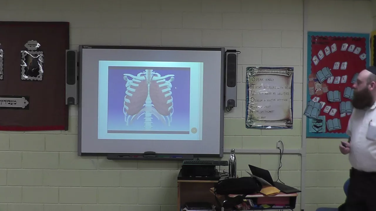
Added:well in this PowerPoint we're going to be thinking about the respiratory system and everyone in any form of healthcare needs to know about the respire tree system and in a minute I'm going to tell you why that is the case or some reasons why that is the case but for now just let's look at a little basic anatomy the bones you can see at the top of the clavicles and then the flat bone which is sawn in half in this model is the sternum and we count the ribs from the top the first rib the second rib third fourth fifth sixth seventh eighth ninth and tenth so there are 10 ribs that are attached to the sternum there's another two ribs at the bottom when you're floating so we have a total of 24 ribs 12 pairs but we notice that there's a blue bit between the bone of the rib in white and the sternum in white and that blue bit represents the costal cartilage so the ribs are not connected directly to the sternum there is this springy flexible costal cartilage making the thoracic cavity relatively springy with the cartilage now between the gaps in the cartilage in this model we can't see the muscles but we can see through to the lung tissue so in reality if you open the chest like that in between the ribs there will be the intercostal muscles but here we're looking directly through into the lung tissue and we see that the lungs are protected by the sternum the ribs the costal cartilage and behind of course there are the thoracic and vertebrae and poking off towards the left there you can see the area occupied by the heart well there are so many reasons why we need to know about the respiratory system when we're looking after patients but let's just think about two main points to begin with and they both relate to the rate of respirations and two things can go wrong with the rate the rate can increase or the rate can decrease now an increase rate of respiration is called a tacky Pinilla fast breathing tacky fast Pinilla means to do with air or breath now a kind of normal rent for an adult at rest would be twelve to sixteen breaths per minute now when children is going to be faster so remember that's the adult rate and if the rate is greater than twenty rest per minute I think we can call that our tachypnea that would be faster than normal so why might the patient be breathing faster than normal if we see a patient breathing faster than normal we see a patient with tachypnea what could this mean clinically why is this observation important well it could be normal of course for example exercise emergencies excitement anything that causes anxiety is going to increase sympathetic activity there's going to be more sympathetic autonomic nervous system activity and that will increase respiratory rate but tachypnea can also be a feature of infections now when the patient is developing an infection very often the first clinical feature if you can spot it is an increased respiratory rate the increased respiratory rate can demonstrate itself before the increased heart rate before the pyrexia so keeping a good eye on the patient's respired to rate if it's increasing they may be developing an infection and of course that can develop on to sepsis so a respiratory rate of greater than 20 breaths per minute it is a set as criteria and patients can be breathing particularly quickly if they have a infection of the respiratory system as in pneumonia where there is infection at the level of the alveoli but having said that any systemic sepsis or infection can cause attacking paneer now pain is another course and pain also often causes gasping respirations and sometimes the patient swallows quite a lot of air when they're in pain and the stomach can blow up a bit but then of course tachypnea can be caused by various lung diseases asthma springs to mind immediately where the patient can have great difficulty breathing out and they have a fast response rate although it's very shallow respirations pulmonary edema where there's a collection of pathological fluid in the lungs is another cause of rapid breathing and then we can think about lung injuries for example there might be a pneumothorax where air gets into the pleural space and one of the lungs collapses or stars to collapse or contusion is quite common where there is bruising to the lung as a result of a chest injury Takapuna could be a sign of pulmonary embolism usually what happens here is a blood clot breaks off from a deep venous thrombosis and lodges in one of the pulmonary arteries or tachypnea can be a sign of various types of heart disease such as heart failure so when we notice an increase respired rearrange we have to note that that is unnormal and that will give us a lot of assessment diagnostic information when we look at that in the context of our overall patient's condition but the first thing is to notice that the respire tree rate has increased we don't want to miss that so Bradley paneer is clearly another essential observation that we mustn't miss brandy means slow slow brandy paneer is the converse of tacky paneer Bradley paneer is slow breathing so why might a patient's breathing slow down well a common one is that we've given them opioids for example morphine or diamorphine these have the ability to depress the response recenter in their brain stem and slow breathing down or virtually slop it sometimes in fact they can stop it they can cause respiratory arrest so if patients are on opioids is particularly important that we monitor their ventilation rate other drugs such as alcohol and barbiturates can also be respond to any presence and in hospitals we sometimes give paralyzing drugs so the patient's being ventilated or operated upon we might give paralyzing drugs and these don't always wear off straightaway so patients who have had muscle relaxing paralyzing drugs might have a degree of respiring paralysis for a period of time after that now another cause of Bradley paneer is chronic obstructive pulmonary disease particularly in patients who are presenting predominantly with the Branka tick form so patients with chronic bronchitis can actually have quite low response rates now guillain-barre is a disease of the nerves as inflammation of the nerves and that can stop patient's breathing sometimes or certainly depress the response rate and there's another neurological disease called myasthenia gravis where respirations can be inhibited now external compression is a cause it's surprising how many people died because someone sat on them if there's someone carving of their abdomen all their chests or sometimes if people have been crushed for example after an earthquake in this heavy objects on their chest and that stops them from breathing an obesity obesity can actually limit respiratory efforts as well particularly if the abdomen is very fat if the other abdomen contains a lot of adipose tissue another cause of brandy paneer can be brain injury especially if this raised intracranial pressure hypothyroidism now it's the thyroid hormone which controls the metabolic rate of the body and the more thyroid hormone the higher the metabolic rate the thyroid hormone stimulates metabolic activity so if someone's hypothyroid their metabolic activity is going to be low and their oxygen demand is going to be less and the amount of carbon dioxide they are producing is going to be less so there can be a brandy paneer in hypothyroidism now hopefully you won't see the next two it depends on where you live but elapids are poisonous snakes like Cobras mambas crates of all elapids and these types of snakes produce paralysis that their venom produces paralysis they are neuromuscular blockades they block the synapse between the motor nerve and the muscle causing paralysis so after people have been bitten by these types of poisonous snakes they might need ventilator support for a few days until the toxin can wear off now polio myelitis are really really hard we don't see this in your working career but it used to be relatively common and this is a viral infection that affects the nerves and was renowned for causing respiratory paralysis and as well as the brandy paneer we all see changes in breathing rhythm and there are many of these so I'm just going to mention to change Stokes is is a diminishing respiration followed by the gasp patient gasps after a period around you so the patient's breathing can die down and die down and die down you think they've stopped breathing and all of us burn or start going through another cycle and that very often occurs terminally when people are are dying and crucial our respiration is another column one this is deep sighing ventilation and the time we see this is in a metabolic acidosis such as diabetic ketoacidosis is a classic feature this deep sighing ventilation and we get in DKA so brandy paneer again make the observation make sure you don't miss it if you do miss it of course your patient can stop breathing altogether so make sure you don't miss it and again interpret this in the context of the entire patient's condition now the first thing we need to understand is the process sort of ventilation and this really is in two parts the first is inspiration breathing in and of course the next is exploration breathing out so so ventilation is talking about ventilating the lungs with air how does air get in and out of the lungs this is what we need to study to understand this process of ventilation well here we see some of the anatomy of the thoracic cavity and I think we'll start with the bones so if we look at the top left there's a bone labeled C and then on the right there's one which is the the same as that of course when I say the left it's the patient's right hand side so that big C is actually on the right clavicle and then you can see the ribs are numbered one two three four and it's the same on both sides so what I've labeled here is the first second third and fourth right rib and they go all the way down to ten which is attached to the ribs via costal cartilages and then there's two smaller floating ribs at the bottom so that's the bones now thinking about the muscles between one and two you can see there's I M this stands for intercostal muscle so between each of the ribs there are intercostal muscles costal is the word which means you rib so the intercostal muscles are between the ribs and if you've ever eaten barbecue spareribs this is the muscle you're eating the intercostal muscle and then at the bottom you can see a large D that is the domed muscle of the diaphragm so dome the muscle of the diaphragm and is the diaphragm that separates the thoracic cavity above from the abdominal cavity below and it's a bone chute of muscle and at the top left that is the patient's right you can see SM that's the sternomastoid muscle now the intercostal muscles and the diaphragm are described as the primary muscles of respiration you use those all the time in normal ventilation but then if you're breathing particularly hard like if you're running and you need extra air there's so called accessory muscles of respiration as well and these connect to the top ribs into the sternum and that's during the mastoid muscle as one of those it attaches to the sternum and then it goes on around the neck and attaches to the mastoid bone behind the ear and these just give the thoracic cage a bit of extra pull when he needs it to increase the rate and volume of ventilation well we can see two colors of blood vessels in this model the blood vessels painted blue are carrying deoxygenated blood and the blood vessels painted red are carrying oxygenated blood so we notice vici to the patient right that's the vena cava and this is in fact the superior vena cava and as you can see it's draining blood back from the top part of the body of course the heart has been removed from this model but we know that the superior vena cava carries blood into the right atrium now to the left that is to the patient's left we have a which is the aorta and this is the main artery carrying blood into the systemic circulation the aorta derives from the left ventricle and it's bright red because it's carrying oxygenated blood and we can actually see that the aorta curves round and goes down now as a bit of an aside there's a structure labeled oh there and that is a muscular tube that goes down through the thoracic cavity and this of course is the esophagus or the food pipe taking food down through the thoracic cavity to the stomach now if we look in the patient's left lung we can see that there are blue and red vessels now because this is the pulmonary circulation the blue vessels are the artery is carrying the deoxygenated blood and the red vessels are the veins carrying the oxygenated blood back to the heart and it's the same on the patient's right lung I think you can see the a for an artery they're in cross-section that's carrying deoxygenated blood from the heart to the lungs to be oxygenated above that you can see a red vain and cross section carrying the oxygen oxygenated blood back from the lung to the heart so remember in the systemic circulation it's the arteries carry the oxygenated blood and the veins carry the deoxygenated blood whereas in the pulmonary circulation the arteries are carrying deoxygenated blood and the veins are carrying oxygenated blood and this is quite straightforward if you remember the simple definition that an artery is any vessel carrying blood away from the heart and a vein is any vessel carrying blood towards the heart now the red background we can see in the lungs is the tissue of the lung or the parenchyma of the lung and this is made up mostly of alveoli millions of alveoli and in each lung if you're young and healthy if you open the alveoli out that will give you about 70 square meters of internal surface area now you can see the large T in the middle there that's the chuckya and the Blues bands the blue stripes around it our rings of cartilage and if you look up you can see that the trachea is going through the neck with extremes of cartilage and the rings of cartilage are to keep the main airway is open and the trachea divides into the left main bronchus and the right main bronchus now the left main bronchus divides into two loba bronchi because the left lung has two lobes whereas the right main bronchus divides into three lower bronchi because the right lung has three lobes now if you look down you can see a pea there on the patient's right lung that is the pleural membrane but you can see I've labeled P twice now the pleura membrane is completely surrounding the lung so as we'll see later there's actually two pleural membranes as an outside one called the parietal and an inside one called the visceral well each lung is completely surrounded independently by a visceral pleural membrane and we'll look at what these do in a later slide so there's some basic background of the thoracic cavity now this light gives the little more background detail we can trace the line of the diaphragm that is the blue line between the thoracic cavity above and the abdominal cavity below and with its main mass towards the right we can see the liver located below the right costal margin and we can't see it but below the left costal margin is the spleen we can't see it because it's further back now the costal margin is the bottom of the ribs as we've said costal means and means ribs under the liver anteriorly at the front there we've got the stomach and then we can also see the colon further down in the abdominal cavity and in this model we've left the heart in situ and I think you can see the PA there that's the pulmonary artery and it's blue so it's carrying deoxygenated blood to the lungs the pulmonary artery emerges from the right ventricle and quickly divides into two with one branch going to the left lung and one branch going to the right so the pro mariachi they're carrying deoxygenated blood and then you can see what's labeled P V pulmonary vein and the pulmonary veins are carrying blood which is oxygenated from the lung back to the left atrium to go through to the left ventricle to be ejected out of the aorta into the systemic circulation and we can see red and blue vessels going over the surface of the heart these derive from the aorta so they are systemic and these are the coronary arteries coronary means crown they've supposedly form a crown around the heart and these are the vital arterial vessels carrying blood to the myocardium itself and we can see the blue veins blue because these are systemic of course carrying deoxygenated blood back from the myocardium this just gives us a little more detail on the lungs we see the main airway the trachea carrying air to the right and the left main bronchus we notice that the left lung has two lobes left superior and left inferior lobe but the right lung is divided into three lobes right superior right middle lobe and right inferior lobe and these lobes are very important if there's a infection in the lung so for example of a patient develops Loba pneumonia the anatomical arrangement of these semi separated lobes means that the infection is less likely to spread around the whole lung so it's sort of compartmentalizes the areas of the lung and the heart is towards the left so if you think about it there's three structures on either side three lobes on the right lung two lobes on the left lung but then we add the heart and that makes three now this diagram shows part of the right chest wall and we can see that the thoracic wall is made up of ribs and intercostal muscles so the dark areas are cut sections of the rib and in between the ribs we see that there are intercostal muscles and we notice that there were two sets of intercostal muscles the external intercostal muscles pointing out the way and the internal intercostal muscles pointing in the way so we have the ribs and the intercostal muscles now figure 1 shows the diaphragm on the bottom and the chest walls in the expired position and figure 2 shows the position of the diaphragm and the ribs when we've taken a deep breath in so I'm going to need your help here if you put your hands on the front of your chest and take a deep breath in look at your hands which direction are they moving in well I think you can see that when you breathe in your hands move up and out and when you breathe out your hands move down and in so during inspiration the external intercostal muscles particularly contract and that pulls the thoracic wall the intercostal muscles on the ribcage up and out so inspiration the ribs move up and out but now if you put your hand on your tummy for me and take a deep breath in and you look at your hand which way is your hand moving when you take a deep breath in well I think you can see when you take a deep breath in your hand is moving up and then when you breathe out your hand is moving down this is because during inspiration the diaphragm moves down and flattens therefore it's pushing onto the abdominal contents which is why your tummy moves up during inspiration so the intercostal muscles contract actively contract pulling the ribs up and out now the diaphragm is at rest when it's domed up so to breathe in the diaphragm actively contracts and when the diaphragm contracts it flattens and moves down the way so inspiration is active muscular contraction of the external intercostal muscles active muscular contraction of the diaphragm and both of these things are going to increase the volume because the ribs are moving up and out and the diaphragm is moving down so that's going to increase intrathoracic volume it's going to increase the volume in the chest and if you increase the volume what are you going to do to the pressure well as you increase the volume the pressure is going to decrease so the increased intrathoracic volume actually generates a negative intrathoracic pressure it reduces the pressure and then in order to equalize the pressure the air is sucked in from the external atmosphere where the air is at higher pressure so we are negative pressure ventilator x' we increase the volume thereby generating negative pressures in the thoracic cavity and this sucks air in so remember we are negative pressure ventilator x' air is sucked into air is ventilated into the lungs by increasing the volume thereby decreasing the pressure thereby sucking air in to equalize that pressure let's think about the process of breathing out or expiration so in order to expire the intercostal muscles will relax and that allows the lips to move down and in the diaphragm will also relax and when the diaphragm relaxes it domes of the way so we notice here that the ribs have gone down and in and the diaphragm has gone up both of these things are going to do what to the volume of the thoracic cavity I think you can see they're both going to reduce intrathoracic volume now if you reduce the volume what does that do to the air which is in the thoracic cavity well if you decrease the volume that's going to pressurize the air molecules and increase the pressure so the ribs move down and in the diaphragm moves up that's going to increase intrathoracic pressure and it will increase that pressure above atmospheric pressure and that will cause the air to be blown out so to use these terms precisely air is sucked in and air is blown out so we are negative pressure ventilator x' we breathe in because we generate negative pressures inside the thoracic cavity and we blow air out we expire because we generate positive pressures in the thoracic cavity and it's the chest wall and the diaphragm working together altering the volume thereby altering the pressures that facilitate this process of inspiration and expiration now these diagrams are representing the parietal and visceral pleural membranes the pleural membranes are the membranes that surround the lungs and we see that there's two of them so diagram one is illustrating the parietal membrane now parietal is around the outside think of P for perimeter and the parietal pleura membrane is lining the inside of the thoracic cavity and it's also lining the superior surface of the diaphragm so if you are to open a chest and pull out a lung the parietal pleura membrane will be left inside attached to the inside surface of the thoracic cavity and the superior surface of the diaphragm whereas the visceral pleural membrane is attached to the surface of each look so we actually notice that there's a right under the left pleural cavity now the V saw membrane is attached to the surface of the lung and it is firmly attached to it and that means that if you pull out the lungs the visceral pleura membrane will come with the lungs and the parietal membrane will be left in the thoracic cavity now between these two membranes there's a potential pleural space now actually in life these two membranes are sucked up against each other and there's actually a negative pressure of about four millimeters of mercury between those two membranes so in life the two membranes are slapped next to each other as if you've just clapped your hands together and your hands are still together they are actually in contact with each other apart from a very thin layer of serous fluid which is between them so they are the two pleural membranes the parietal and the visceral now when you want to breathe in your chest wall will move up and out and because the parietal visceral membrane is attached to the chest wall it will move up and out with it and in the same way one when you want to agree then your diaphragm will move down and because the parietal pleura membrane is attached to the superior surface of the diaphragm that will move down with it as well so this means when the lips move up and out and the diaphragm moves down the parietal pleura membrane will move with it but we mentioned that the visceral pleura membrane is sucking up against the parietal pleura membrane with a negative pressure of about four millimeters of mercury so this means that when the parietal pleura membrane moves with the chest wall because the visceral pleura membrane is sucking up against it that too will move with the chest wall but it's moving because it sucked up against the parietal pleura membrane so what this means in health is that the parietal pleura membrane will always move with the visceral pleura membrane because of this negative pressure and this is how the lungs inflate this is why when the ribs move up and out and the diaphragm moves down that the volume of the lungs will increase with the chest wall because the parietal pleura membrane has the visceral pleura membranes up to it and because the visceral pleura membrane is attached to the surface of the lung the surface of the lung will always travel with the chest wall that's an absolutely vital point of physiology to understand because it's got implications in many areas of clinical practice now I think this sectional microscope slide helps us to understand the relationship between the visceral pleura membrane and the lung so this arrow that we see at the top is pointing to the visceral pleural membrane and you can see that's quite a dense tissue and that's lining the outside surface of the lung then we can see this loose connective tissue and under that there is the lung parenchyma made the alveoli which are the air sacs and we can see that these are relatively thin cellular walls by large empty bits in the middle which of course it's filled with air this is to allow the process of gaseous exchange through the walls of the alveoli so we see the intimate relationship between the visceral pleural membrane and the lung so bear in mind that the parietal pleura membrane is going to be above this that this visceral pleura membrane is sucked onto the parietal pleura membrane so when the parietal pleura membrane moves up because the visceral pleura membrane is sub 2 that that moves up with it and I think you can see now why that results in the expansion of the whole lung well in virtually all clinical situations our priorities are ABC airway breathing and circulation and here we see the upper Airways that we have to make sure our patent patent means that they're open and we can see the air can get in through the nice big nasal cavity now there the air is going to be warmed and it's going to be filtered it's warmed because the nose lining the nasal mucosa is very vascular and it's filtered by the large hairs that sometimes you can see growing out of your nose and the smaller microscopic hairs that we'll see later on called the cilia and it's also lined with mucus and if wet as well as that the nose protects is against air that might contain contaminants and toxins because it allows us to smell so the nose is very protective and the oral cavity below air can go through the oral cavity as well and they may meet up at the back of this pharynx a bit of the pharynx behind the nose is the nasopharynx the bit of Farex behind the man or the-- to the oropharynx and the middle of pharynx at the level of the larynx is the lingo pharynx and the epiglottis is that flap of fibrous tissue that protects the glottis which is the top of the airway so the air will go in from the pharynx past the epiglottis into the larynx and the lines contains the vocal cords and then down on into the trachea and we notice that the esophagus which is responsible for swallowing is at the back so the air should go down the airway through the larynx and trachea then the food should go down the esophagus to the stomach here we see a model that shows us the continuation of the airways now TC is thyroid cartilage and that is the Adam's apple the lovely big that you can feel in your neck and below that is the cricoid cartilage now the cricoid cartilage II is a complete ring of cartilage and it's quite rigid and that holds open the top of the trachea is holding open the top of the airway now between the thyroid cartilage and the cricoid cartilage there's a small grey bit called the cricothyroid ligament and one day you might learn to make an opening into that cricothyroid ah to me as an emergency airway procedure the T is the thyroid gland which is located on either side of the cricoid cartilage and the top of the trachea going down we have the main airway which is the trachea and you can see the blue rings of cartilage going across the trachea we see the left main bronchus branching off to the left lung with the two lobe of bronchi and the smaller bronchial passages you can see there the start of the bronchial tree these are the segmental bronchi go into the lung segments and it's the same on the right we see the right main bronchus we see the three lobe of bronchi going to the three lobes of the right lung and then we see them branching up into the segmental bronchi go into the segments of the right lung this diagram is showing us the relationship between the trachea in the front that you care is anterior and the esophagus which is posterior so we can see the incomplete ring of cartilage in the wall of the trachea and that bit behind it is connective tissue and the thing about this is that the trachea is rigid whereas the connective tissue at the back of the lumen of the trachea is flexible and this is important because if the rings of cartilage were complete every time you swallowed the food would up to bump over the joint joint D joint as opposed all the rings of cartilage in the trachea and swallow it will be a very young interrupted process whereas because there's this area of soft tissue what this means is when a bolus of food goes down the esophagus the wall of the esophagus can expand and it can expand very slightly into that soft tissue of the trachea without having an obstruction from the presence of the physical rings of cartilage so the rings of cartilage in the trachea after the cricoid cartilage now the cricoid cartilage on top is a complete ring of cartilage but after that there are C shaped rings of cartilage to allow smooth swallowing down the immediately posterior esophagus in this diagram we're looking at the columnar epithelium which form the respire through epithelium the lining of the respired retract and this situation is much the same from the nasal cavities all the way down to the terminal bronchioles we have this arrangement of columnar cells some of goblet cells that produce mucus and we notice the cilia are sitting on top of the columnar cells so this respired free epithelium is moistening and protecting the lining of the airway and it's an essential barrier to the entry to potential pathogens and to foreign and potentially toxic particles so columnar epithelium with the goblet cells producing the mucus and this is a ciliated epithelium because it contains the the cilia and the cilia in combination with the mucus from what's called the mucosa Lurie elevator or the mucosal eraklyon system because it's the combination of the mucus and the cilia wafting that mucus up towards the oropharynx that keep the airway clear and it's important to note that this mucociliary clearance system works best at 37 degrees centigrade and at 100% humidity so that means if you want to optimize the functioning of our patient's mucosa Larry clearing system we need to give them warm air to breathe which is very humid moistened air so this is how the nose is clear this is how the sinuses are cleared this is how the bronchial tree is cleared from the mucus because the foreign particles the bacteria and any inhaled particles stick the mucus because it's sticky and then their wife dedup to the oropharynx or in the case of the nose of course wafted down to the oropharynx and once them in their aura fabrics you can cough it up clearing your throat then you can spit it out or indeed you can swallow it and estimates show that about 10 to 20 mils a day of fluid from the lungs are wafted up and coughed up into the oral pharynx now we have an infection of course it would be much more than that but in the normal situation 10 to 10 to 20 miles a day and this is this mucociliary clearance system is aided with good airflow which is why it's very important to tell our patients in bed to take deep breaths because it aids the clearance of the lungs but even without too much deep breathing the figures are quite impressive in the trachea the mucosa Lari clearance system can clear mucus at the rate of about 5 millimeters a minute so that means in two minutes a particle can have moved a centimeter up from the trachea towards the oropharynx so it can be cleared relatively quickly now this diagram shows a section of bronchial passage or it could be part of the trachea but imagine this is one of the bronchial passages in the lungs so we notice that there is the respiratory epithelium on the inside with the odd goblet cell the columnar epithelium and the cilia that is the inside and the goblet cells are producing mucus which is going to go onto the cilia anything foreign that's inhaled bacteria or particles will stick to the mucus and be clean fired the mucosa Larry escalator but then there's mucus glands as well which go deeper in underneath the mucosa now the respired from you Kozar is the surface epithelium a mucosa is just a surface membrane which produces mucus it's a mucous membrane but we see these mucous glands em you see us so em you see us is the sticky stuff em you see oh us is the mucous membrane so as well as mucus entering the surface of the mucosa so surface of the mucous membrane from the goblet cells there's also mucous glands as well in the submucosa and then on the outside there's a layer of connective tissue and that's made a fibrous tissue and it also contains cartilage to maintain the patency to keep open the trachea and to keep open the bronchial passages and of course that whole thing is circular and crucially that is leaving a lumen of patent airway in the middle through which the air can go in and out of the large Airways from the outside all the way down to the respiratory bronchioles and the alveoli themselves now here we see the lining of the bronchial passage and we can see clumps of these very fine hairlike structures projecting from the surface of cells the individual cilia now here we see a scanning electron micrograph of the surface of the internal lining of the trachea and we notice the ciliated nature of the surface and these are motile cilia that they move they can raft back and forward and typically each columnar epithelial cell can have about 200 of these small cilia projecting from their cell surface all wafting mucous towards the oral pharynx so we can see that the cilia are in fact organelles a couple hundred of them projecting from the surface of an individual cell now here we see the cilia at much higher magnification and we can see that there are individual particles sticking on to the cilia and that's good because these particles can be wafted up in the mucus and the cilia a motile they move and they actually whacked quite quickly it's been estimated that about 10 to 15 cycles per second so each cilia is rafting 10 to 15 cycles per second to wet this mucus up towards the oropharynx and the size of the cilia they're about 1 micrometer in diameter and about 10 micrometers long these are quite long ones that you look these look quite long others are shorter they could be as short as one micrometer these ones are probably about 10 micrometers and about one micrometer thick and the reason that their contractile is that they have micro tubules inside them now inside each one of these very fine hairs inside each of these cilia there's an arrangement it's called the nine plus two arrangement of microtubules and these microtubules are made of tubulin and it is the arrangement of the tubulin and the protein inside the cilia which make it flexible and cause the contraction and relaxation of the individual cilia now this rather clever micro graph shows the cilia labeled C below an M above is the layer of mucus so what we can actually see here is the layer of mucus that the cilia are wafting up to the oropharynx and in the trachea it can be wafting this at the rate of about five millimeters per minute so every two minutes a centimeter of rafting will move a bacteria for example or smoke particle sticking to the cilia or sticking to the mucus that is being wafted by the cilia will move towards the oropharynx at the rate of about a centimeter every two minutes so very impressive mucosa Larry elevator well the whole point of the respire trisystem is to facilitate gaseous exchange this is the exchange of gases between the air and the blood so we need the oxygen to go from the air in the lungs into the blood and we need the carbon dioxide from the blood to go from the blood into the air in the lungs and Sarah can we breathe out so let's look at the anatomy and physiology of gaseous exchange now the trachea divides into the right and left main bronchus that divides into the mobile bronchi that divides into the segmental bronchi and they keep dividing and from the trachea all the way down to the diagram that we're seeing here is between 20 and 25 divisions into progressively smaller Airways so on this diagram we see the terminal bronchial and that is taking the air into the respiratory bronchial now the respiratory bronchial is the first level where gaseous exchange actually takes place the terminal bronchioles is part of the airway even though it's on a microscopic scale the walls are too thick to allow gaseous exchange to occur so in the terminal bronchi bronchioles the oxygen doesn't go from the lumen into the blood because the walls are too thick but that happens at the level of the respiro tree bronchioles and we notice that the respiratory bronchioles they have an alveolus which would be a singular or several alveoli projecting from their walls to greatly increase the surface area because here the lining is thin enough for gaseous exchange to take place and the majority of gaseous exchange actually takes place in the alveolar air sacs which are big clusters and sacs of our REO line the oxygen going from the air in the alveoli into the blood and the carbon dioxide go from the blood into the alveolar air sacs to be breathed out in the process of expiration here we're looking at a section of lung at the level of the alveoli and we can see the individual air sacs and surrounding the individual air sacs are the pulmonary capillaries so we can see the very close relationship it's not far for the option to go oxygen to travel to diffuse from the lumen of the alveoli through into the capillary and it's not far for the carbon dioxide to diffuse from the capillary through into the air in the alveolar air sac we also noticed that there's a great number of pulmonary capillaries so there's a lot of surface area over which gaseous exchange can take place and apparently if you open out all of the alveolar air spaces within a lung the internal surface area over which gaseous exchange takes place is in a healthy lung 70 square meters so that means in you you've got 140 square meters of surface area on respiratory surface available for the process of gaseous exchange 70 square meters per log this photograph shows a few individual are we all I so we can see the alveoli are air sacs and we notice the walls are very thin so that the gases can diffuse through these thin alveolar walls and we also notice that the large number of our Rio lie form a large internal surface area over which gaseous exchange can take place so blood is going to be discharged from the right ventricle into the pulmonary artery the pulmonary artery we are going to subdivide and eventually the blood will enter a pulmonary arterial carrying blood which was relatively deoxygenated the saturations may be in the order of 70 to 80 percent typically around about 75% so this relatively deoxygenated blood arrives in the pulmonary arterial and that goes into the pulmonary capillaries and in reality they would be more than the three pulmonary capillaries illustrated here and these pulmonary capillaries are in close contact with the air in the alveolus where the process of gaseous exchange actually takes place and then once the process of gaseous exchange has taken place that blood will leave a viral pulmonary venule carrying blood which is typically about 98% oxygenated high in oxygen and that blood is going to drain back into the larger pulmonary veins which in turn drain into the left atria and we notice the respire through bronchial is carrying air into the alveolus and after that it will carry air out again and the terminal bronchioles are continuous with the respiratory bronchioles so the site of gaseous exchange is the alveoli now this slide summarizes quite a lot of the physiology going on at the level of the alveoli at the level of the pulmonary capillaries in the process of gaseous exchange so first of all let's look at the left hand side of the picture then we can see blood from a branch of the pulmonary artery that is blood that is coming from the right ventricle via a branch of the pulmonary artery to a pulmonary arterial into the pulmonary capillary and as this blood arrives it's going to be relatively low in oxygen and relatively high in carbon dioxide but then as it goes past the alveoli there's going to be diffusion of gases between the capillary blood and the air in the alveoli and we noticed that this movement of oxygen from the alveolus into the blood down its diffusion gradient and we also notice that there is movement of carbon dioxide from the blood into the alveolus again down it's diffusion gradient and as a result of that the blood leaving in a branch of a pulmonary vein is going to be high in oxygen but lower in carbon dioxide now the air going in and out of the lungs when it goes in it's going to be relatively high in oxygen and relatively low in carbon dioxide then when it comes out because the carbon dioxide has gone into the blood and carbon dioxide from the blood has gone into the air in the alveolus as a consequence of that the air that's going out is going to be relatively high in carbon dioxide and relatively lower in oxygen what we see here is a blow-up on one of the alveoli and we can see on the left that there's air space in the alveolus and blood in the capillary now to get from the air in the alveoli through to the hemoglobin in the red cells the oxygen must first diffuse into the fluid layer lining the alveolus and this must contain surface active agent or surfactant that reduces the surface tension allowing the oxygen to diffuse in there next it must diffuse through the thin cell which is the alveolar epithelium and then there's a small interstitial space between the walls of the alveolus and the walls of the capillary then there's the basement membrane of the capillary and then there is the vascular endothelium of the capillary and the red blood cell and even though there's a bit to go through there the distance is very small we dealing with very squamous cells here and the movement of carbon dioxide in the opposite direction is through the same structures but in the reverse direction and each gas is diffusing down its diffusion gradient so initially the concentration of oxygen is higher than the alveoli and lower in the blood initially the concentration of carbon dioxide in the blood is higher than it is in the alveolus and the carbon dioxide diffuses down its diffusion gradient this last bit of respire through physiology shows some volumes associated with the lungs and these are the volumes that we'd expecting in fit men female volumes will be less now when the line goes up that's breathing in when the line goes down that is breathing out and that gentle breathing up and down is the tidal volume then if you breathe in all you can or you can all the way in that's the inspiratory reserve volume then if you breathe out all you can and then drive me that a little bit more so you loot Li bring up again that's the expiratory reserve volume and if you breathe in all you can and then breathe out all you can that's the vital capacity but there's a bit of the bottom then you can never quite breathe out that's the residual volume and of course these volumes are go to change in various disease processes I'm sure none of you listening to this talk smoke but there will be those round about you and especially when you travel to poorer areas of the world tragically you will find people smoking so use your initiative and try and work out ways to make them stop because smoking is not good for a lot of things and one of the things it's certainly not good for are the wonderful lungs that we've been considering in this presentation
Up Next

Functional Residual Capacity FRC Measurement and Clinical Importance
@Enzymembbs
124 views•2026-01-25

Taste Matters: Why We Like the Food We Do | Professor John Prescott
@smagforlivet-video
409 views•2015-12-07

COVID-19 Seroprevalence: Antibody Data Analysis Explained
@Campbellteaching
308.8K views•2022-03-04

Bacteriophages: Earth's Deadliest Killers and Future Antibiotics
@kurzgesagt
34.6M views•2018-05-13
Related Study Plans & Knowledge Roadmaps
Structured learning paths in Biology