


















Functional Residual Capacity (FRC) is the volume of air remaining in the lungs after normal expiration, calculated as Expiratory Reserve Volume plus Residual Volume (approximately 2,300 ml). Since residual volume cannot be measured by spirometry, two indirect methods are used: the helium dilution method (which applies conservation of mass principle using an inert gas that mixes with lung air until equilibrium) and body plethysmography (which uses Boyle's law to measure pressure-volume changes in an airtight chamber). FRC acts clinically as an oxygen reservoir during apnea, prevents alveolar collapse, and maintains stable gas tension; increased FRC indicates obstructive lung diseases (emphysema, COPD), while decreased FRC indicates restrictive lung diseases (pulmonary fibrosis, ARDS). Anesthesia and supine posture significantly reduce FRC, potentially causing atelectasis.
Functional Residual Capacity FRC Measurement and Clinical Importance
Added:Hello everyone, welcome to enzyme MBBS where complex medicine becomes simple and exam ready. Okay, without wasting any time, let's get started. Coming to the definition, first write the definition. So it is the volume of air remaining in lungs after normal expiration. How do you calculate it?
Functional residual capacity is equal to expiratory reserve volume plus residual volume. That comes out to be 2,300 ml.
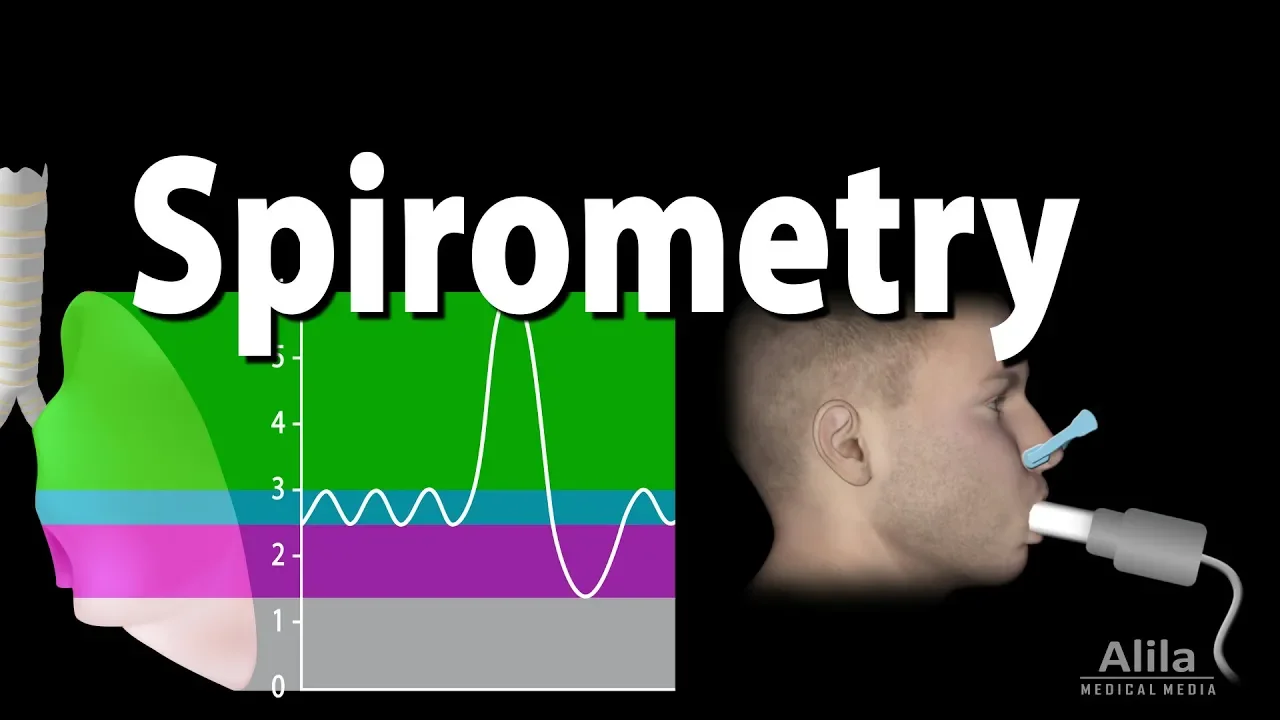
Okay. So this is a definition. While asking the question of functional residual capacity, they ask the definition. They ask how you will measure it and what is the clinical importance of it. So definitions we completed. Now let's go to the measurement part. So now measurement since residual volume cannot be measured using directly by spyometry. FRC also cannot be measured using uh simple spyometry. All the other volumes most of the volumes we measured by spyometry but residual volume cannot be measured. Why it cannot be measured? Because see spyometry measures only the air that moves in and out of the lungs. Okay, if you observe residual volume is the amount of air that remains in the lungs even after a maximum forced expiration.
So since the air never comes out of the lungs. The spyometer has no way to detect or record it. That's why we say we can't measure it using spyometry. So we use some indirect methods here. Okay.
So now what are the indirect methods? So here you have to write two methods. One is helium dilution method and one more is body platismography. I have given the answer format for both. But before reading into the answer here me first.
Okay. So first thing is helium dilution method. How how you use the helium dilution method? First thing you remember about the helium that helium is inert. Okay. It is inert. It does not enter the blood. It only stays in the air spaces. Okay. So here what happens is the subject breathes from a spyometer that is filled with a known concentration of helium after subject rebreathes helium oxygen mixture and equilibriates with the spyometer. The helium concentration in lungs become same as in spyometry uh spyometer. Now I know you didn't understood anything from it. So hear me now. Imagine a situation.
You have a balloon. Okay. Uh correlate that balloon with the lungs. Okay. You have a balloon whose volume you don't know. You don't know how much volume of air you have filled inside that balloon.
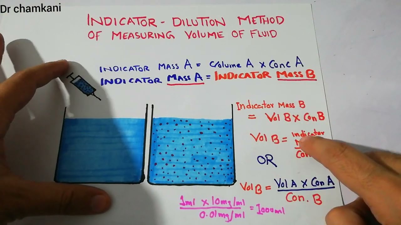
Okay. So now connect that balloon to a box. That box is spyometer. You have connected that unknown volume uh of balloon to a box. Now you know the volume of that box. You know the and that box also has the helium. So you know how much concentration of helium is present inside the box and what is the volume of box. The thing you don't know is the volume of uh eight that is present inside the lungs. Now what you will do? So you will allow the helium to mix between the box and the balloon. The same thing happens here. So patient what he does he starts breathing from a closed circuit like you connect uh you uh the lungs are connected to the spyometer here. Right? He is breathing from the spyometer. You know the volume of spyometer. You know how much helium is present inside the spyometer. What is the concentration of that helium? Now when the patient starts breathing from that closed circuit that helium moves into the lungs and it mixes with the air already present there. Okay. So after sometimes what happens the equilibri equilibration takes place. What is equilibri sorry equilibration.
Uh just um again in simple words saying that spyometer has helium plus oxygen.
Now there is high helium concentration whereas lung just contains the air. It has no helium. Right? So helium concentration in spyometer is not equal to the its concentration in the lungs.
So when he starts breathing he breathes in and out from the same glow circuit helium moves from spyometer to lungs as well as from lungs to spyometer. So this happens again and again. So helium keeps mixing. So after some time the key word that we use that equilibriates equilibration is complete mixing. Now the helium is evenly distributed. So helium percentage is same in spyometer as same as in lungs. So this is what that sentence meant. If you correlate this with an analogy imagine a glass which has a very strong juice and another glass has plain water. Now you keep pouring from one glass to another.
Then what happens after some time both glasses will have same juice concentration. That is equilibration.
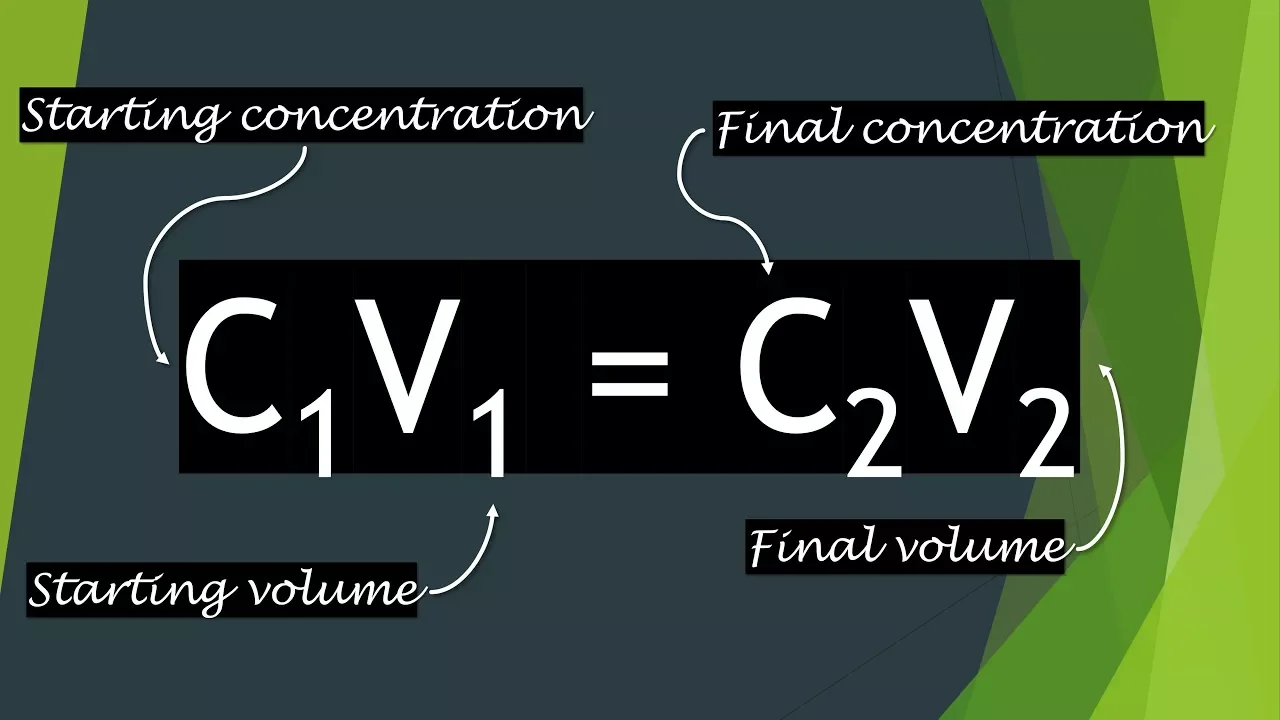
Okay. So this is what happens. Why this equilibriation is important? because only then you can apply the conservation of mass principle. I definitely know that you would have studied the conservation of mass in your chemistry classes, right? Uh so a mass can neither be created nor be destroyed. It can be transferred from it's it's something like that comes now. So the same thing so conservation of mass principle you can only apply after the equilibration happens. So here helium amount is constant only its distribution have been changed. So using that we use a formula C1 V_sub_1 is equal to C2 into V_sub1 + V_sub_2. What is V_sub_2? V_sub2 is unknown volume in LS. What is V_sub_1?
It is the initial volume of helium oxygen mixture. C1 is initial concentration of helium and C2 is helium concentration after equilibrium. And after mathematical calculations we derive this formula because we need the unknown volume in the lungs. Right? So that's how you get the formula. You can just uh like the formula you can memorize it. The key point here is you have to understand what we are basically doing right. Uh I hope that analogy helped to understand it. Okay. So next if this test begins at the end of a normal tidal volume it is a normal expiration. The volume of air that is remaining in the lungs is functional residual capacity. The one limitation of this method is helium cannot reach poorly ventilated or trapped areas. So what happens? It basically underestimates lung volume in those cases. So for that we use one more method which is more accurate method that is body platismography. What you do in body plethmography? It is also known as boil law principle. Again you would have studied about boy's law in the gaseous state chapter of your chemistry.
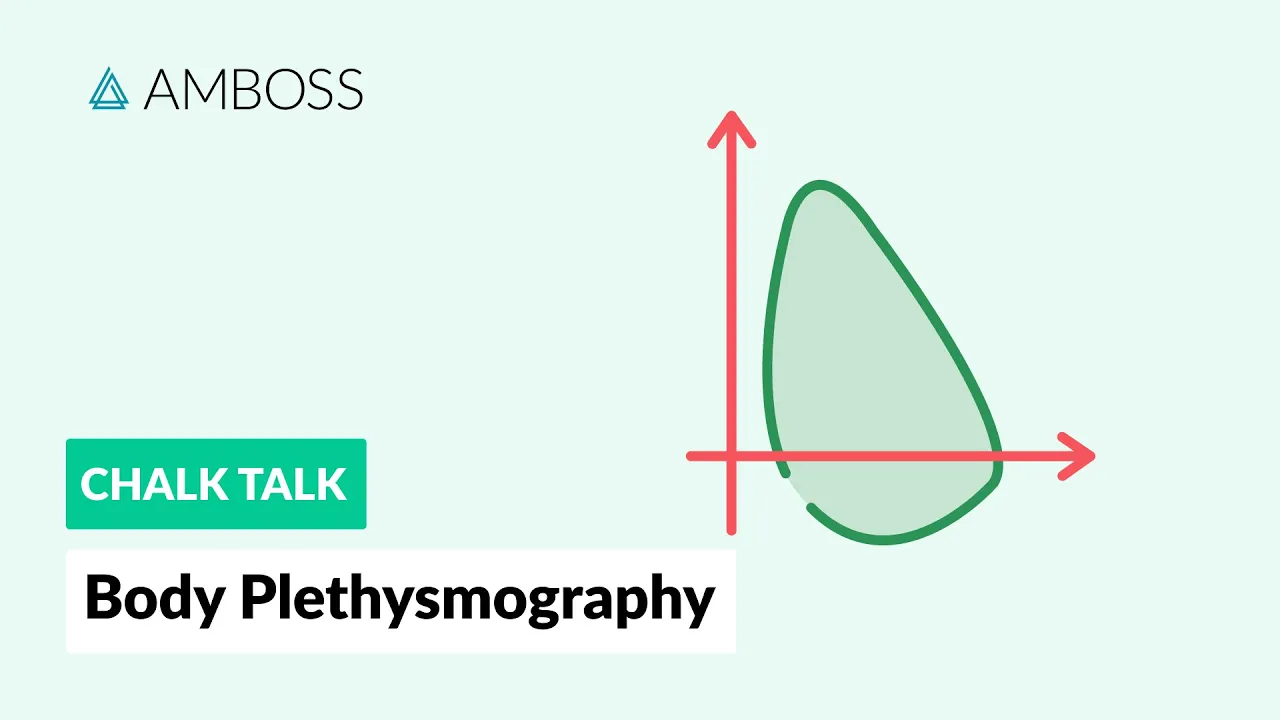
Right? So body platismography in this patient sits inside a airtight box.
Okay, he's already sitting inside a airtight box. Now that box contains air around the body. Imagine you you are sitting inside a box. So around your body inside that box the air is present, right? And your lungs is also containing the air inside the chest and there is a mouthpiece whose shutter is closed so that no air enters or leaves the lungs.
Okay. So now what happens when the person tries to inhale when he tries to inhale the chest expands while chest expanding the rib cage is moving outward. So what happens lung volume is increasing. So pressure inside lung is decreasing. Okay. So air uh what happens even though air does not leave or enter the lung volume still increases because pressure inside lung is falling. Now body volume increases because chest expansion makes the whole body slightly bigger. A body occupies more space inside the box. If the body is occupying more space inside the box, the volume of box is set to be decreased. Right? The body the volume of box is decreasing. So body uh the volume of the our body is increasing. Volume of box is decreasing. Similarly pressure inside our lungs is decreasing but the pressure inside the box is increasing.
Now you apply the boil's law.
what it says P1 V1 is equal to P2 V_sub2 right uh you can like one you can give for box or for lungs of course you know you can measure the pressure inside the box you know the volume of box or you can measure the volume of box uh it's it's like that's how you calculate you know the three points one you missing use the formula you calculate it so this is the most accurate method and it includes even nonventilated lung areas is the exception or the limitation of helium not exception the limitation of helium dilution method is solved here.
So it includes even nonventilated lung areas. So it can measure the trapped gases also. So that is the two methods used for measuring the body uh sorry used to measure the functional residual capacity. Coming to the clinical importance first thing it acts as a reservoir of oxygen. So imagine during expiration or apnea. What is apnea? It is a temporary sessation of breathing.
You imagine you stop breathing for a for a short period of time. Maybe you are play like sometimes we play a breathing challenge or something like that. I don't know or not. Yeah. But any other reasons accidental reasons there is a temporary sessation of breathing. Okay.
So at that time if there is no uh volume inside the lungs the there will be a large fluctuation in the arterial oxygenation.
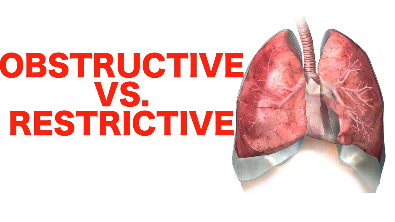
So during that time to avoid this this functional residual capacity comes into the role. As soon as the expiration or uh I mean if if there is any apnea the oxygen in the FRC continues to diffuse into the blood preventing large fluctuations in arterial oxygenation between breaths. Next prevention of alvolar collapse. If there is no residual volume present inside the lungs then the alvolar wall will adhere. It will collapse right? Then it would be very difficult for the patients to recover. So that is one of the main role by maintaining a volume of air in the lungs FRC helps keep alvoli open and reduces work needed for next inspiration. The third point is maintaining stable gas tension in the lungs. So all times enough air is available for gas exchange along the alvolar capillary membrane. It prevents sudden alteration in gas tension due to any brief interruption or respir of respiration. It's is almost similar to the first point. The last point is clinical alteration. What do you mean by like what is the conclusion if there is increased F FRC? If there is increased F F F F F F F F F F F F F F F F F F F F FRC it occurs when there is some obstructive lung disases. there is some obstruction in the airway uh in the diseases like emphyma COPD you'll learn about this diseases in your future classes but yeah in these conditions there is increased FRC then where when you can see the decreased FRC you can say in the restrictive lung diseases where lung compliance is reduced what is lung compliance if you know what is lung compliance please comment what is lung compliance well it is actually the measurement It is actually the measurement. Repeat with me because uh lung compliance is a separate fa question they can be asked you. So it's uh I can say in a simple word the definition of lung compliance is it is the change in lung volume produced per unit change in transpulmonary pressure. In simple words I can say lung compliance is a measure of how easily the lung expand. Okay. So in restrictive lung diseases this lung compliances is reduced. So it can be seen in diseases like pulmonary fibrosis, plural eusion and acute respiratory distress syndrome. Okay. And one more important point that you have to mention here is anesthesia and supine posture. Okay. I mean even if you don't mention it will be chala but the thing is that if you mention this and the examiner sees it like you mention it and make a cloud around it so that examiners can look into it then you'll get definitely get those extra mark because this is a very important point if you're going to become an anesthesian anesthesist or whatever it's very important because see if uh the anesthesia and supine posture both significantly reduce the fr FRC And during surgery the patient most of the patient will be in the supine posture itself and the anesthesia is given. So it can reduce the FRC which which may lead to the atiletasis. What isis? It is the collapse of alvolar membrane. Then the patient may undergo a lot of distress. So it's important to track this. It's important to monitor the functional residual capacity during the surgery and all this you learn in anesthesia too. But yeah that is one important point. Mention it at last. to a cloud and that will be a very good answer for this question and yeah just write with the headings boxes with a good handwriting and all that you'll definitely get a very good marks on this question. So that's all for today's video. If you like this video, please comment and subscribe.
Up Next
Lung Volumes and Capacities Explained | Respiratory Physiology
@armandohasudungan
915.6K views•2018-02-10

Taste Matters: Why We Like the Food We Do | Professor John Prescott
@smagforlivet-video
409 views•2015-12-07

Enteric Nervous System Explained: The Gut's Brain | Neurobiology Lecture
@alumniu6029
438 views•2018-09-12

Bacteriophages: Earth's Deadliest Killers and Future Antibiotics
@kurzgesagt
34.6M views•2018-05-13
Related Study Plans & Knowledge Roadmaps
Structured learning paths in Biology