


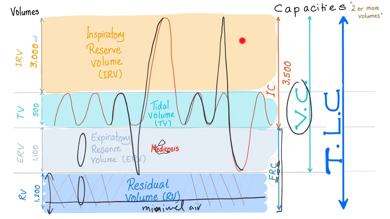













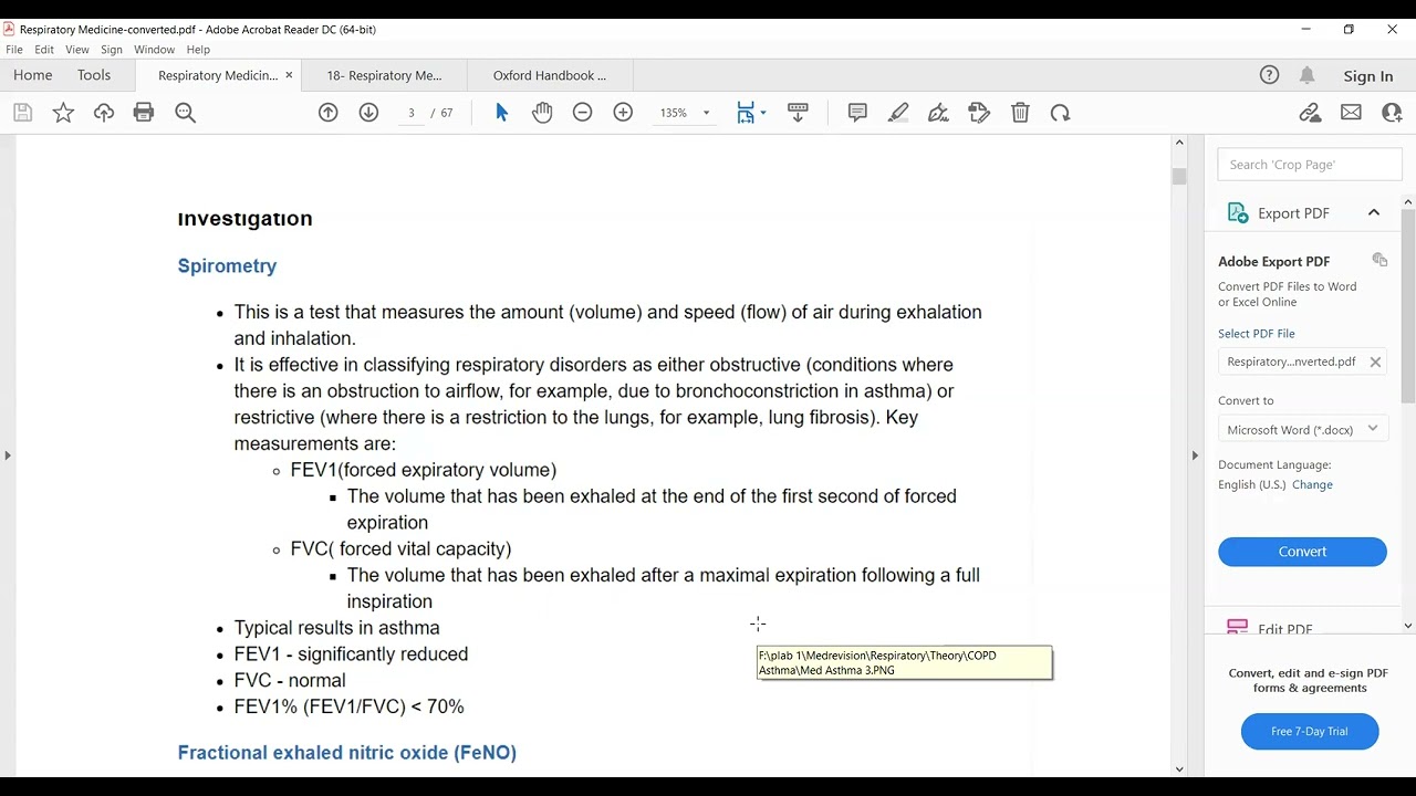







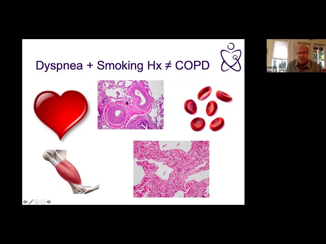



This video explains how to interpret pulmonary function tests (PFTs) using a step-by-step algorithm: First, check if FVC is greater than 80% predicted—if yes, no restriction; if not, proceed. Then, calculate FEV1/FVC ratio: if greater than 0.7, no obstruction; if less than 0.7, obstruction is present. To differentiate between obstruction with air trapping and restriction, check total lung capacity (TLC): if TLC is greater than 80% predicted, it's obstruction with air trapping; if TLC is less than 80% predicted, it's restriction. For obstruction severity, use GOLD stages based on FEV1% predicted (Stage 1: 80-100%, Stage 2: 50-80%, Stage 3: 30-50%, Stage 4: 0-30%). Bronchodilator reactivity is confirmed by FEV1/FVC change greater than 12% and 200ml absolute change. DLCO assessment helps distinguish extrinsic (DLCO/VA >80%) from intrinsic (DLCO/VA <80%) lung disease.
Pulmonary Function Test Interpretation: A Step-by-Step Guide
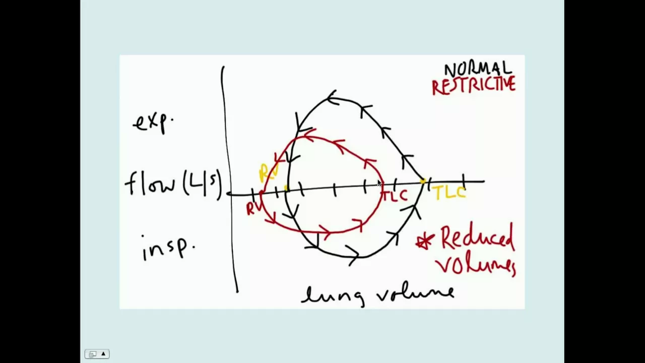
Added:okay so based on everything that we know now we're going to go ahead and show you how to interpret PFDs the first thing you want to do is you want to look at the F V C and you want to see whether or not it's greater than 80% of predicted that's this is the first thing that I look at and you really want to look at just any FEC if any FEC is greater than 80% of predicted the answer is yes or if the answer is no this is the algorithm that we're going to look at here if the FEC is greater than 80% of predicted then what you can say here is that you have no restriction no restriction that's our first diagnosis that we can come up with right off the bat if however the FEC is less than 80% of predictor or the answer is no then we can say that we either have restriction or obstruction with air trapping okay so it's one or the other restriction or obstruction with air trapping the next thing you do in either one of these cases is you move on in this case you're going to ask the question is the fev1 divided by the F V C greater than 0.7 and again you're going to have a yes and you're going to have a no if the answer is yes then there is no obstruction so by definition if the fev1 divided by the FEC is greater than 0.7 there is no obstruction if however it's less than that then you have obstruction okay the next thing let's go back here if the FEC is not greater than 80% predicted then you either have restriction or obstruction with air trapping the next question that you ask in this situation is is the total lung capacity greater than 80 percent predicted if the answer is yes then you have obstruction with air trapping if the answer is no in other words if it's less than 80 percent of predicted then you have guess what restriction now there's something you should know about in terms of obstruction and the severity there's different there used to be gold classifications in terms of Roman numerals they now used severity and also symptomatology but if they're ever talking to you about the gold stages there's gold stage 1 stage 2 stage 3 and stage 4 and for that we look at the fev1 only the FEV 1% predicted 1 is 80 to 100 2 is 50 to 80 3 is 30 to 50 and 4 is 0 to 30 it's also you go right to stage 4 if there is respiratory failure with an elevated P co2 level the other thing that you should know about when you have obstruction is you really should be able to classify if there is reactivity or not and the ATS criteria for reactivity is fev1 or fvc change in prutte in post bronchodilator in comparison to pre bronchodilator of greater than 12% and 200 milliliters if you don't have either of those two or if you have neither of those two then it is non reactive this doesn't tell you whether or not you should give bronchodilators it is helpful though when telling you whether or not this has if there is reactive Airways disease the other thing to look at is the DLC o dl c-- o and whether or not is greater than 80% of predicted okay if the dlco is greater than 80% of predicted then you have normal membrane surface area if it is less than 80% of predicted then of course it's abnormal surface membrane or membrane surface area okay however the next thing to look at is the dlco divided by the alveolar ventilation and if it is greater than 80% of predicted now run what we're looking at here we're seeing whether or not the dlco divided by the Alvar ventilation is still pretty good this is an indication and distinguishing characteristic between extrinsic and intrinsic lung disease of course for yes it would be x trinsic and for no it would be in trinsic so what are some examples this would be like scoliosis or Gyan bray syndrome and this would be for instance pulmonary fibrosis or for instance COPD okay so let's go over this again if you've got a forced vital capacity of greater than 80% of predicted no restriction you can say that right off the bat then you look at the fu v1 divided by the FEC if it's greater than 0.7 then no obstruction you're done you've got no restriction you've got no obstruction if the FE v1 divided by the FEC is less than 0.7 however then you've got obstruction no restriction let's go back to the very beginning if you don't have an FEC of of greater than 80% are predicted then you could have restriction or obstructive with air trapping the way you tell the difference between the two is by looking at spirometry if your total lung capacity is greater than 80% of predicted then you know you don't have restriction going on but it's more of an air trapping situation and that's where the obstruction comes in if your total lung capacity is less than 80% of predicted then there's a good chance that it's restriction that's causing both the total lung capacity to be low and the FEC to be low once you've diagnosed obstruction then you can break it down to different severity and say whether or not those reactivity or no reactivity in a separate situation you've got the dlco if the dlco is normal that's great if it's not it could be because of extrinsic disease or intrinsic disease by looking at the dlco divided by the alveolar ventilation you can make a distinguishing characteristic once you know if this is extrinsic disease or intrinsic disease you can put it together with your other diagnosis up above which are in double squares and figure out which way your diagnosis goes so keep this written down we're going to go over some actual examples and we'll be able to tell what the diagnosis is by interpreting the PFDs correctly you
Up Next
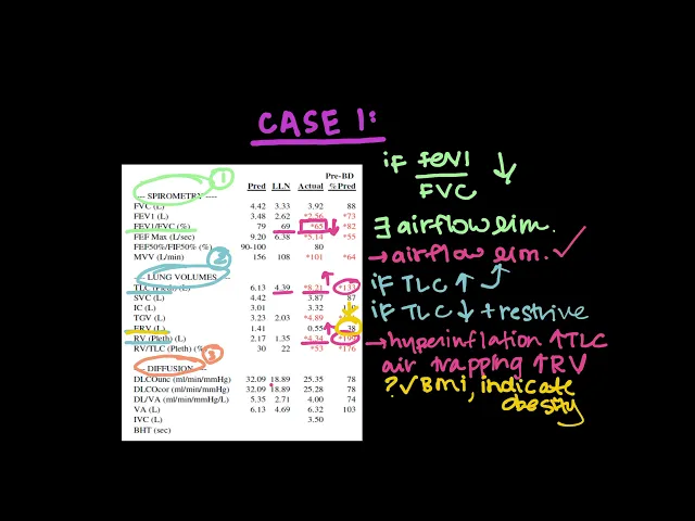
PFT Interpretation: Real-Life Cases for Spirometry & DLCO
@meredithgreer
7.7K views•2019-02-09

Memory and Long-Term Potentiation | Physiology | MBBS Exam Prep
@doctutorialsmbbs
307 views•2025-08-27
Mechanical Ventilation Explained: Settings & Modes
@Medcram
2.6M views•2014-12-06

Stages of Labor and Vaginal Birth | Childbirth Animation
@nucleusmedicalmedia
52.1M views•2017-08-18
Related Study Plans & Knowledge Roadmaps
Structured learning paths in Medicine