Rheumatology: Autoantibodies & Workups
Learning Goal: Navigating Rheumatological Workups: Interpreting Autoantibodies and Diagnosing Systemic Autoimmune Diseases
Prerequisites
- Basic understanding of general biology, cellular structures, and fundamental medical terminology.
- Familiarity with anatomical systems (musculoskeletal, vascular, and integumentary systems).
Estimated Study Time
- Total Study Time: ~14 hours (includes video lectures, note-taking, diagnostic clinical case reviews, and self-assessment).
Module 1: Foundations of the Immune System and Autoimmunity
This module establishes the core immunological framework required to comprehend rheumatological pathology. You will explore how the healthy immune system differentiates self from non-self, the key functional differences between the rapid innate response and the highly specific adaptive response, and the exact cellular mechanisms that lead to a breakdown in self-tolerance (autoimmunity).
Recommended Videos
-
Why this video: This video provides an elegant, structured breakdown of the dual-action architecture of the human immune system. It clearly differentiates the mechanical barriers and non-specific cellular actions of the innate immune system from the antigen-specific, memory-driven mechanics of the adaptive immune system. Understanding this fundamental division is vital because systemic autoimmune diseases often feature dysregulation in both pathways.
-
Knowledge Checkpoint:
- Understand the non-specific defense barriers (skin, mucous membranes) and innate cells (neutrophils, macrophages).
- Explain how the adaptive system leverages specificity and immunological memory to mount targeted attacks.
- Describe the basic role of antigen-presenting cells (APCs) in bridging the innate and adaptive systems.

-
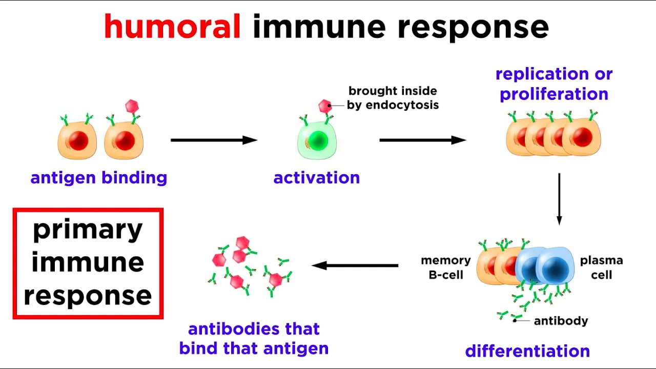
Why this video: Because autoantibodies are the primary diagnostic biomarkers in rheumatology, a granular understanding of antibody structure is mandatory. This lecture explains the anatomy of immunoglobulins, focusing on heavy and light chains, variable and constant regions, and the exact site of antigen binding. This molecular foundation is critical for understanding clinical assays like ELISA and Indirect Immunofluorescence.
-
Knowledge Checkpoint:
- Identify the structural components of a Y-shaped immunoglobulin (Fab region vs. Fc region).
- Differentiate between the variable regions (antigen-binding sites) and the constant regions that determine the antibody's isotype class (IgG, IgM, IgA, etc.).
- Understand how humoral immunity utilizes B-cell differentiation to produce target-specific antibodies.

-
Why this video: This video transitions from healthy physiology to pathology, explaining the precise immunological breakdown behind autoimmune diseases. It details how the immune system loses central and peripheral tolerance, mistaking host antigens for foreign threats. This is critical for understanding why and how pathogenic autoantibodies are formed in patients with rheumatological conditions.
-
Knowledge Checkpoint:
- Define clonal selection and how the failure of clonal deletion in T and B-cells leads to autoimmunity.
- Explain the concept of central and peripheral tolerance.
- Describe the genetic (MHC/HLA mutations) and environmental triggers that can initiate a persistent autoimmune response.
Module 2: Clinical Overview of Major Systemic Autoimmune Diseases
This module transitions from basic science to clinical presentation, detailing the classic signs, symptoms, and disease courses of the major systemic rheumatological conditions: Systemic Lupus Erythematosus (SLE), Rheumatoid Arthritis (RA), Sjogren's Syndrome, and Scleroderma (Systemic Sclerosis).
Recommended Videos

-
Why this video: SLE is the quintessential systemic autoimmune disease, with highly diverse organ involvement. This Osmosis video provides a premier, highly visual breakdown of SLE's pathophysiology—detailing UV-induced apoptosis, nuclear antigen exposure, immune complex formation (Type III Hypersensitivity), and subsequent multi-organ damage.
-
Knowledge Checkpoint:
- Detail the pathophysiological steps from UV-light exposure to nuclear antigen release and deposition of immune complexes.
- Identify classic multi-organ presentations of SLE, including the malar rash, lupus nephritis, and arthritis.
- Describe why SLE is classified as both a Type II and Type III hypersensitivity reaction.
-
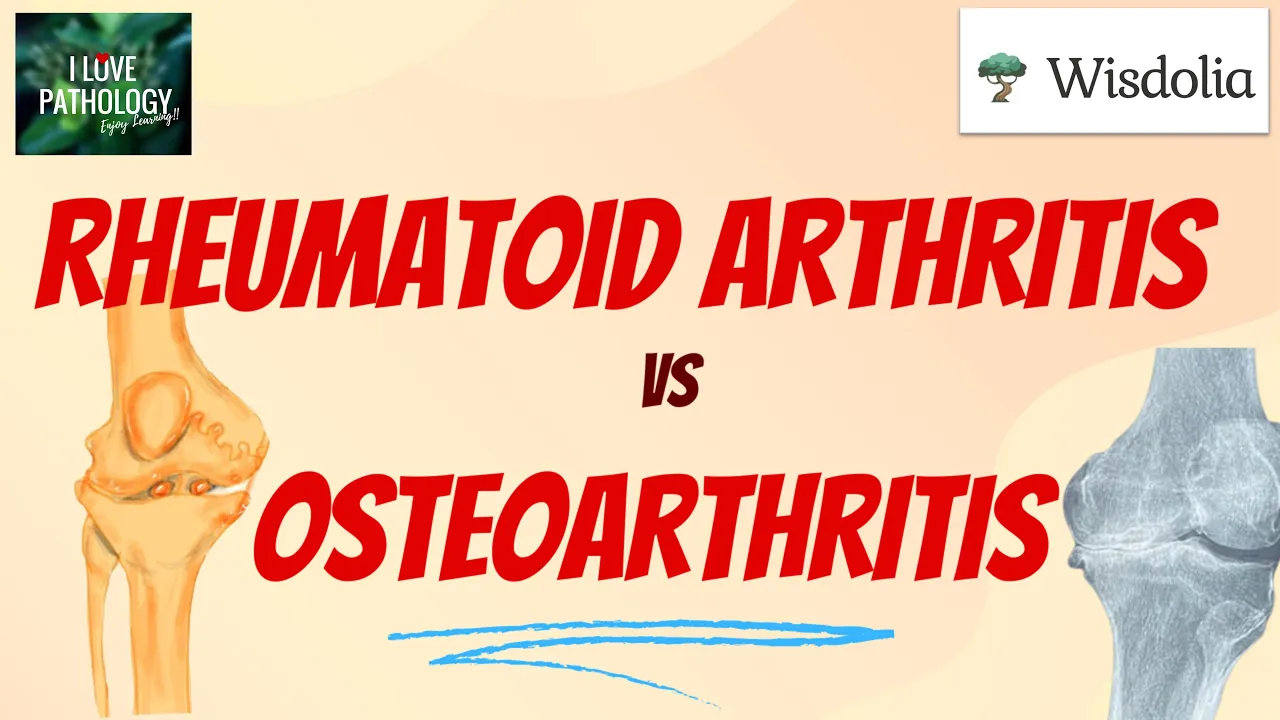
Why this video: Clinicians must confidently distinguish inflammatory arthritis (Rheumatoid Arthritis) from degenerative joint disease (Osteoarthritis). This video systematically compares their underlying pathologies, classic symmetric small-joint distributions, synovial membrane changes (pannus formation in RA), systemic manifestations, and key diagnostic clinical characteristics.
-
Knowledge Checkpoint:
- Compare the underlying etiology of RA (immune-mediated synovitis) with OA (mechanical breakdown of cartilage).
- Contrast the joint presentation patterns (RA: symmetric, small joints, PIP/MCP involvement, morning stiffness >1 hour vs. OA: asymmetric, large weight-bearing joints, DIP involvement, morning stiffness <30 minutes).
- Describe the histological presentation of a rheumatoid joint, highlighting pannus tissue and bone erosion.

-
Why this video: Address the critical curriculum gap identified in Scleroderma education. This professional-grade medical lecture explains Systemic Sclerosis (Scleroderma) pathogenesis. It maps out how vascular damage, CD4 T-cell cytokine release, and subsequent myofibroblast activation lead to widespread skin and internal organ fibrosis.
-
Knowledge Checkpoint:
- Differentiate between Limited Cutaneous Systemic Sclerosis (CREST syndrome) and Diffuse Cutaneous Systemic Sclerosis.
- Explain the pathophysiology behind Raynaud’s phenomenon, digital ulcers, and skin thickening.
- Describe the major systemic complications, specifically focusing on pulmonary arterial hypertension and interstitial lung disease.

-
Why this video: Delivered by clinical experts at Johns Hopkins, this short and highly targeted video identifies the primary manifestations of Sjögren’s Syndrome. It highlights the chronic lymphocytic infiltration of exocrine glands, which causes xerophthalmia (dry eyes) and xerostomia (dry mouth), alongside systematic extraglandular manifestations.
-
Knowledge Checkpoint:
- Understand the primary pathology of Sjögren's: lymphocytic infiltration of salivary and lacrimal glands.
- Identify clinical signs such as persistent dry eyes, dry mouth, dental decay, and parotid gland swelling.
- Distinguish between primary Sjögren’s and secondary Sjögren’s (occurring alongside RA or SLE).
Module 3: The Initial Workup: Inflammatory Markers and Basic Labs
This module introduces the primary laboratory tools used during an initial rheumatological workup. It details the physiological mechanisms behind acute phase reactants (ESR and CRP) and helps clinicians interpret basic hematology and chemistry panels to guide diagnostic reasoning.
Recommended Videos

-
Why this video: This clinical video reviews the debate between ordering Erythrocyte Sedimentation Rate (ESR) versus C-Reactive Protein (CRP). It provides highly actionable clinical practice guidance on which test serves as a superior indicator for acute inflammation, monitoring disease flares, and tracking therapeutic responses.
-
Knowledge Checkpoint:
- Explain why CRP is typically preferred as a first-line marker for acute inflammatory changes over ESR.
- Understand how CRP is synthesized by the liver and how its rapid response profile differs from ESR.
- Learn when it is clinically appropriate to order both tests versus CRP alone.

-
Why this video: To master lab workups, you must understand the underlying physiology and confounding variables of these tests. This medical lecture explains how ESR measures the sedimentation rate of erythrocytes driven by fibrinogen and hepatic cytokine pathways (IL-6, IL-1, TNF). It details the physiological factors that can cause falsely elevated or depressed ESR values.
-
Knowledge Checkpoint:
- Describe how cytokines (IL-6) trigger hepatic production of acute-phase proteins (like fibrinogen), neutralizing negative erythrocyte surface charges (zeta potential) to cause rouleaux formation.
- Identify non-inflammatory factors that elevate ESR (e.g., anemia, female sex, pregnancy, advanced age, macroglobulinemia).
- List factors that can decrease ESR, such as abnormal red blood cell morphology (sickle cells, spherocytes).

-
Why this video: This video highlights a vital clinical nuance: the unique inflammatory profile of SLE. While most systemic inflammatory conditions trigger elevations in both ESR and CRP, SLE flares often show an elevated ESR with normal or minimally elevated CRP. This presentation helps differentiate lupus activity from concurrent bacterial infections.
-
Knowledge Checkpoint:
- Explain why SLE flares typically present with high ESR but normal or near-normal CRP levels (due to the type 1 interferon-mediated pathways rather than the IL-6 pathway).
- Formulate a clinical action plan when an SLE patient presents with a significantly elevated CRP (e.g., evaluating for an underlying bacterial infection rather than a simple lupus flare).

-
Why this video: This expert-led review bridges the gap between basic inflammatory markers and disease-specific antibody testing. It outlines the standard five-test rheumatological screening panel (ESR, CRP, RF, Anti-CCP, and ANA), laying a clear baseline of what to look for when inflammatory arthritis is suspected.
-
Knowledge Checkpoint:
- State the screening panel components for inflammatory joint disease.
- Recognize why non-specific markers (ESR, CRP) must always be interpreted alongside highly specific antibodies (like Anti-CCP).
- Identify common CBC findings in active inflammatory diseases (such as anemia of chronic disease or reactive thrombocytosis).
Module 4: Interpreting Autoantibodies: ANA and ENA Panels
This module explores specific rheumatological serologies. You will master the clinical interpretation of positive Antinuclear Antibody (ANA) results, decipher staining patterns and titers, and learn how to utilize Extractable Nuclear Antigen (ENA) panels and autoantibodies (like Anti-dsDNA, Anti-Sm, Anti-CCP, and RF) to narrow down diagnostic possibilities.
Recommended Videos

-
Why this video: This clinical lecture details ANA testing methodology. It emphasizes why Indirect Immunofluorescence Assay (IFA) utilizing the HEp-2 substrate remains the diagnostic gold standard, and discusses the importance of interpreting staining patterns (homogeneous, speckled, centromere, nucleolar) alongside titers.
-
Knowledge Checkpoint:
- Explain why IFA is the gold standard for ANA testing compared to ELISA screens.
- Link common ANA immunofluorescence staining patterns to their associated systemic autoimmune diseases (e.g., centromere pattern in Limited Scleroderma).
- Define the clinical significance of a positive ANA in a healthy population.

-
Why this video: Interpreting ANA titers is a major clinical hurdle. Dr. Amigues provides a practical, real-world framework for evaluating titer levels (e.g., 1:40 vs. 1:160 vs. 1:640 and higher), helping clinicians determine when a positive test warrants an extensive workup versus when it is likely an incidental finding.
-
Knowledge Checkpoint:
- Explain the dilution mechanism of ANA titers (1:40, 1:80, 1:160, 1:320, 1:640).
- Identify the cut-off dilution titer at which a positive ANA becomes highly clinically significant (typically 1:160 or higher).
- Formulate a clinical pathway for managing an asymptomatic patient with an incidentally detected low-titer ANA (1:40 or 1:80).

-
Why this video: This concise, high-yield board review segment focuses on anti-dsDNA and anti-Sm antibodies. It underscores how these antibodies fit into the EULAR/ACR classification criteria for SLE, highlighting their diagnostic sensitivity, specificity, and prognostic value.
-
Knowledge Checkpoint:
- Identify which autoantibody is most specific for SLE (Anti-Smith/Anti-Sm).
- Recognize which antibody levels fluctuate with disease activity and correlate with lupus nephritis (Anti-dsDNA).
- Name the entry criteria for SLE classification according to the 2019 EULAR/ACR guidelines (positive ANA titer 1:80 on HEp-2 cells).

-
Why this video: This video resolves the clinical challenge of evaluating joint pain by comparing Rheumatoid Factor (RF) with Anti-Cyclic Citrullinated Peptide (anti-CCP). It explains why anti-CCP has become a vital diagnostic test due to its superior specificity for Rheumatoid Arthritis.
-
Knowledge Checkpoint:
- Compare the diagnostic specificity of Rheumatoid Factor (RF) (~50-80%) with Anti-CCP (~95%).
- Identify why RF can be positive in non-rheumatic conditions (e.g., chronic infections, hepatitis C, advanced age).
- Understand how the early presence of Anti-CCP antibodies predicts severe, erosive joint disease.
Module 5: Clinical Reasoning: Synthesizing Labs and Symptoms
This module teaches you how to synthesize medical histories, physical exams, and lab panels into a cohesive differential diagnosis. You will learn to think like a rheumatologist—avoiding the classic trap of "treating the lab result" instead of the patient—by working through mock patient cases and expert clinical reasoning exercises.
Recommended Videos

-
Why this video: Designed for medical trainees, this clinical reasoning exercise models a step-by-step diagnostic workup for a patient presenting with joint pain. The host physicians demonstrate how to combine patient history with basic hematology panels and targeted antibody testing to build an accurate differential diagnosis.
-
Knowledge Checkpoint:
- Observe how a senior clinician takes a targeted rheumatological history (focusing on morning stiffness, constitutional symptoms, and systemic signs).
- Synthesize physical exam findings (joint swelling and warmth) with laboratory results to narrow the differential.
- Recognize how and when to order advanced serologies versus monitoring with basic labs.
-
Why this video: This update focuses on diagnostic pattern recognition and identifying disease mimics. It outlines how to recognize rheumatological patterns while screening for malignancy and infection—two dangerous conditions that often present with inflammatory joint paint and positive markers.
-
Knowledge Checkpoint:
- Master the pattern-recognition method for multi-system autoimmune diseases.
- Identify red flags that suggest an autoimmune mimic (e.g., unexpected weight loss, night sweats, or localized bone pain pointing toward malignancy or occult infection).
- State why a comprehensive physical examination is always required before diagnosing a patient with an autoimmune disease based on bloodwork alone.

-
Why this video: This medical board-style review video presents clinical case scenarios. It is an excellent tool for testing your clinical reasoning, walking through presentations of giant cell arteritis, reactive arthritis, systemic sclerosis, gout, and other rheumatological conditions.
-
Knowledge Checkpoint:
- Analyze clinical case vignettes, pull out key diagnostic clues, and rule out competing diagnoses.
- Apply diagnostic criteria (e.g., classification guidelines for SLE, RA, Spondyloarthritis) to complex clinical scenarios.
- Formulate initial therapeutic steps based on the diagnosed condition.
Course Map
Below is the recommended sequence of modules. Each module provides the foundational knowledge necessary for the subsequent diagnostic steps.
Key People Index
- Dr. Eli Miloslavsky & Dr. Ana Valle (@clinicalproblemsolvers): Internal medicine and rheumatology educators specializing in case-based clinical reasoning and diagnostic medicine for medical trainees.
- Dr. Zainab Vora (@cerebellumacademy): Medical licensing board specialist who teaches high-yield clinical correlations and rheumatology test-taking strategies.
- Dr. Andras Fazakas (@drandrasfazakas): Clinical lecturer focused on systemic pathophysiology and connective tissue disorders.
- Dr. Amigues (@Rheumatology101): Practicing clinical rheumatologist specializing in patient-centered education on interpreting antinuclear antibodies and evaluating autoimmune flares.
Final Self-Assessment
Test your clinical competency by completing this comprehensive self-assessment checklist based on the entire curriculum:
- Can you explain the physiological difference between central tolerance and peripheral tolerance?
- Can you detail the molecular mechanisms that cause systemic sclerosis (Scleroderma), including the role of CD4 T-cells and fibroblasts?
- Do you understand the biochemical differences between C-Reactive Protein (CRP) and Erythrocyte Sedimentation Rate (ESR), including why their levels might diverge during an active SLE flare?
- Can you list at least four non-inflammatory factors that can falsely elevate a patient's ESR?
- Can you define the dilution titer values of an ANA test, identifying the threshold where a positive result becomes clinically significant?
- Can you map out the diagnostic significance and disease associations of the following ENA panel antibodies: Anti-dsDNA, Anti-Sm, Anti-SSA (Ro), Anti-SSB (La), Anti-Scl-70, and Anti-Jo-1?
- Can you explain why Anti-CCP is a much more specific diagnostic marker for Rheumatoid Arthritis than Rheumatoid Factor (RF)?
- Are you able to list the entry criteria and subsequent clinical domains used to classify a patient with Systemic Lupus Erythematosus under the 2019 EULAR/ACR criteria?
- Can you identify the classic "red flags" (e.g., weight loss, localized bone pain) that help clinicians rule out rheumatological mimics like occult infections or malignancies?
- Can you outline the step-by-step diagnostic pathway for an asymptomatic patient who presents with an incidentally detected ANA titer of 1:40?