





















This video demonstrates a systematic approach to diagnosing inflammatory arthritis through comprehensive history-taking using the LIDS mnemonic (Location, Inflammatory features, Duration, Systemic symptoms), physical examination focusing on joint distribution and enthesitis, laboratory testing (rheumatoid factor, CCP, inflammatory markers, ANA, HLA-B27), and imaging modalities (X-ray, dual energy CT, MRI). The case illustrates that no single test confirms a diagnosis; instead, clinicians must weigh pretest probability and integrate multiple findings, as demonstrated by the diagnosis of psoriatic arthritis in a patient with asymmetric arthritis, DIP involvement, and skin findings despite normal inflammatory markers and negative HLA-B27.
Master Rheumatology Differential Diagnosis: Case-Based VMR | Joining the Academy
Added:Basic clinical differentiation between inflammatory and mechanical joint pain (e.g., morning stiffness vs. pain with use).
Pathophysiology of common arthropathies, specifically the mechanisms behind crystalline deposition (Gout) and systemic autoimmune joint inflammation (Psoriatic Arthritis).
Interpretation of baseline rheumatologic laboratory tests, including acute phase reactants (ESR, CRP), serum uric acid, and autoantibodies (RF, anti-CCP).
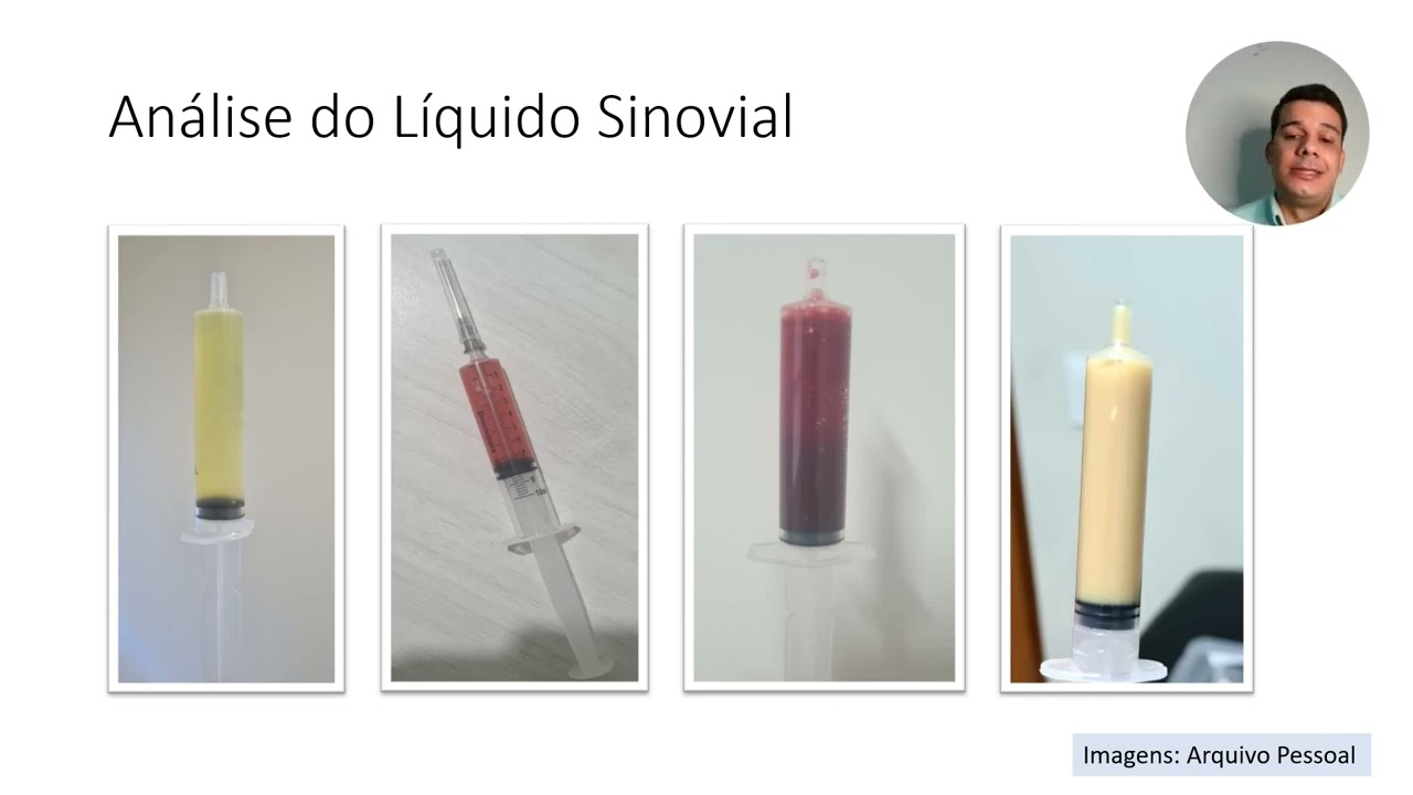
Principles of synovial fluid analysis, including cell count, crystal identification under polarized light microscopy, and Gram stain/culture.
Evidence-based pharmacological management strategies for acute and chronic Gout versus Psoriatic Arthritis (e.g., DMARDs, biologics, and urate-lowering therapy).
Utilization of advanced imaging modalities, such as musculoskeletal ultrasound (MSUS) and MRI, in detecting subclinical enthesitis and early joint erosions.
Systemic and extra-articular manifestations of rheumatologic diseases, focusing on cardiovascular comorbidities, renal implications of gout, and dermatological management of psoriasis.
Applying clinical reasoning and cognitive heuristics to complex, atypical presentations of multi-system autoimmune diseases like systemic lupus erythematosus (SLE) or vasculitis.
397 views12likes1:00:27Original Release: 2025-07-09
All right. Hello. Welcome back clinical problem solvers to another Raffle Medina subsp specialty VMR. My name is Leia.
I'm one of the academy uh team members and I'm really excited that we have a rheumatology subsp specialty VMR today and our guests are uh Eli Miloslavski and Anna Vali. And I will just briefly introduce them um in the beginning before I pass the mic on to the two of them. So our case discussion today is Eli Moslavski, a rheatologist and associate professor of medicine at the Mass Massachusetts General Hospital and Harvard Medical School. Eli's career has been focused on medical education across all stages of training and he serves as a curriculum phase director at Harvard Medical School, firm chief in the MGH internal medicine residency program and a social pro associate program director at MGH uh rheumatology fellowship and also leads the MGH clinician educator track for department of medicine subsp specialty fellows. And our case presenter today is Anna Vali um MDA um and MHS and is a rheumatology fellow at Bigham and Women's Hospital and she attended John's Hopkins Hopkins University and the John's Hopkins Bloomberg School of Public Health where she studied public health and during her undergraduate and graduate years afterwards then she worked as a research coordinator which introduced her to the field of rheumatology and instilled a passion for clinical research and then she obtained her medical degree at Zucker School of Medicine at Hostra Northwell and completed residency at Mont Fiora Medical Center in the Bronx, New York, where she was awarded the intern of the year and ambulatory chief.
And now she's pursuing a clinical research career um and dedicates time to address health inequities within rheumatology and further understanding of idiopathic inflammatory myopathies.
And those of you who've been here a few um months before will know the two of them. They have been guests here already. And to kind of um start us off, I wanted to ask the two of you what was actually the reason that you chose to go into rheumatology and what is um the what do you really enjoy about it? And maybe Anna, if you want to go first.
Certainly. I just want to say thank you for having us again. I'm very excited to be here. Um, I became interested in rheumatology pretty early in my medical career. Um, because I could see how women of all ages were impacted by these rheumatic conditions. We did not know as much about these conditions like we did about other um subsp specialties in internal medicine. Um, I saw women being affected from all ages, all walks of life. They remind me of my own friends and family and I wanted to be able to help them achieve their own goals and passions in their lives uh without having to um kind of have their lives shortened or be limited by rheumatic conditions. And so what I love about my uh about rheumatology is that um we get to have really uh special relationships with our patients through time. These are usually really close longitudinal relationships. Um, and we know each other really well because we're having really important risk benefit discussions about medications, about what to look for in the future and how to prognosticate their disease. And um, there, as I mentioned, there's always still a little bit more space for clinical research, too. So there's always plenty of times to have, you know, be inspired by a patient and their specific clinical condition and then be able to apply that um in clinical research.
Yeah. And so I I agree with what Anna pointed out about our specialty. I came to it though in a totally different way because unlike Anna, I had no idea that I wanted to be a rheatologist until midway through residency. And I think the way I started to think about it was that I really loved internal medicine, but I wanted to to be a specialist in some aspect, but I felt like a lot of the specialties were so single organ focused that I really didn't want to lose like the breadth of what internal medicine had and the kind of the need to really know and think about all of the organ systems. Um, and that's kind of how I came to rheumatology from that and kind of this love for diagnostic puzzles which are so frequent in our specialties and then that ability to like walk with a patient throughout their entire journey and build those relationships that Anna talked about. And you know since becoming a rheatologist I learned many other perks but I'll just highlight one. Our medicines are much better than they were and you could make almost everyone better which is just like an amazing feeling to have when someone walks into your office.
That's amazing and I can only echo um what is here. I'm a of course a bit biased as myself going into the field of rheumatology but um I feel like it's such a diverse field and also the community of people that I've met so far were so um enriching um it's a really really cool field and excited to learn more rheumatology today. So Anna if you're ready um feel free to start the case.
Certainly. So I will go ahead and start with the chief complaint. Uh so Mr. Zeppelin is a 48-year-old man who presents due to one month of right large toe pain.
Okay. And starting with the history of present illness. Uh one month ago he woke up with toe pain in his right big toe. As I mentioned uh the night before he had been playing a gig at one at a private party. So, as a bass player, he was on his feet for multiple hours. And when he finished playing, in order to reach his audience, he hopped the fence um in order to join the party. And once he got to the other side, um he was having multiple sweets um that he wouldn't normally uh indulge in on a on his normal afternoon. Um and um then and but those were the only other abnormalities that he noticed was different on the night prior to the start of his toe pain. Now he describes the toe pain as an 8 out of 10. It feels like a strong stinging sensation. Um it does not radiate. Um it gets significantly worse when he bears weight or tries to move his toe. Uh he has noticed his uh toe is hot and swollen to the touch. Um and also has been uh quite red. Um he has tried some ibuprofen um about 400 mg uh about two times per day which did help his symptoms but it seemed like these symptoms would always return um and the swelling and redness never fully went away. Uh this led for him to present to urgent care where uh he was told that he has gout. Uh they gave him colultine which took the edge off of his symptoms but once again the pain never really resolved. So he went back to the urgent care where he was given what we refer to as a medal dose pack uh and he was handed a referral to rheumatology uh which is when he saw me.
Okay. Thank you so much for the first aloquat Anna. So Eli, we had a lot of information. Now what are your first thoughts that are coming going through your head and how are you structuring this information? Mhm.
Well, I think my first thought probably like everybody else's here is that wow, this sounds a lot like gout, a toad that's like painful and perhaps inflamed. But, you know, I think that level one thinking um or system one thinking I should say is u you know, it's really important to kind of get a sense of what what it could be. But it's always important to kind of take a step back and just think about this as you know a monoarthritis or you know pain in a single joint and think about the buckets here. So this is something that could be mechanical or it could be inflammatory and that's kind of the first branch point. Um, and that's probably the more important branch point than even thinking about well which inflammatory condition could it be? Um, but it's important to to recognize that you know gout is an inflammatory arthritis just like many other inflammatory arthritities including rheumatoid arthritis and spondaloarthopathies etc. Uh, and while this doesn't fit that right now, um, it's kind of important to consider this in the kind of the the bigger bucket of inflammatory arthritis.
Now, thinking specifically about gout, you know, we often spend our time thinking, well, what, you know, here's my working diagnosis. What doesn't fit?
Um, and I think we could apply that lens. So, gout, especially the first episode, often self-resolves. So 1 month of symptoms is not unheard of, but it's like a little long for what for what I would expect is a typical gout flare.
Um, and it has not responded to treatment that, you know, gout might typically respond to, although certainly not all gout will respond to cultine um or a short course of predinazone. So I think those little tidbits just open this up to other possibilities.
Okay. And so when I asked him additional questions, he mentioned he has never had an episode like this in the past. This is his first episode. He does not believe he had drank uh any alcohol uh before it started. Doesn't recall any seafood either. Um no meats such as a steak. Um, and when I asked him about other joint involvement, he said his toe is by far the worst joint, the most severe joint, but he has noticed that over the last two months, he has woken up with the sensation that his hands are stiff and first thing in the morning, it's difficult for him to make a fist.
It feels good to take a hot shower and his hands seem to improve. Um and um and this doesn't seem to last all day, but it seems to come back day after day. And um let's see. And he did not mention that the that um any of this joint pain wakes him up overnight. Um and he said that in the morning when the uh hand pain is at its at the hand stiffness I should say is at its worse it does last for about 2 to three hours.
Okay. So we heard now some additional information. Um you talked in the beginning about differentiating inflammatory from degenerative um pain in a patient. How does this information help or add additional information? Why is it important to ask about the duration of the stiffness?
Yeah, that's kind of a key point. Then I think if there was one thing that we would love for folks listening to take away is how do you differentiate inflammatory from non-inflammatory joint pain because the differentials are are are so different. Um, and I think there's actually a pneummonic that I like to to think of when thinking about inflammatory joint pain, which is I am not great at pneummonics, and this is just something I made up myself, so it may not be the best, but um, it's lids, lid ds. Um, and these this is just to kind of help me remember what are the key features that I want to ask about when differentiating inflammatory versus non-inflammatory joint pain. Um so one is so L is location and in this case that's super important right in the beginning it was a monoarthritis the single joint that that we learned about um which has a you know high chance of being mechanical um but could be inflammatory. Once multiple joints become involved, the likelihood of an inflammatory process goes up. And this is where specific locations of joints are really important. So like I'm very curious about the hand stiffness and where in particular what joints are particularly affected. For example, the uh MCPS and the PIPs. So the metacarpalangial joints and the proximal interfallangial joints. If that's the pattern, then that um is a pattern that really suggests an inflammatory process as opposed to if in the hands if it was like the PIPs and the DIPs that might be seen more commonly in osteoarthritis for example. Um I is for inflammatory features and what Leah what you mentioned about the morning stiffness that's kind of a key question we ask because inflammatory joint pain often presents with pretty prominent morning stiffness. We think of it as longer than 30 to 45 minutes. Um I think sometimes people ask that question in a way like is your pain or stiffness better in the morning or at night? And I find that question a little bit challenging because people with inflammatory arthritis have a lot of pain at night. So the way I usually ask that question is when you wake up in the morning, do you feel stiff? Yes. Does that stiffness get better as you get going? Yes. How long does it take on an average day to feel as good as you're going to feel that day? Um and hours of morning stiffness is definitely suggestive although not diagnostic of an inflammatory arthritis. Um similarly presence of swelling would fit into this inflammatory symptoms um category. D is for duration. So um you know acute versus chronic just kind of helps change our differential. And then S is for systemic symptoms, right?
So symptoms that people might have outside of the joints that might help us think about is there more than just an inflammatory arthritis going on. Is there systemic disease that's affecting other um organs or causing other symptoms? So certainly the distribution and the morning stiffness here are definitely points for an inflammatory process.
And as I continued my review of systems, um he mentioned he's also had pain on the bottom of his right heel and over the posterior um aspect of his right ankle towards his Achilles tendon on and off over the past seven years. He also mentioned that he's had more uh like it's acutely worsened plantar fasciitis in his words um over the last four months. Um he thinks his nail in his left pinky toe um has been changing a little bit. He's noticed it looks a little bit odd. Um and he al has a very small rash on his bilateral anterior shins which you know he doesn't um quite know what it is. His BCP may have told him it's eczema and um and but it responds very well to a steroid cream.
So, he actually doesn't really think about it too frequently. Um, he denied any fevers, chills, night sweats, unintentional weight loss. Um, he did not have much fatigue. He did not have a recent uh upper respiratory illness. He did not have dry eyes or dry mouth, nausea, vomiting, acid reflux, diarrhea, hair loss globally or lo or in a specific location, photosensitivity, mucosal ulcerations, reodess. He also denied any history of ocular issues, um any uh times that his um that his digits have looked really swollen like sausages. um any history of low back pain or personal or family history of inflammatory bowel disease.
Now that is interesting new information.
Is there something um that stands out to you as you hear all of this?
Yeah, I think what really stands out is a pretty negative review of systems and that's really helpful. Um, you know, I think rheumatologists really pride the themselves on their history taking and you know, I think that old saying that um you get to 90% of the diagnosis just from from the history is often really true in rheumatology. Um, so this is a really helpful and comprehensive history when we think about rheumatic diseases.
Even though there's many of them, I think for the most part they can be classified into kind of three categories. The first category is inflammatory arthritis. Second category is connective tissue diseases. These are the ANA positive diseases like lupus, scleraderma, sugars, myasitis and then the vascularities. Um and actually the first case that Anna and I did was a a case that really touched on all three of those categories. um which is tricky because the more categories the broader your workup has to be. Um but in this case what we're getting so far is that the symptoms are all centered around essentially joint and what sounds like enthal. We'll get to that later probably pain. Uh but there's no suggestion that other organs are involved. So we're squarely in the inflammatory arthritis category. Um, and there's like a set number of conditions that live in that category. Even though all basically all rohematic diseases can cause joint pain, when joint pain is really isolated, then we're really um are kind of thinking about that category alone as where the most likely diagnosis lives. So this so what we have so far in the negative review systems is really helpful in narrowing down the list of possibilities.
Yeah. Amazing. And we heard that there were changes on the skin and also on the nail of the patient too. Is that something that you just note and then continue or um is this helpful in any way for your differential diagnosis?
Totally helpful probably. Right. We don't know exactly what the skin looked like at this point, but um you know, I think as a as a med student, I kind of really enjoyed dermatology and how you could look at something and just have a diagnosis. Um and a really fun part of rheumatology is that a lot of there are a lot of joint skin conditions, right? And kind of we still are able to do some of that. So, this may be an example of that. We'll see.
Um, but maybe it's good to kind of get a list of the p the conditions that live in the inflammatory arthritis category.
Um, which will kind of answer Leah's question of how this might be relevant.
Um, so probably the most common condition that causes inflammatory joint pain um as the only presentation is rheumatoid arthritis. Um, and then the the second category is spondyloarththopathies.
Um, and there's four of them. They're good to think of in terms of like an umbrella category because they do share similar features, although each one's a little bit different.
I think folks in the chat are already thinking about seriatic arthritis and that's definitely on the list here because that can involve the skin and the entheses which are um the insertion sites of tendon onto bone um like plantar and achilles um enhances um ankylosing spondylitis goes in this category uh reactive arthritis and IBD associated arthropathy Um and then crystalopathies are the other category here. Gout and pseudo gout live in that uh under that umbrella term. Um polymyalgia rheumatica think about in the inflammatory arthritis bucket. Um and then the infectious arthritities are good to think of here as well. And that's kind of the universe. um it's not it's not dozens of conditions, right?
And so we could narrow our differential down just by thinking of is this an inflammatory arthritis, connective tissue disease or a vasculitis. Um and so when you think about skin enthuses and joint involvement you and nail involvement, psoriasis and seriatic arthritis certainly comes to mind as a possibility.
And so before we um get to his physical exam as we continue with past medical history, he has allergic rhinitis, hyper lipidmia, hypertension, um eczema is listed on his medical chart, uh rosacea as well, um and a remote history of hepatit hepatitis A while he was traveling many years ago.
Um he also broke his foot in 2015. Um and he takes the steroid creams that I mentioned earlier. Um he also had the medal dose pack that I had mentioned earlier and he's on a hydrochloroio side. Um he does not uh take anything for his rosacea um since it doesn't bother him and it's occurs quite um quite uh sparingly.
His only surgical history is a vasectomy uh when he was uh approximately 35 years old. Um his social history, as I mentioned, he's a bass guitarist. He's married without children. He is a social smoker, he said. Um which, um averages out closer to half a pack per week over the last 20 years. Um he averages 21 drinks of alcohol per week. Um and that correlates more when he has shows and kind of other social engagements. Um so it might be closer to around uh six to seven drinks in three nights of the week. Um daily cannabis use, no other illicit substances, and no known allergies.
Thank you, Anna. So, Eli, we have a bit more of um history and health related behaviors, too. How does the past medical history and um medication change your differential and what weight do you put on family history?
Yeah, it's both great questions. So family history is a little bit tricky because it's kind of more important for some conditions than than than others.
So for example, for seriatic arthritis, a family history probably carries a little bit more weight. Um, and it's helpful to know if there's a strong family history of autoimmunity.
But I must be honest, in terms of thinking about what this patient actually has versus their family history, um the patient's actual history is so much more important. Um so it's pretty uncommon unless we're talking about like uh like monogenic genetic disorders or things like or like connective tissue disorders like Ellis Eller Stanlo etc. that have a really really strong genetic component. Um, a family history is kind of a small point in the greater scheme of things. Um, in terms of this patient's social history, I definitely note that he drinks a fair amount of alcohol, 21 drinks a week, and it might all come in like uh, you know, more episodic concentrated pattern. Um, and definitely we think about that being a risk factor for gout. Um, and so you you have to take that into the broader scheme of things, but already gout was high on the list in the very beginning. Um, and we noted some inconsistencies with that. So I think it's something that I note but I probably wouldn't let it drive my diagnostic decision making.
Oh, moving on in in the end I will just add that even though in medicine we always ask why right and we always try to identify the drivers of what we see so many of our diseases and flares of our diseases we actually can't identify triggers for.
So that's just um something that today is the reality of things and maybe in the future we'll be able to identify drivers more.
So moving on to physical exam, his blood pressure was 116 over 64, pulse 73, temperature 96.6 degrees Fahrenheit. Uh he is 6 feet tall and weighs 200 lb. So he has a BMI of 27. Um he generally appeared well. Um his um his head, ear uh nose and throat exam was completely normal without exidates uh moist mucosal membranes uh full ocular movements. Um he did not have any lympadnopathy in his neck. Um and his chest um his respirations were uh were uh regular and he was cleared of oscultation bilaterally. Uh his cardiovascular exam he had regular rate and rhythm. Uh normal S1 and S2 without murmurss, rubs or gallops. Uh his abdominal his abdominal exam his abdomen was soft, non- tender, non-distended.
Um and his extremities were worn two plus pedal pulses without any edema. Um and his neuroexam was he was a no times 4 um and generally non-focal. Um, in terms of his skin, he did have two 2x 2 cm lesions on his anterior left shin that um seemed overall consistent with um post-inflammatory hyperpigmented changes. I didn't see any specific flakiness or um any kind of active athemma there. They seemed um to kind of be a little bit older lesions. Um um however when I looked on the back of his head I saw a one by one cm lesion on his occiput which was more arythemitus with smild scaling. Um and he also had a similar lesion behind his right ear. Um and when I took a look at his nail on his left pinky toe that he had mentioned um it was a really thickened and yellow nail. Um it seemed con um however that was the only nail that was affected. The rest of his nails had some lines in it but did not have any pitting. Um I did not um find any other nodules. Um any uh areas with missing hair such as alopecia uh any tangentas or pestules over his face. Um, and in terms of his muscularkeeletal exam, um, over when I examined his hands, his left third, MCP, PIP, and DIP were all tender to palpation, even though there wasn't any athemma, uh, really minimal warmth and very minimal swelling um, in these joints. When I squeezed his left MCPS together, he endorsed that that was quite painful. His left I'm sorry, his right hand um did not have any signific significant tender in the MCPS, PIPs or DIPs. And his MCP squeeze on the right hand was negative. Um when I evaluated his hips, um he had really full range of motion. he was able to lean forward and fully and nearly touch his toes without bending his knees. When I asked him to not to um keep his legs um extended and bend from side to side, he also had really good um really good flexibility on with lateral motion. Um, and when I rolled his legs and did a favor test, those were also both within normal limits. Um, and did not elicit any pain.
Um, when I looked to his knees, ankles, feet, and toes, um, he his right first MTP had mild tenderness to palpation, and it was still significantly swollen, although not as athemmitous and not as warm. um he had full range of motion, but kind of the extremes of flexion and both extension did cause some discomfort even though he was generally able to walk on that toe. Um and his MTP squeeze was positive on that right side but negative on the left side and um and so no tenderness to palpation along any of his MTPs on the left.
Wow, that was a lot of information.
Thank you very much.
Um the Yeah. So, first maybe let's start this off. What are your general thoughts um about hearing all of this information and then in your pneummonic that you shared with us in the beginning, the first letter stood for L, so for location and now we have more of an objective exam for the patient. How does um how would you categorize location for this patient?
Yeah. So just maybe start out by by saying that you know the one fun part of rheumatology is that the exam really matters and that's definitely the case here and Anna did a phenomenal exam where she kind of thought to look you know at the scalp and areas where psoriasis might might hide. Right? So the exam here was really hypothesis driven and it's so helpful. Um so I I note kind of I guess three things about the exam that are worth commenting on.
One is that there was definitely a swollen joint being the toe. Um and that's really helpful because inflammatory arthritis almost always has at least one swollen joint. Um and we have that here and that further increases the likelihood of an inflammatory process. Although certainly trauma and fractures can cause swelling as well. Um two is that the lo the locations here I think most people would classify it as asymmetric.
Right? Because you have the the left third MCPIP and DIP that are tender and and the right toad that is tender but other areas are non- tender.
So there's some asymmetry to the involvement. Asymmetric involvement is not a slam dunk for anything but certainly the crystallin and the spondalorthopathies can be asymmetric whereas rheumatoid arthritis PMR um are more typically symmetric um so that's maybe a point for um inflammatory arthropathy uh a spondal arthopathy the fact that the that a dip might be involved is kind of another hint. Um, seriatic arthritis as well as gout can involve the DIPs whereas other conditions very uncommonly do so like rheumatoid arthritis is pretty uncommon to involve the the DIP.
And then probably one of the most important things to note is that there were a lot of joints that were tender and not swollen. And I think this goes this kind of highlights a common misconception that all joints have to be swollen in an inflammatory arthritis and that's just not the case. Um you do want to have at least one swollen joint and sometimes that swelling can be really subtle so you may not be able to pick up on it. And that's why if there is inflammatory joint symptoms by history and you're not necessarily seeing that on exam, it's still important to be thinking about an inflammatory arthritis as a possibility.
So I think the exam here does not absolutely confirm but supports um our kind of hypothesis that this may be an inflammatory arthritis and that you have kind of an an asymmetric small joint arthritis here. Um and there is no involvement of the back or the emphases at least on exam. Um and those are the other features other muscularkeeletal features of spondaloarthopathies that makes it different than rheumatoid arthritis.
So back involvement, DIP involvement, antithesial involvement and asymmetric arthritis are all things that we often see in the spondylarthopathies but not so commonly in rheumatoid arthritis.
There were so many pearls here. I love love the discussion so far. Um maybe an interesting question would be what tests would you order for this patient to kind of um narrow down your differential further and get closer to what the underlying disease might be.
Yeah. So I think it's great to just start with what tests are at our disposal in general, right? there are labs, um there's imaging, and then there is like tissue, right? Those are like the three big joint uh the three big categories of tests. So, we could start with thinking about labs. It's kind of rare to walk into a rheatologist and walk out without any labs. So, um we we we could start there. You know, we always get basic labs, right? because it's important for possible future treatment, you know, in terms of like CBC, complete metabolic panel, and there might be hints on those that alert us to kind of other conditions that we may not have thought about from history. Um, and then it's important to kind of think about, well, what labs might be helpful for this um for inflammatory arth arthritities as a bucket, right? And if you look at those um if you look at the conditions that are in this bucket um you'll see that the only one that really has kind of good corologies is rheumatoid arthritis. So someone mentioned in the chat rheumatoid factor and an anti-CCCP antibbody and we always get that in a case like this really just to make sure that it's not rheumatoid arthritis even though we're not that's not necessarily the high on our list here but because the treatment of rheumatoid arthritis is so different and because the CCP antibbody is really quite specific whereas the rheumatoid factor much less so um that's appropriate to get in this setting and actually is seriatic arthritis criteria require a negative rheumatoid factor as or don't require it but it's it's one of the points for it. Um so for the spondylarththopathies there's not a great test.
HLA B27 is something to consider. HLAB B27 though has better test characteristics for patients with axial disease, those with spinal involvement.
This patient doesn't have that. So, I think it's reasonable to get, but it very well may be negative and it may not change how we think about this patient.
Um, and then um inflammatory markers, ESR and CRP are really helpful for all three of our categories. Um, so we would get that here. Um, and then the question of an ANA comes up frequently, right? Should I get an ANA in this patient? And I think it's important to just highlight that an anti-uclear antibbody is really only helpful for diagnosing the connective tissue diseases, right? That's our our second bucket of diseases. That's lupus, sugar, scleroderma, um, myasitis and mixed connective tissue disease.
And I think sometimes we get an anti-uclear antibbody for a presentation of just joint pain because sometimes lupus for example can present with joint pain as a first manifestation and you might check an ANA to exclude that. But in this case, I find that it's pretty unlikely that that's the case because we're talking about a 48-year-old male who is not a common uh demographic for lupus and you have an asymmetric inflammatory arthritis perhaps with psoriasis on the skin. So all three of those things make lupus um kind of unlikely. lupus has a similar joint distribution to rheumatoid arthritis.
Um, which doesn't seem to be the case here. So, while it's reasonable to get, I would also be perfectly happy not getting it. um as a as kind of a first step. But the kind of the the typical workup for the inflammatory arthritis category in all comers is rheumatoid factor CCP, ESR, CRP plus minus ANA depending on the um uh on the demographic and the clinical situation. And then if gout is on the differential, and I guess it was and still is to some degree, um it would be reasonable to get a uric acid. Just remembering that it's not a diagnostic test on it on its own.
Well, I can help provide some of those labs. So, white blood cells um were 6.9, red blood cells were 4.44, hemoglobin 14.2, 2 hematocr 41.4 uh and his uh CBC had a normal differential. Um hopping over to his CMP. Uh his sodium was 138, potassium 4.2, um chloride 101, uh carbon dioxide 29, blood ura nitrogen 17, creatinin 1.04, 4 GFR 89 glucose 93 annion gap 8.0 uh albamin 4.3 total protein 7.3 uric acid uh 7.3 and I will mention that the reference range for uric acid is 3.4 to 7 and all the labs I've me mentioned up until this point we're all within normal limits uh calcium 9.4 4, ALF 45, total Billy Rubin.88, AS 22 and ALT32 and um the labs after that after the uric acid were all within normal limits as well. Um I can kind of walk through these now or I can go right into our uh more kind of inflammatory arthritis specific labs if you'd like. Certainly.
So, um, the urgent care did obtain an an ANA. Um, so we did have on file that that was negative. Um, we obtained an HLA B27 which was negative as well. A rheumatoid factor returned at 18 with a reference range of 0 to 15. So, it was very mildly elevated. However, CCP was less than 8, which is negative for CCP antibbody. high sensitivity CRP was 1.1 with a reference of 0 to three and the um ariththraite sedimentation rate was 13 with a reference of 0 to 15 and his lime uh antibbody test was negative.
Thank you very much. So to summarize, we have an elevated uric acid, negative HLAB27, and an elevated rheumatoid factor with 18. Um, otherwise pretty unremarkable labs. But what does this mean for our case now?
Yeah, I think this is like one of those situations where the labs doesn't the labs don't give you the answer. Um, and I think that is really common um, in all of medicine and it's really important to keep the pre-est probability in mind.
And so let's just go go back to our pretest probability which was maybe seroriatic arthritis number one. What would we expect? We would expect uh a negative rheumatoid factor and a negative CCP. Here you only have a mildly elevated rheumatoid factor. And it's important to remember that rheumatoid factor is only 70% specific for RA. So we have a clinical presentation that really doesn't seem that consistent with rheumatoid arthritis and then a mildly elevated rheumatoid factor that doesn't move RA up for me that much and the negative CCP is very reassuring. Now if this was a high tighter CCP antibbody I think we would be thinking differently because that has a a much higher positive predictive value. Um the uric acid is notable. I think it keeps gout on the differential um but doesn't necessarily dramatically increase the probability. Um it's important to remember that um gout crystals can this the solubility of ur of urate and is 6.8. So even if you have like uric acid levels in the sixes um which is normal, you could still have gout with levels six or even sometimes at five. Although the higher the level, the higher the probability.
Um and so this may this keeps gout as possible but doesn't necessarily move it up like a uric acid level of 13 might.
for example, um the ANA that was negative is reassuring and makes sense. There's so many consults we get for ANAs that are positive um that have really no clinical uh meaning. Um and the negative HLA B27 is also not surprising for patients with seriatic arthritis who have uh a peripheral peripheral arthritis as the main manifestations. the sensitivity of the test is like in the mid30s. Um, and so it's honestly more likely to be negative than it is to be positive. In a situation like this, a positive HLA B27 would have further increased suspicion for um seriatic arthritis, but a negative test certainly does not rule it out.
Um but I think in a situation like this um you know imaging can be helpful to see if we can have kind of more confirmation of what's happening.
And maybe just a quick last question to the labs. We're thinking about inflammatory arthodities but the inflammatory markers are normal here.
How does that fit into the picture?
Thank you Leah. That is a really key point. Um and you know when I was a resident a a medicine resident before I became a rheatology fellow I used ESR and CRP as my rule out tests like the ESR and CRP are normal this is like not a rheumatic process and then when I became a fellow I realized that by using that approach I was probably missing a third of possible cases right and so when you look sensitivity of inflammatory markers.
They're really good for some conditions.
For example, the vasculitis category usually has high inflammatory markers.
If you're working up giant cell arteritis and you have negative inflammatory markers, that is very reassuring. But if you look at the inflammatory arthritis category and the connective tissue disease category, probably a third of patients with active disease have normal inflammatory markers. And so it decreases probability but not so much so that it dramatically changes our pre-est probability here.
And I do want to remind you all that the patient had received steroids.
So in in our history we also have a reason why those could have uh decreased as well.
That that's right. That's a key point, right? And often we've kind of forget to ask or really think about what meds folks receive before they've gotten to you. And in this case, it may not only change symptoms, but as Anna points out, may change lab interpretation. Love that point.
And so as we felt in the same way when we had this patient that we would really like a little bit uh more information and we thought that imaging might be able to help us out. Um so we started with X-rays of his bilateral feet. Um and uh his he had mild degenerative changes of his bilateral great toe uh in the MTP joint but no specific bindings such as erosions. Um and he did have small non-specific focus of mineralization between the base of his third and fourth MTPs um in the right hand. Uh I apologize. I meant I meant to say he had x-rays of both his hands and his feet. And so we're seeing that a focus of mineralization between the base of his third and fourth MTP MCPS, excuse me, in his right hand. Uh it was uh said to be likely distrophic.
And given this was not too specific, um we followed up with uh one more type of imi uh imaging. Leah, I told you about this one. And then there was I just received the results for the third type of imaging that we ended up doing since the patient wanted a little bit more confirmation. So the second type of imaging that we did was a dual energy CT of the right foot uh which showed uh a single non-specific tiny focus of positive green color uh coding at the plantar aspect of the third MTP head on the CPPD reconstructions which may be artifact or a tiny focus of condroalinosis.
Otherwise, no dual energy evidence of crystal arthropathy or of a gouty arthropathy.
And then because we didn't have any luck there either, we followed up with an MRI of that left hand, specifically the left third digit, where we saw enhancing cineitis uh with uh bone edema um that was consistent with an inflammatory arthritis.
Thank you, Anna. Eli, what are your thoughts here?
Um well first of all I'm thankful for the increasing imaging modalities that we have um in rheumatology right because even probably five years ago this would would be a case where you'd get an X-ray and you'd kind of make a diagnosis based on that. uh but you know gout and CPPD and seroriatic arthritis are treated so so differently right that it makes sense to be as sure as you can be uh of what the the diagnosis is before you go forward um I think I think X-rays were kind of non-specific and as you might expect either in early gout or in early seratic arthritis because it takes years to have erosions um which if present could be diagnostic because the erosions of seriatic arthritis, rheumatoid arthritis, and gout all look different.
Um, but in this case, probably wasn't expecting X-rays to tell to show erosions yet. Dual energy CT is like a wonderful modality that is quite sensitive for gout and may detect CPPD in some patients. It's not yet prime time for the diagnosis of CPPD. Um and then MRI is a great study as is muscularkeeletal ultrasound typically done by rheatologists um that allow you to really look into the joint. So remember earlier I mentioned that a lot of people can have arthralgia in other words pain without presence of like swelling on exam. Um but that arthrology is due to subclinical sinoitis that we may not be able to detect with our exam but we can detect MRI. Um and that's exactly what you saw here. This MRI kind of confirmed that there was involvement in the hand that we suspected even though there wasn't kind of swelling on on exam. Um and so in a case like this it was kind of not not really behaving as gout and there's no confirmatory evidence of gout. CPPD, while I think increasingly recognized as a cause of inflammatory arthritis, I think the spectrum of CPPD and of what it can do has expanded over the last decade. It still really should not cause kind of this like asymmetric um arthritis like this in a 48-year-old man. Um whereas seriatic arthritis, especially with the skin findings, seems uh to be a much better fit. So I think I would to change our pre-est probability I would really want some compelling findings, right? So interpreting the imaging studies in the context of our pre-EST probability and always going back to that initial assessment based on the history. Um so I think I would be pretty comfortable with the diagnosis of seriatic arthritis um in this case.
Yeah, that's exactly what we thought as well. Um and we spoke about kind of different modalities of treatment. Um and he actually uh chose that he wanted to move forward with methtraate. And so um as we were um so our goal is to treat him as seroriatic arthritis. We know that he has risk factors for gout. Um, and so if once we're treating his inflammatory arthritis, if he were to have another presentation more similar to gout, um, then we would revisit that possibility of gout in the future.
Oh, nice. Thank you so much for for bringing this case, Anna. And I think to me what stood out for this case was really this weighing of probabilities and findings throughout the case because there was not one finding that was like a make or break the diagnosis. Um so it was really tricky to see with all of the findings how do I put the different puzzles together versus just having one piece that really uh changes everything.
And I think this case really beautifully illustrated that. And uh before I ask Eli for your reflection on the case too and what you maybe want to give us three main takeaways and I'm curious to hear your reflections and what you learned from this case.
Certainly. So um a few different things.
So the first one um which I learned in residency um is to always trust but verify um in terms of past medical history using my physical exam to verify what the patient endorses um such as the Achilles you know Achilles uh heel tenderness um the presence of uh types of rashes and things like that um and also along with that a physical exam especially for the joints takes a lot of practice. I really think it's one of those things where um you know you really need to put in hours and hours and different types of of skin, different ages of skin um you know kind of different uh patients with different like uh backgrounds and work uh you know and so um and experiences to really become comfortable with that um with that exam. Um, and then also, you know, we we assume it's all a seroriatic arthritis, but that could change in the future. Um, you know, he could eventually develop gout. And so I also think this is a great case to keep in mind that the you know in rheumatology there's always that ability to go back to the drawing board, re-evaluate from another perspective and just keep asking yourself if what you're seeing could be the disease you're thinking of or a secondary disease or to anticipate another disease in the future.
Yeah, thank you so much for sharing your points. Those are really key key points to rheumatology and I think medicine in general. I think it's so cool how in rheumatology we use our hands so much too to examine patients. I think that is um it's not as present in in all specialties and I think it's one really fun and it's really also learning how to use your exam as a skill um which yeah I think is is really cool.
Eli, what are your thoughts?
Yeah, so I mean aside from that, Anna is a great rheatologist. Uh my uh a couple of things come to mind. You know, I think we talked about what doesn't fit earlier in the case. And I think it's just good to kind of take that frame now that we have all of the information. are working diagnosis with seriatic arthritis and the things that don't fit, you know, mildly elevated rheumatoid factor, mildly elevated uric acid, those are things that are easy to kind of explain away because of their non-specificity.
Um, so this is a case where there's nothing that's kind of hanging out there that I can't sleep with at night. And that's kind of like a good rule r rule of thumb to make sure that you're comfortable that there's nothing that's just a really terrible fit. Um, and there's nothing like that here. Um, number one. Number two, um, I think this case was kind of made a little bit easier diagnostically because there was probable psoriasis on the skin and maybe enthitis in the past and that kind of really zeros people in on seriatic arthritis. If those features weren't present, and let's just take out the DIP involvement, too, which is kind of more consistent with seriatic arthritis, you could still move forward with treatment in a case like this. You just wouldn't be able to label it seroriatic arthritis. You would call it an undifferentiated inflammatory arthritis. Um, and you could, so that just kind of speaks to the fact that once you kind of make that distinction that it's inflammatory and that it's non-infectious, you can really help people even in those tricky cases where you don't really know what it is yet.
Yeah. And I will add that uh, you know, a common misconception is that the amount of psoriasis is somehow a reflection of the amount of joint involvement. But there there really is no correlation between the amount of seroriatic arthritis joint involvement or the uh surface area of the psoriasis um um of skin involved with psoriasis.
Um sometimes I have patients that you know initially had psoriasis on presentation and now 10 years later they're like why are you still calling me seriatic arthritis? That was one summer. And so um you know it's always I think it's good to think of our um kind of diagnosis as dynamic um because they can change over time with more information or um have an evolving course as well.
Thank you to the two of you so much for having making this session happen today, for preparing the case, for letting us learn from it too and also sharing your thoughts, Eli, and um help us learn from your clinical reasoning and how you think about a rheumatology case like this. I will definitely take away um this main thought. Okay, what does what does not fit? And then with I think I'm only starting to learn this. Okay, we still treat even though we don't have certainty. But I think rheumatology is a field where you learn to live with not having certainty in many areas, but you still want to go ahead and um and help the patients and um and be able to treat them and relieve them from their symptoms. So, thank you again to both of you very much for the time also um and for everybody who listened in and I hope to see you again soon. Have a nice rest of your day.
Thank you.
Thanks so much for having us, Leah.
Yeah. Thank you. Take care.
Up Next
39:49
MRCP Part 1 Rheumatology: Key Concepts & Exam Practice
@Hassan_Mesherf
100 views•2021-07-07

48:47
Graphic Medicine: Comics for Collaborative Healthcare Communication
@nationalpatientadvocate
189 views•2023-12-04

10:28
Neuroanatomy: Central and Peripheral Nervous System Divisions Explained
@AKLECTURES
136.2K views•2014-09-20

3:05
Stages of Labor and Vaginal Birth | Childbirth Animation
@nucleusmedicalmedia
52.1M views•2017-08-18
Related Study Plans & Knowledge Roadmaps
Structured learning paths in Medicine
medicine
Abnormal LFTs: Diagnostic Workup & Pathology
01Liver Anatomy and Basic Physiology
02Introduction to Liver Function Tests (LFTs)
03Hepatocellular vs. Cholestatic Patterns
+ 3 more learning modules
12h6 modules
Explore Roadmap
medicine
Analyzing Blood Smears: RBC, WBC & Platelets
01Introduction to Blood Components & Smear Preparation
02Normal Morphology of Blood Cells
03Erythrocyte Pathologies & Morphological Variants
+ 2 more learning modules
12h5 modules
Explore Roadmap
medicine
Mechanical Ventilation: Waveforms & Alarms
01Anatomy and Physiology of Breathing
02Introduction to Mechanical Ventilation & Core Modes
03Ventilator Settings and Waveform Interpretation
+ 2 more learning modules
12h5 modules
Explore Roadmap
medicine
Neuro Exam: Localization & Stroke Syndromes
01Foundations of Neuroanatomy and Vascular Supply
02The Neurological Examination: Step-by-Step
03Principles of Neurological Localization
+ 3 more learning modules
18h6 modules
Explore Roadmap