























Systemic sclerosis (scleroderma) is an autoimmune disease primarily affecting middle-aged women, characterized by abnormal activation of CD4 T-helper lymphocytes that release cytokines causing vascular injury and excessive collagen production by activated fibroblasts, leading to widespread fibrosis affecting the skin (causing thickening, tightness, and Raynaud's phenomenon), gastrointestinal tract (esophageal dysmotility, dysphagia), heart (myocardial fibrosis, cardiomyopathy), lungs (pulmonary hypertension, interstitial fibrosis), kidneys (potentially fatal scleroderma crisis), and musculoskeletal system; treatment focuses on managing symptoms through vasodilators, immunosuppressants, and supportive care while monitoring for life-threatening complications.
Scleroderma (Systemic Sclerosis) Pathophysiology Explained
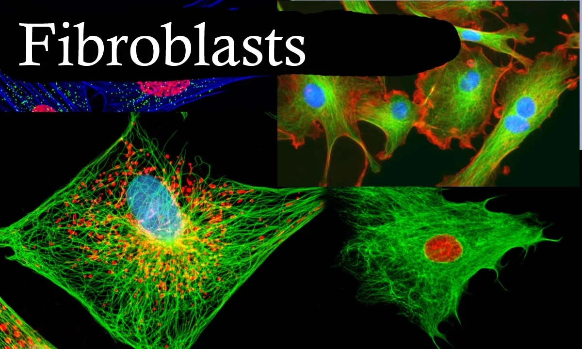
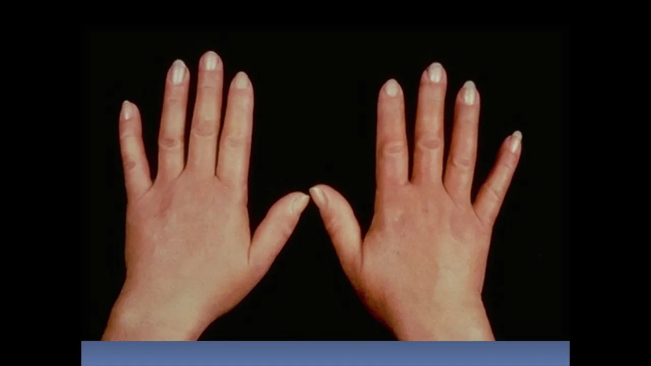
Added:What would you do if your skin on your face started to become thick, tight, and your mouth narrowed to the point where you would look like a mouse? This woman suffered from systemic sclerosis. She was a beautiful, happy woman who sadly turned into a woman with low self-esteem. This disease is affecting mostly middle-aged woman. And it all began when her CD4 T- helpper lymphosytes reacted against some antigens in her body. These lymphocytes started to accumulate and release cytoines which eventually led to vascular injury especially on their small finger vessels. The damaged endothelium produced less vasodilators like the nitric oxide and produced more of the vaso constrictors like endotailing one. You can imagine what this led to. It led to a vessel constriction which caused tissue hypoxia meaning that the oxygen supply decreased. The ma damaged endothelium also released platelet derived growth factors and transforming growth factor beta. And these two agents together with the tissue hypoxia attracted and activated fibrolast. These activated fibrolast started to produce a huge amount of collagen and extracellular proteins. What happens when you have an overp production of collagen? Fibrosis happens. And in her case this means that she got fibrosis in both the visual organs, skin, vascular system and the small vessels in her hand narrowed which led to eskeemic injury. Her symptoms all began with the swollen fingers in her hands in a symmetrical fashion meaning that both hands were affected. She noticed that her skin became firm and thick. It began in the hands and then moved up approximately to involve the upper arms and shoulders and neck and face trunk. So when her hands she noticed that her fingers became white and then blue when it was cold or when she was dressed. Then when she warmed her hands they became red instead. So this is called reos phenomenon which is a vasculitis of the small vessels in the fingers which narrows them. So how do we help her with this problem? We tell her to stop smoking since smoking narrows the blood vessels even more and makes it worse. We tell her to dress warmly, especially protecting the hands and the feet. Staying indoor during cold weather is also an option. Calcium channel blockers like nifodane can help in opening up the small blood vessels and thereby improving the circulation. Some fingers lost blood supply unfortunately which led to fingertip necrosis due to this digital eskemia. It became so severe that ulcers appeared and some fingers even got autoamputated. We have to treat her digital eskemia with some vasodilators that increased the blood flow. For example, the introvenous infusions of al prostadil a prostaglandin E1 or with epoprostanol a prostaglandin I2 also called prostyc. So her fingers also became tapered claw-like also called sclerodactyli which is a flexion of this proximal interfallenial joint. On her nails the capillary lubes dilated and some loops even got lost. This was confirmed with the microirculation microscope. She started to have calcific which is a distrophic calcification of subcutaneous tissue. So these can be found on the hands, face, trunk and the skin above the elbows and knees. The face became tight like a mask with radial furring around the lips which made her mouth opening smaller and this makes it hard for her to take care of her teeth. Furthermore, in systemic sclerosis there's connective tissue damage in the mouth which can lose the teeth. So she also noticed some telangctasia on her face which are small red spots caused by the swelling of these tiny blood vessels beneath the skin that can be eliminated by guided lasers. We can also see epidermal thinning with loss of retipex and dermal thickening because of increased amount of collagen fibers. When too much collagen fibers builds up in the skin, it crowds out the skin appendages like the sweat and sebaceous oil glands. This is what leads to the dry and stiff skin.
So what we can do here is to apply oilbased creams and lotions frequently and always write off the bathing. We should avoid the hot showers as hot water dries the skin. So we can apply sunscreen to protect from sunrays, exercise regularly like the swimming to stimulate blood circulation to these areas. So until now we know that increased collagen production can affect the skin. But what about internal organs? The digestive tract, heart, lungs, kidneys, muscles, and joints can all be affected. In the digestive tract, there can be collagen deposition in the walls. The collagen can damage the walls to the point where air enters causing pneumatosis intestinalis, which are gas cysts in the bowel wall. This can be seen radiologically. What parts of the digestive tract can be affected?
Esophagus, stomach, small and large intestine. In the in the esophagus we can see esophagal strictctrictures in in the lower two3 the collagen deposition in the wall cause absent pristtosis which leads to dysphasia that means difficulty swallowing both solid and liquids are hardly swallowed. Actually the esophagal mucosa may become thin with areas of ulceration. The lower esophagial sphincter may lose its function which is to close the connection between stomach and esophagus. It's important because otherwise the acid of the stomach may enter into the esophagus leading to gastric acid reflux causing pain named heartburn. The wall of the esophagus is not used to be in contact with this acid. So if reflux happens then the wall of the esophagus changes its structure to be able to resist this acid and this change called metaplasia. So squamous espithelium becomes columner epithelium instead and now we call the esophagus bar esophagus which predisposes to adenocarcinoma. What happens with the stomach? The same story here that the collagen deposition in the wall cause less peralysis or also called dismotility of the stomach. This leads to postrandial bloating. You know after eating a meal the sensation of feeling full with a bloated stomach. In the small intestine the same again collagen deposition leading to hypermotility and atrophy of the villi leading to anorobic bacterial overgrowth causing malabsorption of the carbohydrates, fats and proteins. Widemouth diverticola can also happen that can cause leakage of bowel contents into the paronal cavity causing peritonitis in the large intestine. Guess what happens in collagen position wide mouth diverticula and hypom motility. So how do we help her? The reflux esvagitis can be relieved by eating moist soft foods.
Chew them well and eat small frequent meals. Avoiding late night meals, spicy or fatty foods, alcohol, caffeine, that all can worsen the situation. After eat, stand up for at least an hour because gravity prevents food from entering the esophagus. Medically we can give proton pump inhibitors or it can go so far that we have to do a gastroplasty for the esophagures we can use periodic dilation. Malabsorption can be prevented by suppressing the overgrowth of intestinal flora by tetraycling or other broadspectctrum antibiotics. What happens in the heart?
The collagen deposition is the bad guy again. Here it can cause myioardial fibrrosis which is a scarring and weakening of heart leading to cardiammyopathy, paricarditis with diffusion, myioarditis which is an inflammation of the heart, conduction abnormatis like the arythmias which means that your heart beats abnormally and it can go so far that heart failure happens. So as you have realized by now collagen and fibrosis is the key words in this disease. So in the lungs there will be interstitial fibrosis and intimal hyperlasia of the small pulmonary arteries that cause narrowing of the arteries leading to pulmonary hypertension. Pulmonary alvolitis or pleuritis can also be seen which is an inflammation of the alvoli and pleora.
The patient will also complain about exertional dispnnea which means shortness of breath and it can even lead to respiratory failure and death. So it's very important the patient and doctor watching for any signs of lung disease like shortness of breath, fatigue, uh swollen feets. But it's even more important to investigate the lungs with for example high resolution chest CT scan because these tests can find problems even before the patient notice any symptoms. For pulmonary alvolitis and fibrosis we give imunosuppressants like metotres, aatoprine or cytophospamid.
For pulmonary hypertension we give epoprostanol a prostaglandin I2 also called protocycine which is a vasoddilator or we give bosentan an endothelon one receptor antagonist that blocks the vasel constriction usually caused by this endothein one. The muscoskeleletal problems can be myopathy which is a muscle weakness that can be treated with physiootherapy to help preserve the muscle strength. There can be arthralgia arthritis which is joint pain or inflammation. We can do tendon friction rub test where we put a hand on the knees for example and try to feel a leathery crepitus during motion of the leg and these friction rubs may develop over knees, wrists, fingers, ankles and we treat the pain with acetaminophen also called paracetamol which is an analesic pain reliever and antibiotic a fever reducer. So we can use non-steroidal anti-inflammatory drugs for the inflammation.
uh physical therapist and an occupational therapist can help in preventing joint motion loss and to learn performing daily tasks such as lifting objects, opening doors to put less stress on the tender joint. Flexor contractions may develop in the fingers, wrist or elbows. Corpot tunnel syndrome can happen in which the medial nerve is compressed in the wrist causing numbness in the fingers. For example, the kidney can be also affected also which is called scleroderma crisis. This is a malignant hypertension with acute renal failure. Malignant hypertension meaning that she got a sudden increase in systolic and diastolic blood pressure and this leads to hyperlastic arterial sclerosis which is an intimal hyperlasia of the small kidney arteries that leads to fibbronoid necrosis and eventually renal failure which cause death. A very important drug in this case is angotensin converting enzyme inhibitors which have made this disease to be less dangerous as it used to be. Remember to measure your blood pressure frequently to detect any sudden changes and headaches or shortness of breath because these are signs of hypertension. The main message that I want to share with you are that the skin is thick and hard and internal organs are affected uh which is very dangerous and can lead to death. But how do we know that this is systemic sclerosis? We have to make a differential diagnosis because hard skin can be caused by many things. Scarodma means hard skin and it can be localized or systemic. Localized means that it only affects the skin and related to tissues and no other internal organs are affected. Morphia and linear scleroderma are two examples. Morphia refers to local patches of scleroderma and linear scadma also called kubesare in French meaning sword stroke is a single line of thickened abnormal colored skin which can be treated with cosmetic surgery.
Systemic scleraderma or also called systemic sclerosis is affecting the whole body including the internal organs thereby the word systemic and this is what the woman have in our case. The systemic can be divided into three types. Limited cutaneous, diffused cutaneous and systemic sclerosis cena sclerodma. Cena meaning without skin thickening. The limited scroma have limited skin involvement often just the fingers face and the progression to visual organ damage is very slow meaning several decades. And there is a pneumonic for the symptoms of limited scaradma called crest syndrome which means that we can you we can see calcification reos phenomenon esophedial dismotility sclerodactyl telandas. The diffuse in the other hand have a rapid onset progression which occurs every few three to five years. It is a diffuse generous skin thickening affecting fingers, extremities, face, trunk etc. This active phase have all these visual problems that we talked about which is rapidly progressive and deadly and after this active phase it enters into stable phase where the symptoms decrease a bit but remember that thick skin can be caused by many things like diabetes mucosis fingoidis amoidosis adeliac disasema graft versus host disease perfutane tarda aosenophilic fasciitis and many more even Um chemicals can cause sclerodma like people working with polyphenol chloride or epoxy, people taking bloomin which is a cancer treatment or opioid painkiller like pentazosine. Even people eating contaminated rape seed oil or dietary supplements like elptophan. So what we need to do is some lab test to be more confident in our diagnosis. we'll find rheumatoid factors and auto antibodies like the anti-entromeir mainly in the limited cutaneous and anti-topoase one also called sc70 mainly seen in the diffuse cutaneous so to summarize we can say that this is a middle-aged woman who have systemic scerosis she have abnormally high collagen production which cause fibrosis all over her body affecting the skin gastrointestinal tract heart lungs kidney muscles joint so I ask you again what would you you if your skin on your face started to become thick, tight, and your mouth narrowed to the point where you would look like a mouse.
Up Next

Sjögren's Syndrome Symptoms: Dry Eyes, Mouth & More | Johns Hopkins
@JohnsHopkinsRheumatology
241.3K views•2018-07-17

Graphic Medicine: Comics for Collaborative Healthcare Communication
@nationalpatientadvocate
189 views•2023-12-04

Neuroanatomy: Central and Peripheral Nervous System Divisions Explained
@AKLECTURES
136.2K views•2014-09-20

Stages of Labor and Vaginal Birth | Childbirth Animation
@nucleusmedicalmedia
52.1M views•2017-08-18
Related Study Plans & Knowledge Roadmaps
Structured learning paths in Medicine