The NIH Stroke Scale limb ataxia assessment evaluates coordination by having patients perform finger-to-nose tests with each arm and heel-to-shin tests with each leg; a score of 0 indicates no ataxia, 1 indicates ataxia in one limb, 2 indicates ataxia in two limbs, and 9 indicates untestable due to amputation or joint fusion.
NIH Stroke Scale: Limb Ataxia Scoring Explained | Neurology Education
Added:Deep Dive
Basic neuroanatomy of the cerebellum and its role in coordinating voluntary muscle movements.

Anatomy of Cerebellum | Structure & Function | Neuroanatomy
648.4K
The cerebellum, located posteriorly on the brainstem, is responsible for coordinating movements, maintaining posture and balance, and regulating muscle tone. It consists of three main functional divisions: the vestibulocerebellum (flocculonodular lobe) concerned with balance and eye-head coordination, the spinocerebellum (vermis and paravermal areas) controlling axial musculature and limb movements, and the cerebrocerebellum (lateral hemispheres) involved in voluntary movement coordination. The cerebellum receives input through climbing fibers (from inferior olive) and mossy fibers (from spinal cord, vestibular system, and cerebral cortex), processes information through Purkinje cells, and sends output to deep cerebellar nuclei (fastigial, globose, emboliform, and dentate nuclei) that project to brainstem nuclei and thalamus to modify motor commands.
Click to watch →
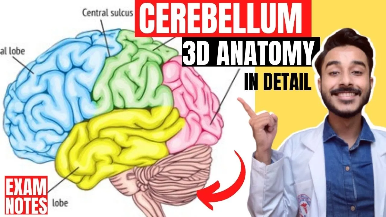
external features of cerebellum anatomy 3d | morphological and functional division of cerebellum
415.4K
The cerebellum, located in the posterior cranial fossa behind the brainstem, is divided into three functional regions based on its evolutionary development: the vestibulocerebellum (flocculonodular lobe) maintains balance and muscle tone, the paleocerebellum (anterior lobe) regulates muscle tone and simple movements, and the neocerebellum (posterior lobe) coordinates skilled voluntary movements; anatomically, it is divided by the primary fissure into anterior and posterior lobes, and by the posterior lateral fissure into the flocculonodular lobe, with the horizontal fissure separating superior from inferior surfaces.
Click to watch →

Plus One Biology | Neural Control & Coordination/Breathing And Exchange Of Gases /Excretory Products
115.5K
The cerebellum is the second largest part of the brain and is located at the back of the brainstem. It is responsible for coordination, balance, and motor control. The cerebellum receives information from the sensory systems and motor cortex and helps coordinate voluntary movements. It also plays a role in cognitive functions and learning.
Click to watch →
![[Neuroanatomia] 5 - Cerebelo](https://i.ytimg.com/vi/0uKjGIByklE/hqdefault.jpg)
[Neuroanatomia] 5 - Cerebelo
107.5K
The cerebellum is a brain structure located dorsally to the brainstem, divided into lobes (vermis, hemispheres, flocculonodular lobe) and zones (vestibulocerebellum, spinocerebellum, cerebrocerebellum) that coordinate motor functions through connections with vestibular nuclei, spinal cord, and cerebral cortex; it maintains balance and posture, controls muscle tone, and participates in motor planning and correction through its four central nuclei (dentate, emboliform, globose, fastigial) and characteristic three-layered cortex containing Purkinje cells.
Click to watch →
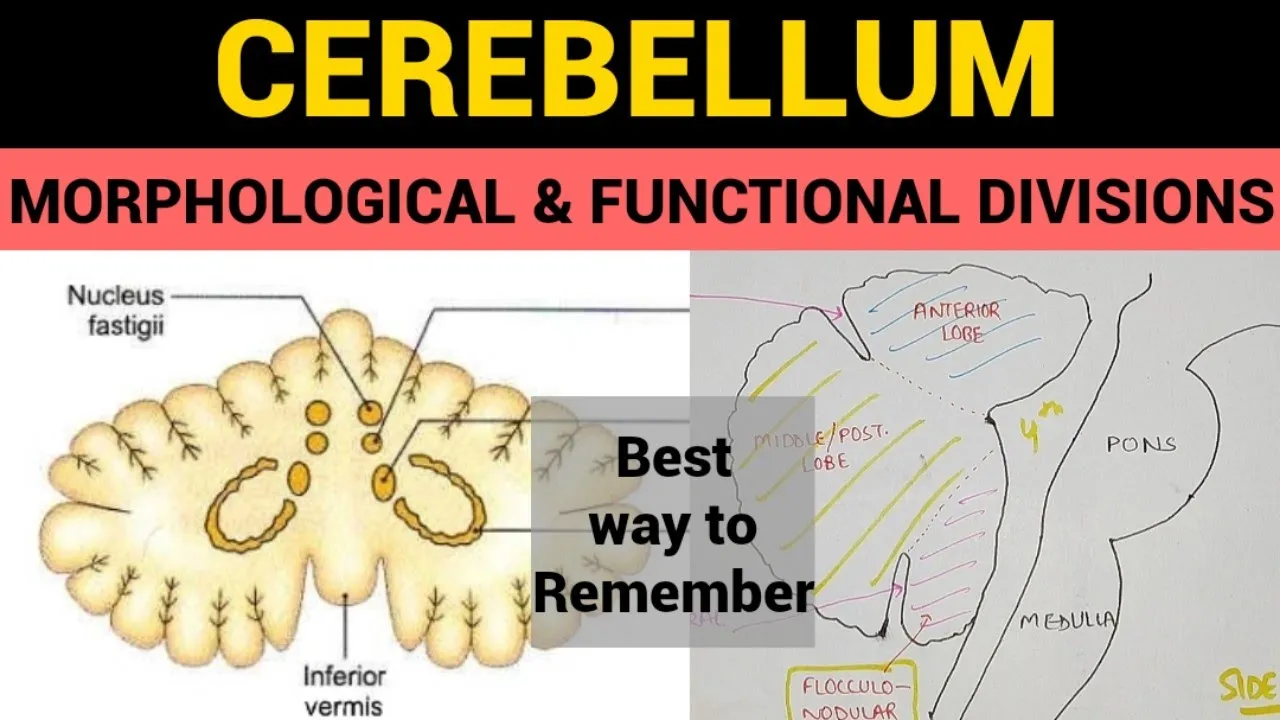
Cerebellum - 2 | Morphological & Functional Divisions
189.4K
The cerebellum is divided into three main morphological zones—the anterior lobe (vestibulocerebellum), intermediate zone, and posterior lobe (spinocerebellum)—each controlling specific motor functions: the anterior lobe plans and programs movements, the intermediate zone controls trunk and proximal limb movements, and the posterior lobe controls distal limb movements; additionally, the cerebellum is divided into three functional zones—the lateral zone (controlling limb movements), intermediate zone (controlling trunk movements), and anterior zone (controlling movement planning)—which work together to maintain equilibrium, coordinate voluntary movements, and ensure smooth muscle activity.
Click to watch →
Deep Dive
An overview of the National Institutes of Health Stroke Scale (NIHSS), including its overall purpose, structure, and clinical utility.

1 NIHSS Concisely
286
The NIH Stroke Scale (NIHSS) is the standard bedside neurological examination used across the United States to determine the severity of acute stroke, with a total score ranging from 0 to 40 where higher scores indicate more proximal occlusions and larger strokes; the scale assesses 11 items including level of consciousness, command following, gaze, visual fields, facial weakness, extremity strength, ataxia, sensory, language, dysarthria, and extinction, with scores of 4 or less indicating minor stroke, scores of 5-25 in the 3-4.5 hour window qualifying for intravenous alteplase, and scores of 6 or greater or severe aphasia/hemiparesis indicating consideration for endovascular therapy, though the scale has limitations in assessing posterior circulation strokes and may be confounded by stroke mimics such as seizures, migraines, or conversion disorders.
Click to watch →
NIH stroke scale assessment overview OT510
284
The NIH Stroke Scale is a standardized neurological assessment tool used to evaluate stroke severity across 11 domains including consciousness, gaze, facial palsy, motor function, ataxia, sensation, language, dysarthria, and extinction/inattention, using a 0-5 scoring system where 0 indicates no impairment and 5 indicates inability to perform tasks; it is designed for patients aged 18-64, takes approximately 6 minutes to administer, and demonstrates excellent reliability (test-retest 0.93, inter-rater 0.95) and validity (0.90), making it valuable for predicting functional outcomes and planning rehabilitation interventions.
Click to watch →

LÂM SÀNG LÃO ĐỢT 17 - NHÓM NỘI THẦN KINH - THANG ĐIỂM NIHSS
239
The NIHSS (National Institutes of Health Stroke Scale) is a standardized 11-item assessment tool with 13 sub-items evaluating neurological function in stroke patients. Scores range from 0-42, with higher scores indicating greater severity. NIHSS serves multiple clinical purposes: assessing neurological status, facilitating provider communication, rapidly evaluating stroke severity, and tracking disease progression. It does not support stroke diagnosis as it cannot determine lesion location. NIHSS thresholds guide treatment decisions, with scores above 22 points serving as relative contraindications for thrombolysis. Specific criteria identify patients at high risk of severe disability, including complete motor deficit, severe aphasia, or complete sensory loss. NIHSS assessments follow standardized timing: initial contact, 2 hours post-onset, 24 hours post-onset, and 7-10 days post-onset. The primary purpose is evaluating treatment efficacy by comparing pre- and post-treatment scores. NIHSS scores can assess recurrence risk, with higher scores potentially indicating greater risk of future stroke events.
Click to watch →

National Institutes of Health (NIH) Stroke Scale Demonstration
91.5K
The NIH Stroke Scale is a 15-item standardized assessment tool used to quantify neurological deficits in stroke patients, ranging from 0 (no deficits) to 42 (severe deficits), with key components including level of consciousness assessment, lateral gaze testing, visual field evaluation, facial weakness examination, arm and leg strength testing, limb coordination assessment, sensation testing, language comprehension evaluation, and assessment for unilateral neglect; the scale helps identify patients eligible for emergent treatment, allows objective measurement of clinical status changes, and identifies those at higher risk for complications.
Click to watch →
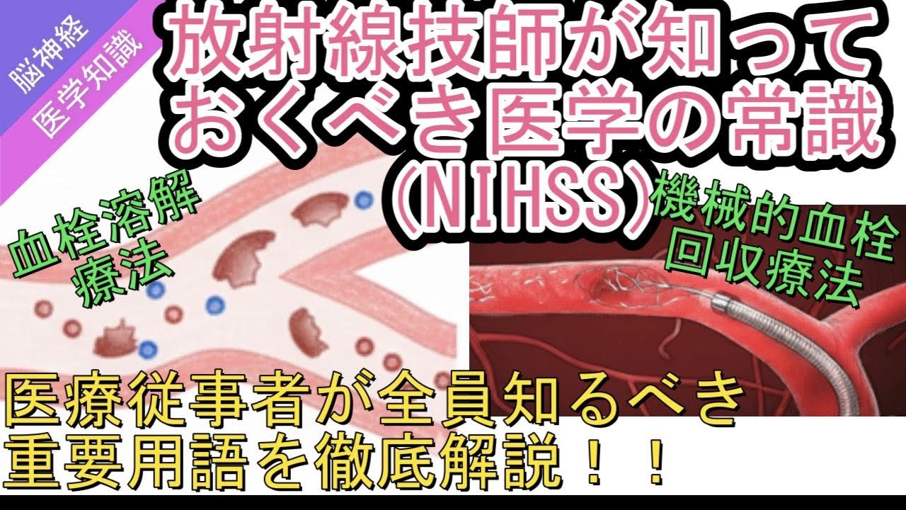
放射線技師が知っておくべき医学の常識(NIHSS)
479
NIHSS (National Institutes of Health Stroke Scale) is a standardized neurological assessment scale developed in the late 1980s for quantifying stroke severity. It evaluates 11 neurological functions across 15 domains, scoring 0-4 points per item with total scores from 0 (no abnormalities) to 42 (most severe). NIHSS prioritizes inter-rater reliability over diagnostic specificity, making it ideal for large-scale clinical trials. Severity classification: 0 (no symptoms), 1-4 (mild), 5-15 (moderate), 16-20 (moderate-severe), 21-42 (severe). Clinical thresholds: ≤5 points predicts 80% home return, 6-13 points requires rehabilitation care, ≥14 points often needs long-term care facility. NIHSS guides treatment decisions: tPA therapy applies for scores 6-25, mechanical thrombectomy requires NIHSS ≥6 for anterior circulation and ≥10 for posterior circulation. NIHSS has limitations including floor effect, posterior circulation bias, and left hemisphere bias. sNIHSS provides rapid assessment for time-constrained situations.
Click to watch →
Deep Dive
The clinical definition of ataxia, particularly distinguishing true coordination deficits from profound motor weakness or paresis.

Ataxia How to differentiate motor & sensory ataxia
600
Ataxia is defined as loss of coordination, clinically presenting with falls or swaying while walking. Coordination is assessed through specific tests: upper limb tests include finger-nose test (modified supination/pronation) and drawing circles in air; lower limb tests include knee-heel test, Romberg sign, tandem walking, and toe circles. The two main types of ataxia are sensory (proprioceptive pathway dysfunction) and motor (cerebellar dysfunction). The finger-nose test differentiates these: inability with eyes open indicates motor ataxia, while inability only with eyes closed indicates sensory ataxia.
Click to watch →
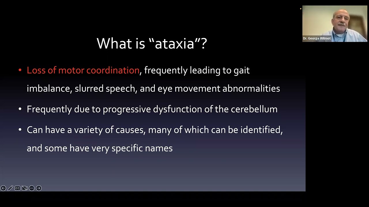
All About Unknown Ataxia Without Family History
5.7K
Ataxia is defined as loss of motor coordination resulting from dysfunction of the cerebellum, which coordinates voluntary movement and balance. Key clinical features include increased path length during reaching movements, abnormal rapid alternating movements with movement delay, and characteristic eye movement abnormalities. Eye movement assessment is particularly important because cerebellar connections to the vestibular and eye movement systems produce distinctive patterns that help confirm cerebellar origin versus peripheral causes like neuropathy or knee problems.
Click to watch →

Ataxies (Partie 1) : Généralités
8.2K
Ataxia is a neurological disorder characterized by impaired balance and coordination, distinct from motor deficits, motor schema perturbations, or abnormal movements like tremors. Clinically, ataxia manifests in three forms: static ataxia (impaired standing balance with widened base of support), dynamic ataxia (impaired walking with elevated base of support), and kinetic ataxia (impaired voluntary movements with hypermetria and terminal tremors). The condition is classified into four syndromes based on lesion location: vestibular ataxia (lesions of the vestibulocochlear system), sensory ataxia (lesions of the sensory system including parietal lobe, thalamus, brainstem, spinal cord, or peripheral nerves), frontal ataxia (lesions of the frontal lobe or its connections), and cerebellar ataxia (lesions of the cerebellum or its connections).
Click to watch →
🔴LIVE PALESTRA INAUGURAL || ORTOPÉDICO X NEUROLÓGICO: COMO DIFERENCIAR? COM DR. HUGO CANELAS
100
Ataxia is uncoordinated movement with three types: proprioceptive (limb crossing during walking), vestibular (circular walking with head tilt), and cerebellar (dysmetria, hypermetria, tremor). Paresis is loss of ability to support weight (weakness), while plegia is loss of voluntary movement (paralysis). Paresis types include paraparesis (hind limbs), tetraparesis (all four limbs), and hemiparesis (one side). Plegia types include paraplegia (hind limbs), tetraplegia (all four limbs), and hemiplegia (one side). Ambulatory paresis allows walking but not weight support, while non-ambulatory paresis shows movement but cannot stand.
Click to watch →
Enregistrement syndrome cérébelleux
851
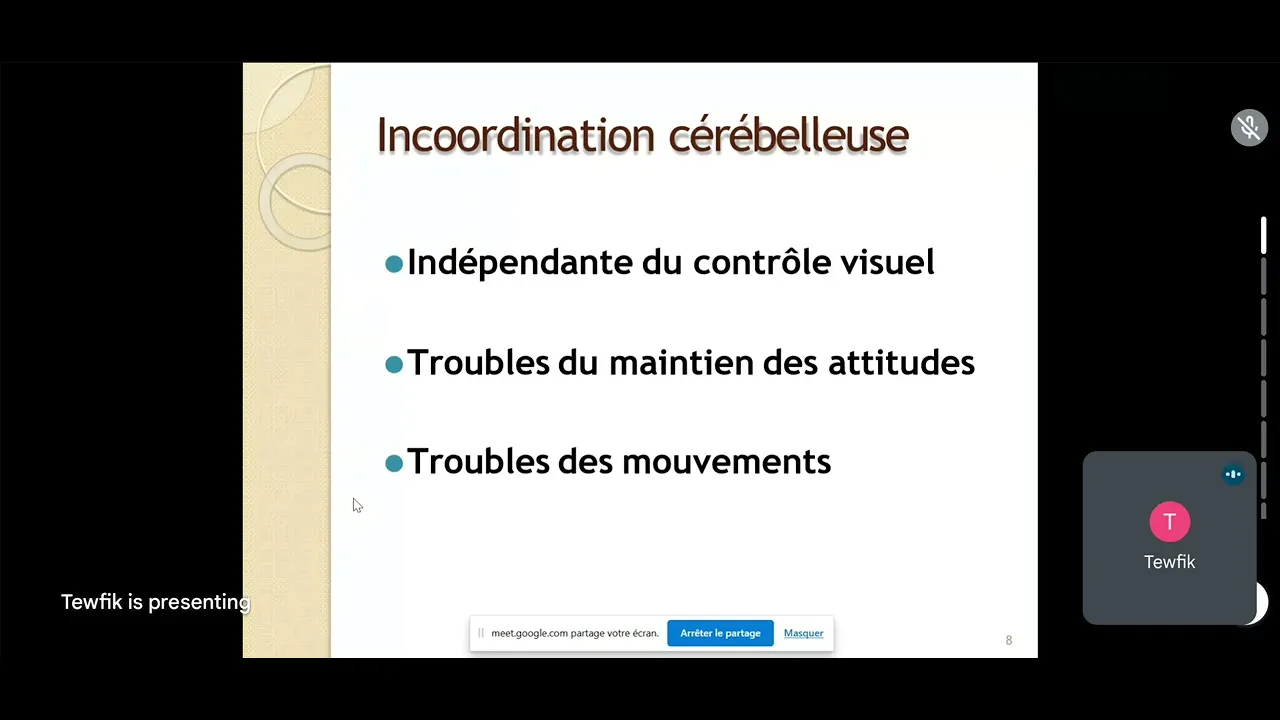
Ataxia is defined as a coordination disorder without motor deficit. Cerebellar ataxia presents with: widened base of support, irregular festooned gait, and difficulty maintaining balance. The cerebellum maintains equilibrium and coordination, so its dysfunction leads to these characteristic gait abnormalities. Different types of ataxia can be distinguished by eye closure: cerebellar ataxia remains unchanged, while vestibular and proprioceptive ataxia worsen with eye closure.
Click to watch →
Deep Dive
Standard neurological examination maneuvers used to assess coordination, such as the finger-to-nose and heel-to-shin tests.
The Neurologic Physical Examination
558.2K
Coordination testing includes finger-to-nose (upper extremity) and heel-to-shin (lower extremity). For finger-to-nose, hold your hand far enough that the patient must fully extend to reach, looking for side-to-side weaving and difficulty hitting the stationary target in cerebellar ataxia. For heel-to-shin, slide the heel up and down the tibia, watching for side-to-side swaying on the shin. Gait assessment includes normal walking and tandem (heel-to-toe) walking. The Romberg test (standing with eyes closed) tests balance by removing visual input, revealing problems with vestibular or proprioceptive systems. A positive Romberg indicates deficit in one of these systems.
Click to watch →
![[ASMR] Real Person Head to Toe Physical Exam Assessment | Soft Spoken Medical Roleplay](https://i.ytimg.com/vi/JbA_DWhjmzM/maxresdefault.jpg)
[ASMR] Real Person Head to Toe Physical Exam Assessment | Soft Spoken Medical Roleplay
1.1M
Neurological examination includes Romberg test (balance assessment with eyes open and closed), finger-to-nose and heel-to-shin coordination testing, vibration sensation testing with tuning fork, and Babinski reflex elicitation (normal response is plantar flexion). These tests assess cerebellar function, proprioception, and upper motor neuron integrity.
Click to watch →

UTEP Advanced Health Assessment -Lomack
663.4K
Finger-to-nose testing assesses cerebellar function by having the patient touch their nose with each finger. Heel-to-shin testing involves sliding the heel down the opposite shin. These tests assess coordination and cerebellar function.
Click to watch →
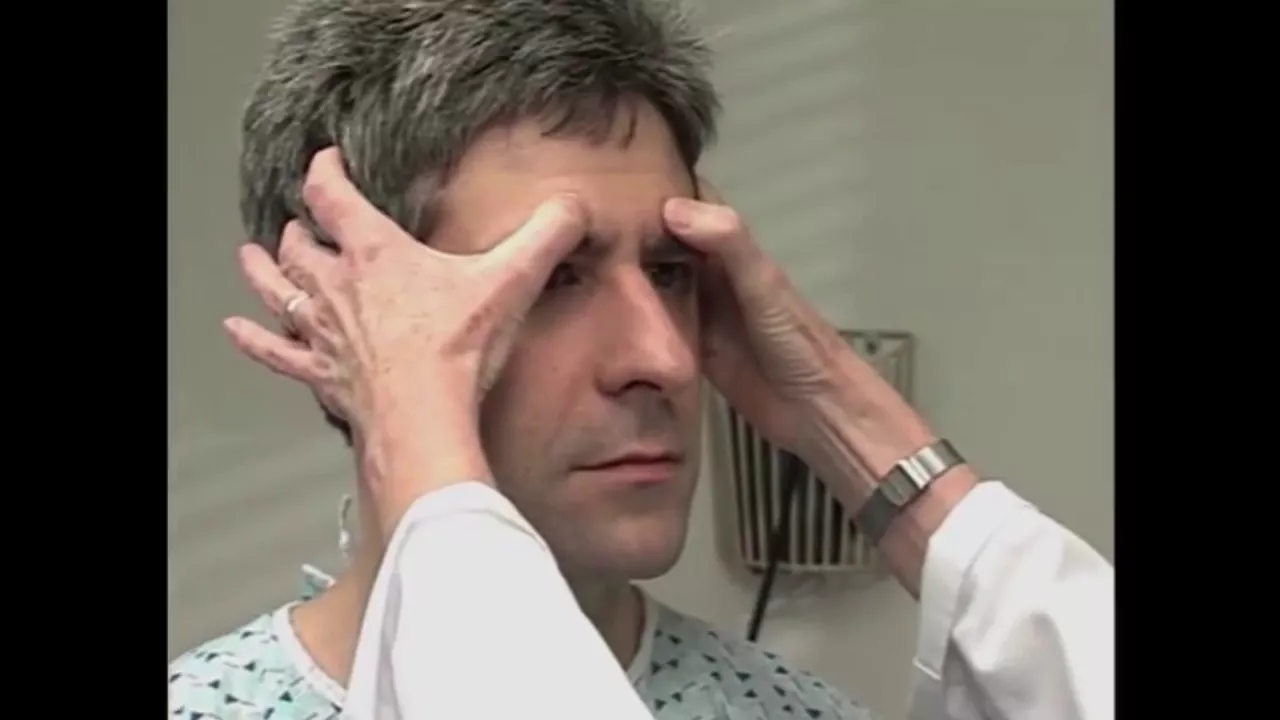
Unintentional ASMR: This checkup is my favourite!
4.2M
Coordination is tested using the finger-to-nose test (touching the physician's finger then the nose repeatedly) and the heel-to-shin test (running the heel down the shin). These tests assess cerebellar function and the integrity of the motor pathways.
Click to watch →

Neurological Assessment
557.7K
Coordination testing includes finger-to-nose test (patient touches each finger as quickly as possible, then touches nose and then examiner's finger), heel-to-shin test (patient runs heel down shin), and Romberg test (patient stands with feet together, arms at sides, eyes closed for 20 seconds while examiner counts). During finger-to-nose testing, the examiner must not move their finger while the patient is reaching toward it, as this would invalidate the test.
Click to watch →
Prerequisite Knowledge
- Concept 01Basic neuroanatomy of the cerebellum and its role in coordinating voluntary muscle movements.
- Concept 02An overview of the National Institutes of Health Stroke Scale (NIHSS), including its overall purpose, structure, and clinical utility.
- Concept 03The clinical definition of ataxia, particularly distinguishing true coordination deficits from profound motor weakness or paresis.
- Concept 04Standard neurological examination maneuvers used to assess coordination, such as the finger-to-nose and heel-to-shin tests.
Subsequent Learning
- Step 01How to score other critical subscales of the NIHSS, such as motor drift, sensory loss, and language deficits, to calculate a complete stroke score.
- Step 02Clinical decision-making in acute stroke management, specifically how NIHSS scores influence eligibility for thrombolytic therapy (tPA) or mechanical thrombectomy.
- Step 03Stroke localization principles, correlating limb ataxia with posterior circulation or vertebrobasilar arterial territory infarcts.
- Step 04Differential diagnosis of acute ataxia, distinguishing acute ischemic stroke from stroke mimics like vestibular neuritis, acute toxic ingestion, or demyelinating diseases.
Zero & One
Two & Nine
Zero & One
0:06Playing Section
- 1
Demonstrates absent ataxia scoring as zero.
- 2
Shows one-limb ataxia scoring as one.
- 3
Covers coma or untestable cases as zero.
Limitations and Confounding Factors in NIHSS Limb Ataxia Scoring
While the NIH Stroke Scale (NIHSS) provides a standardized framework for stroke assessment, its limb ataxia subtest is frequently criticized for low inter-rater reliability and diagnostic confounding. A major limitation is the difficulty in distinguishing true cerebellar ataxia from motor weakness; severe weakness can mimic ataxia (pseudoataxia) or prevent the test from being performed accurately, leading to potential misscoring. Furthermore, because the NIHSS is heavily weighted toward anterior circulation strokes, the crude 0-2 scale for ataxia often underrepresents the severity of posterior circulation strokes. Critics suggest that supplementary assessments, such as the Scale for the Assessment and Rating of Ataxia (SARA), provide a more precise evaluation of coordination deficits.
Deep Dive
How to score other critical subscales of the NIHSS, such as motor drift, sensory loss, and language deficits, to calculate a complete stroke score.
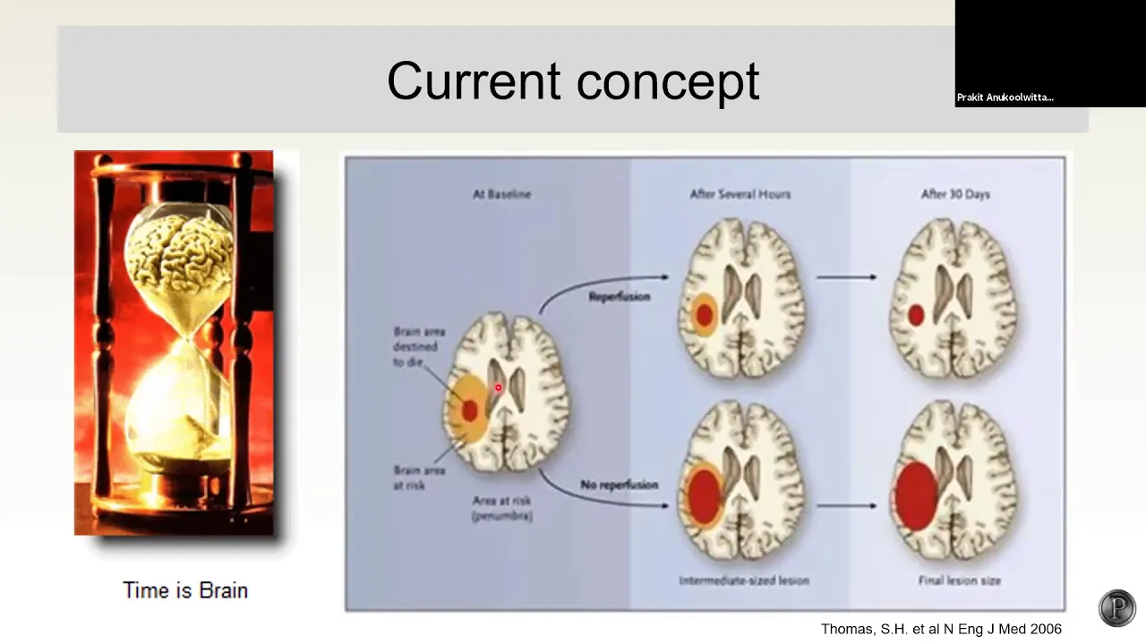
ประเมิน NIHSS ใน acute stroke
6.6K
The NIHSS (National Institutes of Health Stroke Scale) is the standard tool for stroke severity assessment, scoring 0-42. Level of consciousness uses 0-3 scale: 0 (alert), 1 (verbal response), 2 (physical response), 3 (unarousable). Orientation tests age and month knowledge (0-2 points). Command following evaluates eye-opening and hand-squeezing (0-2 points). Visual field and acuity assess tracking ability (0-2 points each). Facial motor evaluates smiling (0-3 points). Motor assessment tests arm (90 degrees, 10 seconds) and leg (30 degrees, 15 seconds) strength (0-3 points). Ataxia uses finger-to-nose and heel-to-shin testing (0-3 points). Sensory assessment uses object identification by touch (0-3 points). Language evaluation tests naming and picture description (0-3 points). Auditory verbal learning tests word repetition (0-3 points). The total score guides treatment decisions and monitors progress.
Click to watch →

NIH Stroke Scale Training - Part 3 - Demo Patient A
215.9K
This video demonstrates the complete administration of the NIH Stroke Scale (NIHSS) on a 71-year-old patient, showing how each of the 11 items is scored: orientation (0), best gaze (0), visual fields (0), facial palsy (1 for decreased right nasolabial fold), motor arm (0), motor leg (1 each for bilateral leg drift), limb ataxia (0), sensory (0), language (0), dysarthria (1 for slurred speech), and extinction/inattention (0). The patient's total NIHSS score is 4, indicating mild stroke severity.
Click to watch →
Introduction To Stroke
166.8K
The NIH Stroke Scale is a standardized neurological exam consisting of 11 domains: level of consciousness, orientation, command following, gaze, visual fields, facial palsy, motor strength of extremities, limb ataxia, sensory loss, language (aphasia), articulation, extinction/intention (neglect). Higher scores indicate larger strokes; a score of 0 suggests normal or near-normal function while 42 indicates very severe stroke. It allows rapid assessment and treatment of stroke patients.
Click to watch →

Acute Stroke - Diagnosis and Management | KGMOA Webinar
241
The NIHSS is a standardized tool for assessing stroke severity. Key components include consciousness level assessment (2 points for correct answers to two questions), gaze deviation (0 points for no deviation), visual hemianopia (3 points for bilateral, 2 points for unilateral), facial palsy (3 points for complete paralysis), motor arm weakness (1 point for mild drift, 3 points for no movement against gravity), motor leg weakness, limb ataxia, sensory deficit, aphasia (3 points for global aphasia), dysarthria, and attention. Total score ranges from 0-42; scores >4 indicate major stroke, and scores of 8-9 suggest large vessel occlusion requiring urgent intervention.
Click to watch →
GSA - NIH e Rankin
5.6K
The NIHSS comprehensively assesses motor, visual, and communication function through standardized tests. Motor command assessment (score 0-2) evaluates ability to close eyes and make fist. Visual pursuit (score 0-2) tests conjugate eye movement to extremes. Visual field assessment (score 0-3) uses confrontation testing for quadrant defects or hemianopsia. Facial motor assessment (score 0-3) distinguishes central (lower face only) from peripheral (entire face) paralysis. Motor function assessment (score 0-3) tests ability to hold arm at 90° for 10 seconds and leg at 45° for 5 seconds. Ataxia assessment (score 0-2) uses finger-to-nose and heel-to-shin tests, scored only when disproportionate to paresis. Sensory assessment (score 0-2) uses thermal and pain stimuli. Language assessment (score 0-2) uses five pre-defined phrases. Dysarthria assessment (score 0-3) uses five words. Extinction assessment (score 0-2) tests for neglect using simultaneous bilateral stimulation. Each assessment requires specific techniques and interpretation guidelines.
Click to watch →
Deep Dive
Clinical decision-making in acute stroke management, specifically how NIHSS scores influence eligibility for thrombolytic therapy (tPA) or mechanical thrombectomy.
放射線技師が知っておくべき医学の常識(NIHSS)
479
NIHSS scores guide critical stroke treatment decisions. For tPA thrombolytic therapy, scores of 5 or below indicate treatment benefits may not outweigh bleeding risks, while scores of 22+ suggest completed infarction with hemorrhage risk. Most facilities apply tPA for NIHSS 6-25 points. For mechanical thrombectomy, anterior circulation occlusion requires NIHSS ≥6 with ASPECTS ≥6, while posterior circulation requires NIHSS ≥10 with ASPECTS ≥6. NIHSS has limitations: floor effect (score 0 may miss significant MRI abnormalities), posterior circulation bias (underestimates vertigo/ataxia), and left hemisphere bias (language items favor left hemisphere strokes). These limitations should be considered in clinical decision-making.
Click to watch →

Manejo do AVC Isquêmico - Episódio 63
6.4K
The National Institutes of Health Stroke Scale (NIHSS) is a validated clinical tool scoring from 0 to 42 points to quantify stroke severity. Scores of 5 or higher typically qualify for thrombolysis, though lower scores may be acceptable if deficits are functionally incapacitating. The scale is reliable and reproducible with proper training. Thrombolysis eligibility requires age over 18, symptom onset within 4.5 hours, and NIHSS of 5 or higher. The risk-benefit trade-off involves potential neurological recovery versus bleeding risk. Each minute of delay reduces potential benefit, creating urgency in decision-making. Consent requirements are minimal: verbal consent suffices for capable patients, and treatment can proceed without family authorization for incapacitated patients.
Click to watch →
Conduite à tenir devant un AVC - Pr.I.KEDIHA
43.7K
Brain imaging is essential: non-contrast CT differentiates hemorrhagic (hyperdensity) from ischemic (hypodensity) stroke. Early ischemic CT signs include loss of gray-white matter differentiation, loss of insular ribbon, loss of lentiform nucleus density, and effacement of sulci. The NIH Stroke Scale (NISS) scores 0-42 points across 11 items evaluating consciousness, motor function, sensory function, visual function, language, and cognition. Scores of 1-4 indicate minor stroke, 5-15 indicate moderate stroke, and scores above 15 indicate severe stroke. Continuous monitoring includes vital signs, blood glucose, temperature, and neurological status. Blood pressure should be maintained above 180/105 mmHg during thrombolytic therapy and above 220/120 mmHg in non-thrombolyzed patients. Intravenous thrombolysis with rtPA is the standard acute stroke treatment, with standard dose 0.9 mg/kg (maximum 90 mg), 10% as initial bolus followed by 90% over 1 hour. Treatment must be initiated within 4 hours and 30 minutes of symptom onset. Age over 80 is no longer a formal contraindication.
Click to watch →

WeUrgences Webinar_AVC Ischémique aux urgences
4K
Basic monitoring includes temperature (fever worsens outcomes), oxygen saturation (hypoxia worsens outcomes), blood pressure (hypotension worsens outcomes), and blood glucose (both hypo- and hyperglycemia worsen outcomes). Stroke mimics include hypoglycemia, postictal states, and migraine aura. Thrombolytic treatment (tPA) is first-line for eligible patients: onset within 4.5 hours, NIHSS score 4-25, no contraindications. Eligibility rate is 7-12%. ASPECTS score must be >7 for favorable outcomes. NIHSS provides objective severity assessment (0-42): 1-4 minor, 5-15 moderate, 15-20 severe, >20 very severe. Each point increase correlates with 17% worse prognosis. Rankin Scale assesses functional outcomes at 3 months.
Click to watch →

Epidemiologia, anatomia vascular, síndromes vasculares e escalas de avaliação em AVC
781
NIHSS score thresholds for clinical decision-making: scores of 3-4 may indicate 'minor stroke' or 'stroke minor' (non-disabling). Scores of 10 or higher may indicate large vessel occlusion requiring thrombectomy consideration. Scores of 15 or higher indicate severe stroke. The NIHSS was originally derived from correlation with infarct volume measured 7-10 days after stroke, but has significant limitations for individual patient assessment. It is most useful for clinical trials and research, not for individual patient management.
Click to watch →
Deep Dive
Stroke localization principles, correlating limb ataxia with posterior circulation or vertebrobasilar arterial territory infarcts.

Stroke Localization
44.2K
Stroke localization involves correlating clinical signs with neuroanatomy and vascular supply to identify the site of infarction; key principles include understanding that lacunar syndromes arise from subcortical perforator occlusions (pure motor, pure sensory, sensorimotor, dysarthria-clumsy hand, ataxic hemiparesis, hemiballismus), cortical syndromes involve specific motor homunculus distributions (MCA supplying face/upper limb, ACA supplying lower limb/trunk), and brainstem syndromes produce characteristic cross-signs due to decussation of pyramidal and spinothalamic tracts, with posterior circulation syndromes potentially causing bilateral or generalized deficits requiring urgent recognition for improved outcomes.
Click to watch →

100% Medicine Part-2 for FMGE Aspirants by Dr. Dilip Kumar
19.5K
Stroke localization is essential for diagnosis and treatment. Anterior circulation strokes (MCA, ACA) cause hemiparesis with arm/face predominance (MCA) or leg predominance (ACA), visual field defects, and cortical signs (aphasia, apraxia). Posterior circulation strokes (vertebral, basilar, PCA) cause vertigo, ataxia, cranial nerve palsies, and consciousness disturbances. Weber syndrome (PCA) shows ipsilateral CN III palsy with contralateral hemiparesis. Lateral medullary syndrome (vertebral) causes ipsilateral Horner's syndrome, ataxia, and crossed sensory loss. Stroke treatment follows a systematic algorithm: non-contrast CT to rule out hemorrhage, thrombolysis with tPA within 4.5 hours, CT angiogram for large vessel occlusion, and mechanical thrombectomy if indicated.
Click to watch →
ACV: Fisiopatología y Presentación Clínica 📚
249K
The brain receives dual blood supply: anterior circulation (carotid system: ACA, MCA) and posterior circulation (vertebrobasilar system: PCA). ACA irrigates medial brain including motor/sensory lower limb representations and supplementary motor area. MCA irrigates cerebral convexity including primary motor cortex, somatosensory cortex, Broca's/Wernicke's areas, and eye movement control. PCA irrigates occipital/temporal lobes including primary visual cortex. Syndromes are classified by vessel size (large vs. small/lacunar) and circulatory system (anterior/posterior). This anatomical-functional correlation allows clinicians to localize lesions and predict clinical manifestations based on vascular territory involvement.
Click to watch →
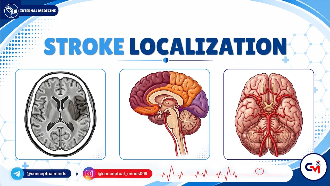
2. Stroke Localization | Neurology Internal Medicine | USMLE Step 2 CK Clinical Neuro Review
8.4K
Stroke localization requires understanding the circle of Willis anatomy, where the vertebral and basilar arteries supply posterior circulation (brainstem, cerebellum, thalamus, and medial temporal lobes), while the internal carotid artery supplies anterior circulation (ACA and MCA territories); ACA strokes cause contralateral cerebral monoparesis, urinary incontinence, and apathy-abulia, whereas MCA strokes produce contralateral hemiplegia, sensory loss, and language disturbances depending on dominant/non-dominant hemisphere involvement, with PCA strokes causing homonymous hemianopia with macular sparing, memory disturbances, and visual agnosia syndromes.
Click to watch →

Neurology Step 2-3 — Stroke II — Boards and Beyond
20.3K
Stroke localization involves distinguishing between anterior circulation strokes (affecting the cerebral cortex via middle and anterior cerebral arteries) and posterior circulation strokes (affecting brainstem, cerebellum, and occipital lobe via vertebral and basilar arteries), with cortical strokes showing preferential motor involvement (face > arm > leg) due to the homunculus organization, while subcortical strokes show equal involvement of all limbs; lacunar strokes are small subcortical infarcts associated with hypertension, presenting as pure motor stroke (most common), pure sensory stroke, ataxic hemiparesis, or dysarthria-clumsy hand syndrome, and require MRI for detection since CT may be negative.
Click to watch →
Deep Dive
Differential diagnosis of acute ataxia, distinguishing acute ischemic stroke from stroke mimics like vestibular neuritis, acute toxic ingestion, or demyelinating diseases.

ICU 'uncut' academic discussions #4: How to manage acute ischemic stroke in ICU & ED; Dr Sushant
36.2K
Stroke is defined clinically as an abrupt onset neurological deficit attributable to a focal vascular cause. It comprises two main types: ischemic stroke (85% of all strokes) from clot obstruction, and hemorrhagic stroke (15%) from bleeding. Critical to accurate diagnosis is recognizing stroke mimics that must be differentiated: hypoglycemia is the most common mimic and should be first investigated; seizures can present with residual weakness; migraines (hemiplegic type) can cause hemiparesis; venous sinus thrombosis and hypertensive encephalopathy also present similarly. The fundamental principle is that any acute focal neurological deficit should be considered stroke until proven otherwise, requiring systematic evaluation to avoid missing treatable conditions.
Click to watch →
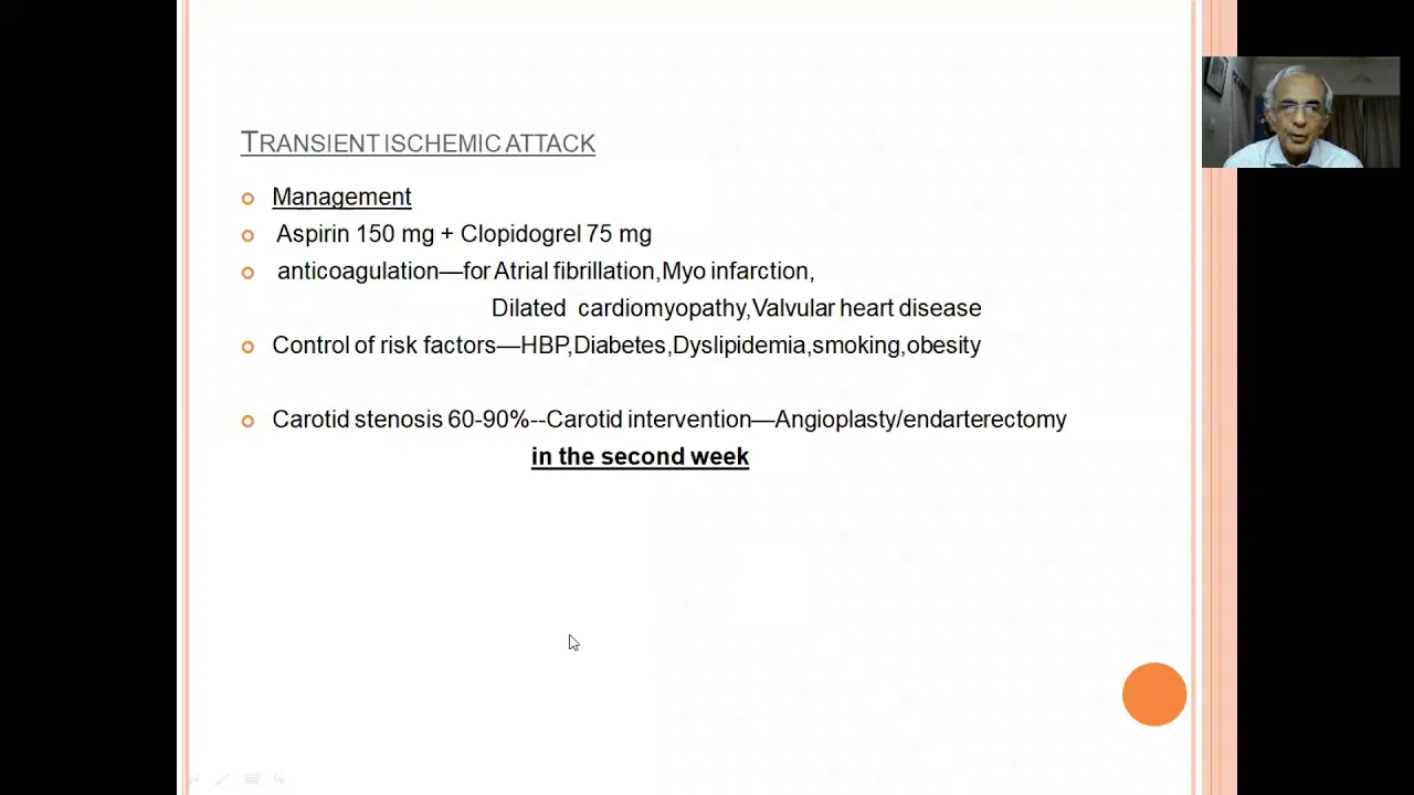
Ischemic stroke--Diagnosis and management
4.6K
Stroke is fundamentally a clinical diagnosis based on history and examination, not imaging. Key stroke mimics include hypoglycemia (resolves with glucose), postictal paralysis (following seizures), and hemiplegic migraine. Subdural hematoma from minor trauma can also mimic stroke and is eminently treatable surgically. Recognition of these mimics prevents unnecessary investigations and inappropriate treatment while ensuring appropriate management of true stroke cases.
Click to watch →
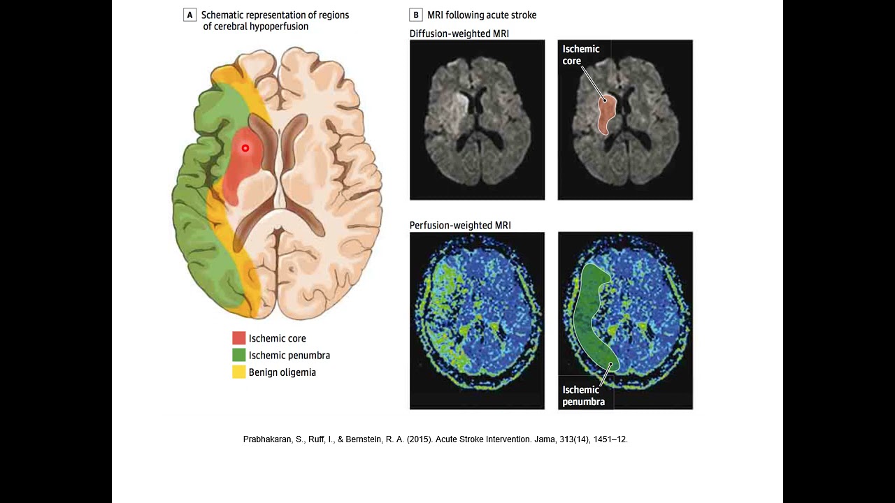
Enfermedad Cerebrovascular
262
Several conditions can mimic stroke symptoms: metabolic disorders (hypoglycemia, hyponatremia, vitamin deficiencies) typically cause altered consciousness without focal deficits. Non-vascular focal deficits include focal seizures (Todd's paralysis), migraine aura, brain tumors, abscesses, and demyelinating lesions. These conditions lack the characteristic vascular distribution pattern of true stroke. The absence of vascular distribution is a key differentiating feature. Approximately 20% of patients with acute focal deficits may have stroke mimics rather than true stroke.
Click to watch →

Acil Serviste İskemik İnme Yönetimi – Part 2 | Tanısal testler ve Ayırıcı Tanı
651
Stroke mimics include hypoglycemia (can present with all neurological symptoms), space-occupying lesions, CNS infections, metabolic disorders (especially sodium abnormalities), and drug intoxication. Hypertensive encephalopathy (BP >200/130 mmHg) presents with headache, nausea, vomiting, and confusion. Wernicke encephalopathy results from chronic thiamine deficiency, commonly in alcoholics or malnourished patients. All these conditions must be considered in the differential diagnosis of stroke.
Click to watch →
ACCIDENTE CEREBROVASCULAR (ACV)
21.4K
Stroke mimics include meningiomas, subdural hematomas, hypoglycemia, syncope, seizures, and hypertensive encephalopathy. Hypoglycemia must always be ruled out before thrombolysis. Diagnostic workup includes glucose, ECG, toxicology, and cultures. CT scan is essential to rule out hemorrhage. MRI helps confirm ischemic stroke. The goal is to distinguish true stroke from mimics to guide appropriate treatment.
Click to watch →
Zero & One
Two & Nine
Zero & One
0:06- 1
Demonstrates absent ataxia scoring as zero.
- 2
Shows one-limb ataxia scoring as one.
- 3
Covers coma or untestable cases as zero.
Limitations and Confounding Factors in NIHSS Limb Ataxia Scoring
While the NIH Stroke Scale (NIHSS) provides a standardized framework for stroke assessment, its limb ataxia subtest is frequently criticized for low inter-rater reliability and diagnostic confounding. A major limitation is the difficulty in distinguishing true cerebellar ataxia from motor weakness; severe weakness can mimic ataxia (pseudoataxia) or prevent the test from being performed accurately, leading to potential misscoring. Furthermore, because the NIHSS is heavily weighted toward anterior circulation strokes, the crude 0-2 scale for ataxia often underrepresents the severity of posterior circulation strokes. Critics suggest that supplementary assessments, such as the Scale for the Assessment and Rating of Ataxia (SARA), provide a more precise evaluation of coordination deficits.
Basic neuroanatomy of the cerebellum and its role in coordinating voluntary muscle movements.
An overview of the National Institutes of Health Stroke Scale (NIHSS), including its overall purpose, structure, and clinical utility.
The clinical definition of ataxia, particularly distinguishing true coordination deficits from profound motor weakness or paresis.
Standard neurological examination maneuvers used to assess coordination, such as the finger-to-nose and heel-to-shin tests.
How to score other critical subscales of the NIHSS, such as motor drift, sensory loss, and language deficits, to calculate a complete stroke score.
Clinical decision-making in acute stroke management, specifically how NIHSS scores influence eligibility for thrombolytic therapy (tPA) or mechanical thrombectomy.
Stroke localization principles, correlating limb ataxia with posterior circulation or vertebrobasilar arterial territory infarcts.
Differential diagnosis of acute ataxia, distinguishing acute ischemic stroke from stroke mimics like vestibular neuritis, acute toxic ingestion, or demyelinating diseases.
55.1K views183likes3:24@rhicsimulationcenter2975Original Release: 2020-06-11
now for number seven we're going to assess lim ataxia and we're going to start off with showing what a zero what a zero is for absent limitaxia so sandy can you raise up your left arm and touch your finger to your nose and then back out and back to your nose and back out okay and you can go ahead and put that one down and now can you do the same thing with your right arm finger to your nose back out and finger your nose back out excellent so we would give both of her arms a zero for that no ataxia but we're also gonna check her her legs so now sandy i'm gonna have you take your right heel and move it up your left shin just like that thank you excellent so that's a zero no a taxi in the right now can you do the same for your left move the heel up okay very good so she demonstrated zero or absent ataxia now if she was in a coma or unresponsive we would also rate this as a zero because we would not be able to assess it otherwise now to show a one that means one limb has ataxia so it could be one of her upper extremities or one of her lower extremities and if that was the case we would give that a one so sandy we're going to do the same thing again and demonstrate a one all right can you go ahead and raise up your left arm and touch your finger your nose back out again finger to your nose and back out again okay now can you do the same thing with your right raise up your right arm and your finger to nose okay all right very good and now i'm going to have you check your legs can you can you um take your right foot and move it up your shin on your left very good now can you take your left heel and move it up the shin on your right excellent so she has a partial or one limb that has ataxia so we would score that as a one for present in one limb now if it's present in two limbs we would give it a two so noataxi is a zero one limb is a one and two limbs is a two so we're going to do the same thing again sandy can you raise up your left arm for me and touch your nose very good and back out and touch your nose with your finger excellent and back down again okay now how about with your right arm can you raise up your right arm and touch your nose okay all right now can you take your left heel and move it up your right shin for me very good down again now try to see if you can move that right heel okay all right and you move that right peel up nope okay thank you so we would give that a two so she's got some hemiparesis on this right side and that's two limbs so we would give that a two okay so that was limb ataxia now if the patient has an amputation or a joint fusion of one of her extremities or both we could we could score that as a nine and a nine means untestable alrighty now we're ready to go to the next section
Up Next

6:15
NIH Stroke Scale Assessment: Scoring a Zero for CVA Signs
@drewlandau7097
235 views•2026-04-17

48:47
Graphic Medicine: Comics for Collaborative Healthcare Communication
@nationalpatientadvocate
189 views•2023-12-04

10:28
Neuroanatomy: Central and Peripheral Nervous System Divisions Explained
@AKLECTURES
136.2K views•2014-09-20

3:05
Stages of Labor and Vaginal Birth | Childbirth Animation
@nucleusmedicalmedia
52.1M views•2017-08-18
Related Study Plans & Knowledge Roadmaps
Structured learning paths in Medicine
medicine
Abnormal LFTs: Diagnostic Workup & Pathology
01Liver Anatomy and Basic Physiology
02Introduction to Liver Function Tests (LFTs)
03Hepatocellular vs. Cholestatic Patterns
+ 3 more learning modules
12h6 modules
Explore Roadmap
medicine
Analyzing Blood Smears: RBC, WBC & Platelets
01Introduction to Blood Components & Smear Preparation
02Normal Morphology of Blood Cells
03Erythrocyte Pathologies & Morphological Variants
+ 2 more learning modules
12h5 modules
Explore Roadmap
medicine
Mechanical Ventilation: Waveforms & Alarms
01Anatomy and Physiology of Breathing
02Introduction to Mechanical Ventilation & Core Modes
03Ventilator Settings and Waveform Interpretation
+ 2 more learning modules
12h5 modules
Explore Roadmap
medicine
Neuro Exam: Localization & Stroke Syndromes
01Foundations of Neuroanatomy and Vascular Supply
02The Neurological Examination: Step-by-Step
03Principles of Neurological Localization
+ 3 more learning modules
18h6 modules
Explore Roadmap