





















Chronic diarrhea (>4 weeks) requires systematic evaluation distinguishing it from acute (<2 weeks) and persistent diarrhea; key diagnostic approaches include classifying diarrhea by mechanism (secretory vs osmotic), site of origin (small bowel vs large bowel), and pattern (fatty, watery, inflammatory), while abnormal liver function tests require pattern recognition based on bilirubin metabolism (pre-hepatic, hepatic, post-hepatic) and enzyme profiles (ALT/AST for hepatocellular injury, alkaline phosphatase for cholestatic injury).
Chronic Diarrhoea & LFT Interpretation | ISG GAPIO Lectures
Added:Basic anatomy and physiology of the human gastrointestinal tract, focusing on fluid absorption and motility.
Fundamental liver physiology, including the metabolic and excretory functions of hepatocytes and the biliary tree.
The basic biochemistry of liver enzymes (ALT, AST, ALP, GGT) and synthetic function markers (bilirubin, albumin, INR).
Clinical distinction between acute and chronic diarrhea, including basic pathophysiological mechanisms (osmotic, secretory, inflammatory).
Differential diagnosis and management of specific chronic diarrheal diseases, such as Celiac Disease and Inflammatory Bowel Disease (IBD).
Clinical algorithms for evaluating drug-induced liver injury (DILI) and non-alcoholic fatty liver disease (NAFLD/MASLD).
Advanced diagnostic modalities in gastroenterology, including endoscopy, colonoscopy, and hepatobiliary imaging (MRCP, ERCP).
Interpretation of complex, mixed liver injury patterns and chronic liver disease complications like cirrhosis and portal hypertension.
4.4K views85likes1:06:12Original Release: 2023-08-14
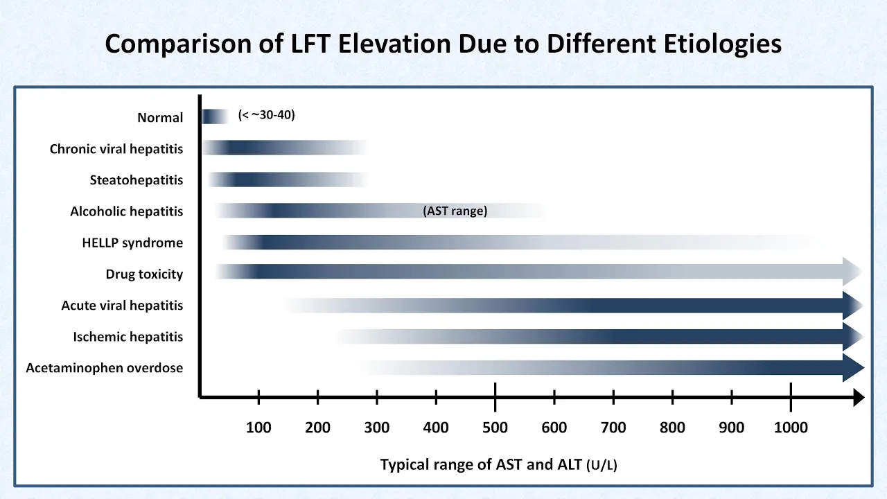
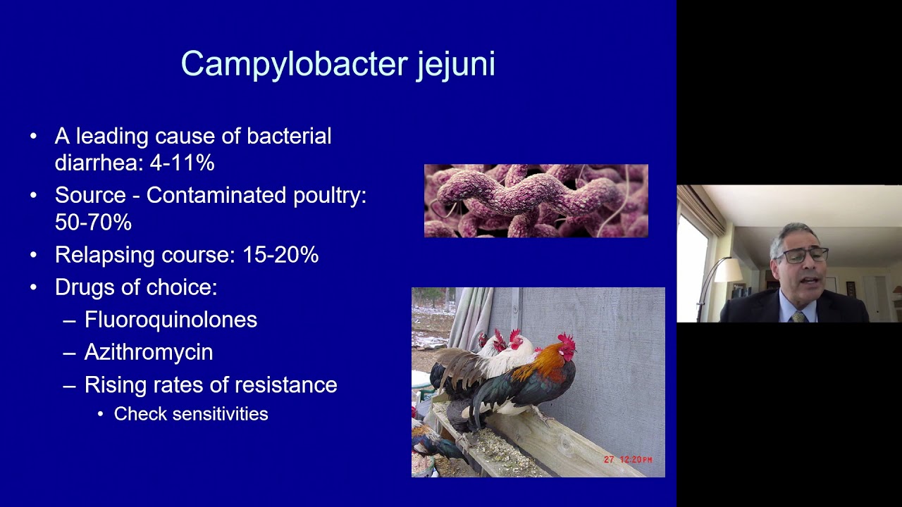
taking on how we manage patient with chronic diarrhea Dr Usha is also joined so I'll ask her to actually say a few words about the session thank you Dr nijavan it's uh we are all very fortunate to have the president of Indian Society of gastroenterology address the Gathering and it is wonderful to connect at a global platform with the Physicians of Indian origin it is always a pleasure to be in Connect I have been on this program earlier on also and today we have two very important sessions which are simple practical approaches to two common conditions one is of a luminal pathology and one is of a liver pathology so chronic diarrhea is quite often a vexing issue so we have Dr Vishal Sharma who's an associate professor who's soon to become an additional professor in the department of gastroenterology at PJ Chandigarh he is keenly interested in the area of luminal gastroenterology and works on tuberculosis and IBD and following that we have another lecture on abnormal lft how to approach it from Dr Amit goyal over to Dr Vishal Sharma to speak to us on approach to chronic diarrhea Dr Vishal please thank you madam and uh thank you Professor Javan sir for the kind words of introduction uh I hope my slides are visible yeah they are so uh as Madam said that chronic diarrhea is an important clinical problem and quite often it is a waxing issue uh for gastroenterologists and Physicians and in fact it is diarrhea is a universal Human Experience in fact it is uh it is a bit of a surprise that we don't have diarrhea much more often than we have in fact daily around 8 to 10 liters of fluid enters our intestines and this is largely through secretions by various organs and also dietary fluid intake that we have and it is remarkable that our intestinal tract can absorb almost all of that and it is a very small amount of fluid that is excreted now when there is a dysfunction or imbalance in GI function that results in either an increased tool frequency or lose consistency and usually both of them which we term as diarrhea now anything which is shorter than two weeks is is usually termed as acute diarrhea and anything which is more than four weeks is usually chronic and in in between what we have is persistent diarrhea which mostly is due to some of the Infectious causes which I have listed here one one miss a diagnosis of chronic diarrhea it is important to be sure that we are indeed dealing with diarrhea and there could be some things which could actually mimic diarrhea one of this is fecal incontinence which patients are often not very willing to come out with this history and they will call it diarrhea because of the staining of clothes and therefore it is important that clinicians inquire into it there's another term known as pseudo diarrhea which is basically more frequent passage of stools although there is no actual increase in volume and there are a number of causes which have been listed as uh resulting in pseudo diarrhea one of which is hyperthyroidism another entity is Overflow diarrhea which actually is uh in elderly individuals who might have chronic constipation with fecal impaction and there is basically an overflow because of bacterial digestion of the of the obstructed fecal products and this again can easily be sorted out with a per rectal examination now once we have uh we have the we have the diagnosis of chronic diarrhea then I think it is important to have some clinical strategy to evaluate these patients and usually uh what we have is clinical classification which utilizes uh one of these uh pointers so either one could classify the diary on the basis of mechanism site of origin presence of pain or the pattern of diarrhea now if we talk about the mechanism of diarrhea then broadly people talk about two dist distinctive patterns one is secretary and one is osmotic in Secretary either there is an excess secretion of electrolytes and water into the Lumen which may be because of certain toxins or there is a problem with these receptors which results in increased fluid in the lumen but usually because this these solutes are the usual solutes like sodium and potassium the stool osmolal Gap here is low on the other hand in the osmotic diarrhea which is usually because of certain unabsorbed solutes which may be something like unabsorbed fructose sorbitol or even carbohydrate malabsorption there is an osmotic load in the GI Lumen which results in increased fluid and because the solutes here are other than the usual solutes you have an increased or small Gap and we will come to it so basically secretary versus osmotic in secretory we know the etiologies could be stimulant laxatives bile acid diarrhea certain endocrine or uh functional neuroendocrine tumors which can result in excessive secretion of water and electrolytes and usually this diarrhea continues even when individuals fast on the other hand osmotic is could be due to multiple things like magnesium containing antacids carbohydrate malabsorption sorbital intake and this draws fluid into the cologne resulting in diarrhea and usually this abates with fasting so as you can see the osmotic Gap the fecal osmotic gap which is 290 minus 2 times sodium and potassium in the stool is actually high in the osmotic Diaries but practically one can understand that usually it is a combination of these things and there are only a very few etiologies which have either Pure secretory or osmotic diarrhea coming to the next uh people talk about small bowel versus large bubble diarrhea now the defining feature of these two entities is actually uh involvement of the rectum and involvement of the rectum in uh certain etiologies of diarrhea uh disturbs the reservoir function of of the of this of the large intestine and this results in passage of small volume of multiple stools associated with rectal symptoms of tenesma urgency and frequency now on the contrary diseases which usually impact the small bowel or the right colon do not have this more frequent passage of stools but they have much larger volume of stool and the rectal Reservoir function is actually preserved malnutrition or malabsorption is actually more common with small bowel diarrhea but of course as we understand certain etiologies of colonic origin like microscopic colitis could also be similar to what is traditionally recognized as small bowel diarrhea and therefore people often use the term large volume diarrhea for small bowel and small volume diarrhea for large bowel diarrhea in fact this these are often the preferred terms these days now coming to pattern of diarrhea now one could be what is known as fatty diarrhea which typically is with uh described with chronic pancreatitis where there is maldization because of loss of pancreatic exocrine function and this results in uh lack of absorption or lack of digestion of fat and increased fat in the stool and therefore the stool could be oily on the other hand other etiology other type would be the watery diarrhea where typically there will be lot of voluminous tool and in fact here malabsorption may occur across various macronutrients and this is typically uh described with endocrinopathies and even osmotic diarrhea would typically be a watery diarrhea one another entity is the inflammatory diarrhea anything that has blood or mucus and fecal leukocytes in stool examination and often these etiologies also have underlying pain and examples for this type of diarrhea would be inflammatory bowel disease gitb ischemic colitis colon cancer and diverticulitis but if we actually look then many of these uh patterns overlap for example diseases like celiac disease or Crohn's disease which have ideal involvement extensive uh intestinal involvement would practically result in uh virtually all of these things happening together some amount of inflammation some amount of secretory component and of course malabsorption of various macronutrients including fat so again there is a lot of overlap but again in some of the situations one could have a specific pattern now pain is not typically a common feature of chronic diarrhea but when present it could be helpful in pointing to the underlying etiology and typically epigastric pain could be part of pancreas chronic pancreatitis or Zollinger Alison syndrome pain of small bowel origin could happen in diseases like Crohn's disease tuberculosis lymphoma mesentric ischemia and complicated celiac disease whereas a hypogastric pain which is typically described as a cramping kind of a pain and which is a lead with defecation is largely a component of diarrhea predominant IBS but could also occur in patients with ulcerative colitis now before we go further I think it is important to understand how various nutrients especially the macronutrients are absorbed so we have the ability largely to absorb only the monosaccharides so the dietary carbohydrates which could be in the form of mono die or polysaccharides need to be digested first in the intestinal lumen through various enzymes and convert it to monosaccharides which actually enter the anthrocytes and then are transported to the portal blood on the other hand protein absorption is again initially through digestion into smaller peptides and amino acids and this is mediated by proteases and peptideases once these small peptides and amino acids are formed our enterocytes they will have the ability to absorb both of these and eventually these are converted to amino acids in the anthrocytes and then transported to the portal blood now the digestion of triglycerides is more complex and in fact the fats typically these are digested through a combination of mechanical and enzymatic processes which involve lipases and then there is formation of mycenes through the bile acids because these are insoluble and these myself help in transportation of monotriglycerides and fatty acids into the endocytes again inside the anthrocytes triglycerides are formed and they go into the to form the chylomicrons which are absorbed into the lectials and one important thing is to understand that the medium chain triglycerides can actually bypass this entire process and can directly be absorbed into the blood so they can be used when these uh these mechanisms of digestion are somehow compromised so if we look in the proximal intestine in the middle part of the small bowel most of the carbohydrate fat and peptide absorption happens proximal small intestine is again an important site for absorption of iron copper zinc and foliate and in fact diseases which impact this region are associated with deficiency of these minerals now helium distill allium especially has a very important role in absorption of bile acid and vitamin B12 when in fact in I'll terminal ideal resections actually these could be compromised and these are functions which actually no other part of the bowel can take up all of the intestine is involved in absorption of water and electrolytes alone usually does not have much of an absorptive function but it does absorb electrolytes and in situations like short Bobble syndrome it becomes a very important source of uh obtaining energy through the digestion of actually dietary fiber or the uh or the so or the carbohydrates which are actually digested into short chain fatty acids and which are absorbs and absorbed and provide energy in these patients so if we understand this there are two important processes that happen one is digestion that is break a breakup of the digested nutrients into absorbable form and then is the process of absorption so the problem could happen at either of these two levels mild digestion is when there is an interluminal defective hydrolysis and malabsorption when the mucosa cannot absorb the digested uh products so typically it's not a clinical of much clinical relevance to distinguish this except probably when we talk about fatty diarrhea now fatty diarrhea could be because of problems with the pancreatic enzyme which is in quarter chronic pancreatitis and then the triglycerides are not typically absorbed and these are excreted in this tool so the stool is bulky and usually well formed but let's say if this part is okay the pancreatic enzymes are okay energy this results in formation of fatty acids that actually and if that is not absorbed through the Lumen so fatty acids and then there are malabsorption of other nutrients through the uh through the Lumen so that results in more liquid stools and actually fatty acids can also result in increased colonic water output kind of a secretory diarrhea so this is the difference largely between fatty fat malabs option which is associated either with chronic pancreatitis or with mucosal diseases so how do we approach chronic diarrhea the first and foremost it is important to establish the severity and the pattern of chronic diarrhea so one could establish whether there is a significant volume loss on the basis of symptoms of of a volume of slow uh the stool which is passed presence of thirst dryness or even orthostasis now relationship of uh diarrhea with food could potentially distinguish osmotic from secretory passage of undigested food which is uncommon may represent either mild digestion or intestinal hurry features of passage of blood urgency frequency or 10 SMS could suggest underlying etiology and involvement of the rectum one important part of this entire conundrum is to separate the organic from the functional presence of a recent history upon sort of diarrhea older age nocturnal symptoms weight loss blood in stools abnormalities and blood tests especially hemogram and elevated fecal protecting would usually point to an organic cause of diarrhea now there could be other Clues like a recent new drug which has been started associated temporally with diarrhea sexual history could point to underlying immunodeficiency past history of surgery could suggest underlying short bubble if there have been multiple resections of the small bowel or a bacterial overgrowth which could be because of strictures or blind Loops source of uh Waterway eating out could potentially be associated with infective causes like giardia and then excessive intake of sugar containing juices syrups gums Etc could suggest you know osmotic diarrhea because of malabsorbed or unabsorbable entities which are present in these products now coming to drug induced diarrhea which is often missed and not thought of some of the drugs actually are very important causes of of chronic diarrhea and in fact some of the drugs can cause diarrhea in more than 20 percent of patients including ohas certain other drugs would cause in around 10 to 20 percent like antibiotics or ssris and then there are some infrequent causes of drug-induced diarrhea like Olney certain or iron preparations so these are important to consider in every patient where we are evaluating for chronic diarrhea now historically certain features could point to malabsorption looking for carbohydrate malabsorption usually because this results in formation of acids by bacterial digestion of malabsorbed carbohydrates the features are flatulence bloating gaseous distension and explosive diarrhea on the other hand passage of oil or stools which float on water could suggest underlying fat malabsorption of statoria and false smelling stool which is usually because of sulfide containing gases it usually suggests an underlying criteria or protein malabsorption coming to Clues on examination so one could find features which are suggestive of malabsorption of macronutrients but certain findings could suggest underlying diagnosis for example clubbing could potentially be suggestive of entities like celiac disease where it could be in around five percent of patients and then even ipsid which is a non-common cause of diarrhea presence of lymph nodes could potentially point to lymphomas or underlying gitb then there are very important cutaneous Clues which are uncommon but if present they can point to an underlying etiology so for example dermographism could point to make underlying uh underlying his underlying etiology macroglossia for example could point to emelodosis Flushing and wheezing could point to carcinoid syndrome dermatitis her petiformis may suggest underlying gluten hypersensitivity could be part of underlying IBD and then there could be Clues to vitamin Mal absorption which may manifest as cutaneous manifestations now coming to blood test it so a routine battery of tests would suggest sometimes the underlying etiology for example anemia with a low MCV suggesting iron deficiency could potentially suggest celiac disease deficiency then HIV serology positivity may suggest underlying immunodeficiency elevated hba1c could suggest diabetic diarrhea but then there are more specific tests for underlying etiology so typically antibodies to tissue transglutaminase typically done with the associated you know IGA levels may suggest underlying celiac disease now hormone secreting syndromes especially hyper functioning neuroendocrine or endocrine tumors would be picked up with the appropriate marker which is measured in the blood so something like chromogranin AMA just suggests any team whereas in a carcinoid syndrome one could have an elevated hia both in Blood and in the urine in certain etiologies like immunodeficiency IGA deficiency or cvid the immunoglobulin levels may also be low now Imaging is actually not commonly used for all of the cases of chronic diarrhea but if there is significant pain or underlying suspicion of inflammatory causes it may be relevant to take up Imaging investigations and then something like a judgmentation and you can see that something like volvolay started so this could suggest underlying celiac disease or malabsorption now thickening of the bowel wall could potentially suggest IBD tuberculosis eosinophilic gastroenteritis or it said then underlying cause of bacterial overgrowth could be picked up with imaging like strictures divertically or mesenteric ischemia and again Imaging could help in the diagnosis of chronic pancreatitis in patients who have steatoria fecal tests are again extremely important and here actually the infectious routine examination or cultures and in fact nowadays Multiplex PCR could pick up unuses infectious causes of diarrhea and may be needed when the cause is not clear now a simple test like looking using a sudan dye would pick up underlying steatoria fecal protection could be elevated in inflammatory causes in pH would suggest a carbohydrate malabsorption fecal elastase one is suggestive of exocrine in sub pancreatic exocrine insufficiency and occasionally when one suspects that there may be a patient may be taking laxatives one may want to do a stool analysis for various laxatives now breath tests are actually very important for diagnosis of certain conditions especially bacterial overgrowth and the principle is that one administers substrates for example let's say glucose so glucose would typically be absorbed in the small intestine but let's say if there is a bacterial overgrowth then rather than a normal absorption actually the bacterial metabolism will happen which will result in formation of acids and hydrogen and this hydrogen can be detected in a breath test now lactulose is usually used to look for Oro SQL Transit because it is typically metabolized in the cecum but in patients who have a bacterial overgrowth there would be an a peak of hydrogen which will happen much earlier and again these tests can be used for a plethora for the conditions like lactase deficiency fructose malabsorption there are certain tests for even exocrine pancreatic insufficiency not available in India and the c-cat test for bile acid diarrhea again not routinely available the next step of course is Endoscopy in fact which test to do first is often uh a very difficult question and usually the clinical pointers suggest how to go forward for example if somebody has an iron deficiency with diarrhea one would want to do an upper GI to look at the duodenum and then one could find scalloping uh you can see scalloping here and then reduce fold height typically described with Celiac but which could also occur with the number of other conditions like tropical sprue grds is a melodices and eosinophilic gastroenteritis the yellowish discoloration of mucosa could suggest underlying lymphangesia or even Whipple's disease which is again uncommon or mycobacterium AVM into a cell where again in in immunodeficient patients thickened folds could occur in conditions like eosinophilic gastroenteritis ipsid Crohn's disease amyloidosis Etc now histology can give us a diagnosis in some of the conditions [Music] sir I can finish in maybe one minute or maybe a couple of minutes so histology can again be conclusive in some of the situations like Ripple's disease giardia so in Whipple's disease one could have foamy macrophages and past positive inclusions foamy cells with AFB positivity in avium infection then lymph injectasia could suggest either a primary or a secondary which could be due to tumors or uh or TB eosinophilia could suggest underlying stronger doses or even used gastroenteritis and one could also pick up parasites now Villas atrophy again is uh is not very specific but typically seen with celiac disease tropical sprue and again a number of other conditions now since I have maybe a minute before I go to this algorithm I'll just show quickly a few cases so this was a young female with diarrhea anemia which was iron deficiency so we did an endoscopy you can see scalloping and grooving IGA TTG was actually undetectable and the IGA levels were low so we did IG gdgp and this was celiac disease in setting of 5G and efficiency this was another patient similar age recurrent episodes of diarrhea stool examination Giardia multiple times and you can see this kind of lesions nodule nodular lesions in the in the deuteronum and this was nodium lymphoid hyperplasia the immunoglobulin levels were low and this was cvid with duodenal nodal lymphoid hypoplasia another case 62 year old male with multiple comorbidities who came with decent prawns at diarrhea and there had been the igtc and all the negatives there is scalloping and actually the patient was an old me certain which had been started recently and this improved once this drug was stopped so this is all Miss art and sprue in another case elderly lady came with three months of diarrhea and weight loss change of taste alopecia and these nail changes and you can see polyps everywhere stomach this is a trophy in the deutenant you can see multiple polyps in the cologne and these were all inflammatory polyps and this was Cronkite Canada syndrome uh this is another case 28 year old male treated as ulcerative colitis no improvement even with steroids so you can see thickened poles in the duodenum and this is the ideological area again thick and folds all of them showing amelodosis and eventually at the age of 28 year old the age at this young age this patient had myeloma so I come to the algorithm again it is very difficult to have an algorithm for an entity as complexes chronic diarrhea but largely you establish whether it's a chronic diarrhea with history for persistent you try to rule out infectious ideologies first an easy step would be looking at uh whether there are any alarm symptoms in their absence you diagnose dibs or functional diarrhea recent intake of or new drug which have been started one could diagnose drug induced diarrhea one looks for malabsorption using clinical Clues as well as various tests and if it is present then you try to determine the etiology also and again what is the first test to do is usually suggested by the underlying clinical manifestations for example if siren deficiency one would want to you know start off by excluding celiac disease if there is a history of surgery one would want to exclude bacterial overgrowth or a short bowel syndrome if there is pain and bloody diarrhea usually inflammatory causes and one might want to go ahead with a colonoscopy and if there is a suggested history one may want to rule out HIV and opportunistic infections if it is unclear then it often demands an extensive workup and look looking up for uncommon causes thank you for your attention thank you Dr Vishal for a masterly lecture on a very complex condition and quite often a vexing problem as I suggested earlier uh just to reiterate for our audience the Communist causes if I say for diarrhea which we should exclude before we move on to others is celiac disease which is very simple to exclude secondly is inflammatory bowel disease Crohn's as well as ulcerative colitis where Cal protectant can help us thirdly is chronic diarrhea due to grdss so always look for it otherwise we will end up missing it and as Dr Vishal suggested drug history is important to exclude drug induced diarrhea irritable bowel syndrome anyway remains always in the back burner in these situations and pancreatic insufficiency small bowel bacterial overgrowth and lactose malabsorption unless we look for it we will not find it so a careful history a systematic examination and a systematic approach to investigation will often give us the clue as to which way to proceed forward and we have a few questions I think we'll take quickly four or five questions [Music] at the end uh Dr Vishal please stay on till we finish the next talk uh with this it is my pleasure to invite Dr Ashish goyal from CMC Vellore who will be delivering a lecture on approach to abnormal liver function test it is such a common thing sitting in the OPD to find and we have Dr Ashish goel to deliver this lecture on a very important problem over to Dr Ashish please thank you ma'am thank you to the organizers for the kind invite or am I Audible and my sites visible yes very much so the Mandate for today is approach to abnormal lfts and uh what we will do in next 15-20 minutes is actually do a part in recognition how do we understand our pattern and then how do we uh uh make a differential diagnosis for the sale so let me just start with the the first case and we will go back to these cases towards the end of the lecture first case that we see commonly in our OPD is a 17 year old boy who presents with incidentally uh raised bilirubin level and you can see the normal values here given uh this is of total bilirubin direct bilirubin alanine transaminase alkaline phosphatase and albumin and uh so normal values are provided and you will see here there is a high bilirubin but the other values are normal the second case that we usually see in again in outpatient is a short history in a young person a 15-day history of low grade fever nausea who presents with high bilirubin of 15 uh alanine transamination which is very high and but otherwise the preserved albumin the third patient that we will see is a elderly patient who presents with jaundice priorities again or short duration with high grade fever and a mass in the abdomen with a predominantly alkaline phosphatase which is fine the case for that we see usually in our outpatient is a patient who presents with hematinesis and uh is is can be a diabetic with high BMI a 50 year old lady who presents with mildly raised bilirubin mildly high alanine transamination but a low alkyl case five is of a patient who is again a very short history of five days after a toxic intake presents with sick with very sick with altered sensorium so bilirubin may not be verified but enzymes are high and INR is grossly prolonged so during this lecture what we will do is by the end of lecture we should be able to list out what are the causes of jaundice let's start what are the differential features in this major causes in each type and approach to case is that we have discussed so coming to the bilirubin metabolism as we are all aware that bilirubin is formed by degradation of hemoglobin and this bilirubin which is unconjugated goes in the circulation and reaches the hepatocyte where it is taken up by this receptor oatp and is undergoes gluconidation and is ultimately converted into a conjugated bibliography which is excreted out into the Wilder some of this conjugated bilirubin if it is in excess can reflux back into the plasma through the receptor on the basolateral membrane so if the problem is in the uh pre-hepatic phase where the hemoglobin degradation or bilirubin obtain is a problem this is this will be a pre-hepatic jaundice which leads to unconjugated hyperbilirukulenium if the problem is in the hepatocide this predominantly will lead to conjugated hyperplicopinemia because the rate limiting step in the repetocide is the excretion of conjugated bilirubin so the preservation of conjugation is there in hepatocyte injury so a hepatic injury predominantly causes conjugated hyperbilirubinium again in the post hepatic cause like bile duction there is a buildup of conjugated bilirubin inside the hepatocytes which refluxes out and therefore leads to conjugated hyperplative so we know is measured by a diazo Vandenberg reaction which is a calorimetric reaction and a direct fraction is equivalent to conjugated fraction action whereas the indirect building is what's known as unconjugated so to come to the classification of jaundice the jaundice can be classified into three major types I think one is isolated disorders of bilirubin metabolism that we have described it could be because of liver disease where the hepatocyte injury is there and all it could be because of the bioduct obstruction as far as liver disease is to understand again as per part and recognition we can recognize a predominantly hepatocellular pattern or a predominantly cholestatic pattern and I will come to it in further slides so coming to isolated disorders of bilirubin uh metabolism these disorders have believed in metabolism just rate results in high bilirubin levels but a normal liver function otherwise so your enzymes like alanine transamin is asparted from awesome it is objective functions both from mid time all of them are normal now jaundice then can be in the isolated within metabolism disorders can be either determinantly or unconjugated with more than 80 percent is indirect this could be because of increased production of bilirubin uh which is usually seen in hemolysis or decreased update which can be seen in drugs like refumbacing which affects the oltp receptor it could be more commonly seen with defective conjugation and this is very commonly what we see the Gilbert Syndrome which is indirect normal otherwise liver function test if there is a conjugated linear which is more than 50 percent is direct it is more likely a rare uh syndromes like Dubin Johnson water syndrome which is defective exhibition of the conjugated now uh coming to the more common causes of liver disease and wiled up obstruction and I deal with more or less both of them together and these are this is how we interpret the lft so if there is a alanine transaminase or as part A transaminase which is high this indicates injury to hepatocytes aspartate transaminase is a more non-specific enzyme can be present in muscles and myocardium as well but allylene trans aminase is more hepatocide specific uh enzymes so it can increase is indicating hepatocyte injury increase in alkaline phosphatase indicates injury to the biliary cells and so-call angiocytes predominantly so if there is a predominant alkaline phosphate is usually we are looking at delivery engine whereas albumin decrease our growth form in time increase indicates synthetic dysfunction of the liver these are the proteins which are produced from the liver and they have a half life so it indicates synthetic dysfunction and a more chronic injury in the hepatocytes so coming to the patent recognition in the divertices predominantly hepatocellular or predominantly cholestatic so if I look at hepatocellular patterns uh acute hepatocellular injury this patient usually presents with a short history with or without protrome and uh when we look at the lft bilirubin will be high but if the predominant feature is the high ALT and AST which is up to five to ten times the upper limit of normal alkaline phosphatase generally is normal or may be slightly high now these are the causes most likely what we are dealing with are viruses like acute hepatitis virus like hepatitis A E or B toxins like drugs or alcohol uh and these are the causes and once we recognize this pattern we have to evaluate for the causes these patients are generally symptomatic for more than six months with non-specific symptoms or weakness or portal hypertension related symptoms uh bilirubin maybe may be mildly high but enzymes are not that high so it is around two to five times upper limit of normal a history duration of more than six months again we have to look for common causes here which is hepatitis BC alcohol autoimmune liver disease and others so these are the common causes for acute hepatocellular dysfunction and you can see hepatitis A E and B drugs like intertuberculosis drugs for us complementary and Alternate medications again toxins for us it is rodenticide but it could be any toxin uh locally prevalent and we have to look for systemic mimics like malaria and leptospar energy species as far as the chronic more common causes are concerned these are Hepatitis B and C alcohol and more like more recently it is non-alcoholic fatty liver disease with metabolic syndrome the other common other not so common causes are Wilson's disease again autoimmune liver disease we have to look out for most in most of these patients vascular divertices can also present us ultrate lfts the other pattern that sometimes is seen in in our patient is a patient who is presenting with systemic symptoms say low grade fever weight loss or chronic puff has a predominant raised alkaline phosphatase and when we do an Imaging there is normal bile dumps this is a pattern which we then say it is may be suggestive of granular matters liver disorders or infiltrative liver disorders like tuberculosis sarcoidosis or rheumatological malignancies if the patient is presenting with significant providers and Clay colors tools uh this is a pattern and with very high alkaline this is a part of static liver injury and here we have to look for causes with predominal polystasis like drugs and primary limits as far as bile duct box function is concerned again uh obstruction this is uh this can be both it can be the obstruction can be interluminal it could be in the wall or it could be because of the extrinsic compression uh idea is to recognize a pattern of bile duct obstruction and then look for causes of these uh ultrasound or a further Imaging should be able to clarify uh what is the cause of biological Construction now type of bilirubin doesn't differentiate the site of a site of jaundice here because both hepatic and post hepatic or obstructed jaundice both of them have conjugated hyperperia but what will help you differentiate is history taking uh and we have pointers like a programmable illness in patients with hepatocellular jaundice if the patient has Associated prioritis you're looking at polystatic liver disease either it could be because of the bile of obstruction or the infiltrate infiltrative liver disease uh if there is a suggesting toxins like rodenticide intake we are looking at repeal which on the same some of the history things history pointers like biliary call it hybrid fever suggesting cholingitis or prior biliary surgery all right all point out towards the possible bile duct obstruction on physical examination anything to suggest portal hypertension like ascitase splenomegaly or any chronic liver disease stigmatode like spinal angioma all this suggest hepatocellular disease whereas the presence of high grade fever abdominal mass or a star which is biliary obstruction biliary surgery suggests a possible bile duct obstruction as far as the liver function is concerned as we have already said there is a kind of overlap and predominant enzyme elevation if it is alt AST then it is a more hepatocellular pattern and a liver disease whereas a predominant enzyme is more alkaline phosphatase you are thinking of ruling out bile that's obstruction in this patient now programming time uh you is elevated even in deficiency of vitamin K which could be because of chronic bile duct obstruction so if we are giving vitamin K in these patients and Prothrombin time corrects this suggests a possible cholestatic liver disease the further test to be done once we recognize a pattern and this may give you a clue so if you have hepatitis B C or a into e one of these tests positive then you are looking at liver disease whereas if the ultrasound shows dilated by products you are looking at a bioduct obstruction so after the history and physical examination and once we have your lfts we will have a general understanding whether we are dealing with isolated disorders of biliructing metabolism or we are dealing with bile duct production if we are dealing with bile duct obstruction we are going into Imaging route where we are doing ultrasound to start with and if needed a CT scan or an MRI scan and then according to the course we intervene on the management issue if we do not have a bile duct obstruction then we have to look for underlying Capital cellular disease uh so if there is ultrasonic by showing bind UPS uh we will go ahead and do an ercp or other management as per the cause but if there is no bind up dilutation but still you are highly suspicious of uh biliary obstruction we will go ahead and do a further Imaging which is like MRCP or endoscopic ultrasound the cause I'm not dealing with uh here and the the cause will be depending on the cause or the treatment will be depending on the cause of the disease that is identified now I am going to uh deal in the next five seven minutes about the cases that we had discussed this is the first case we had brought up which is a 17 year old boy which was who was found to have raised bilirubin incidentally now what we have looked at is this person as normal Alena in transaminases normal alkaline phosphatase and normal albumin below them which is then for putting us into a category of isolated disorder of bilirubin metabolism uh this is again when we look at the pattern this is this is predominantly indirect hyperbilirubinemia so what we are looking at is either hemolysis or more likely Gilbert Syndrome here because the person is asymptomatic but we have to rule out demolishes the second patient is a short as a young patient who presents with a short history of jaundice with a low grade fever and nausea suggesting broad Rome and uh when we look at the lft or the predominant pattern here is which is around which is more than 10 times the upper limit of normal and alkaline phosphatase which is not very high which is almost on the upper limit of normal range we have got direct hyperbilirubinemia here so conjugated hyperpillarupinemia so this suggests a possibly hepatocellular jaundice a low grade fever and a short history suggests the acute viral hepatitis like illness so we have to look for acute hepatocellular injury here and look for viral causes like hepatitis or hepatitis B [Music] uh case three was a patient elderly guy who presented with uh with the short history of jaundice again alkaline phosphatase which is very high almost 10 times the upper limit of normal with no almost uh not very high rain in transformation so and significant priorities so we are looking at a polystatic pattern here but the patient has high grade fever suggesting cholingitis with epigastric Mass so we are suspecting a biliary obstruction here so the the test to do here is an ultrasound abdomen and proceed from there and see the cause and evaluate and treat the cause the case 4 is a patient who presents with limitations and uh if you look at the patterns here you this hematomasis suggests possibility of viruses and total hypertension and there is a mild hyperbiliruthinemia with a mild alanine transamine is high and uh this also suggests a chronic the possibility of cirrhosis under present with as a patient is diabetic with a high BMI in a 50 year old person most likely what we are dealing with is chronic Capital cellular dysfunction and circulosis secondary tool cartilage disease a young girl who presents with the five days after intake of rotenticide and altered sensorium and if you look at the pattern here altered sensorium suggests presence of any kephlopathy and uh hyper European with very high alanine transaminase and a very high Prothrombin time all these suggests presence of mkflopathy and severe cobalopathy such as acute liver failure and uh and in the history it suggests a rodenticide or toxic related acute liver failure so uh to summarize here the liver function is basically a pattern recognition but we have to rely on our history taking skills physical examination and very relevant investigations to assess the cause of liver dysfunction we have to eliminate one by one various causes and to then evaluate for specific causes and treatment accordingly thank you thank you Dr Ashish for a splendid lecture on an approach to abnormal liver function test as you all noticed that he kept on using a word pattern and it is this pattern identification which is very important in interpreting liver function tests so if you have the setting if you have the lft and if you have the ultrasound I think 99 of the situations we will get a approach to how to move further so I would think the combination of the setting lft whole lft pattern and the third thing is an ultrasound I noticed that he did not mention about sgot and or AST as we call it here uh increasingly we were moving towards using only alt however ASD to alt ratio sometimes has certain Clues elevated AST to alt helps in diagnosing cirrhosis as well as Wilson's disease as well as alcoholic liver disease so and infiltrative liver disease so in such a kind of setting OTP ratio may be of use otherwise alt alone is sufficient and with this we would now move on to the chat box and take up the questions we start with the questions to Dr Vishal Sharma on chronic diarrhea and uh would you like to take up the questions or shall I go ahead okay okay so you can maybe do the liver thing I will uh address these questions due to uh to talk to people Dr Vishal Dr ashindeep has asked what is the duration of fasting to distinguish osmotic and secretary literature says 24 hours but that's practically difficult what is your view yeah I think uh Dr ashnip has raised a very important question because uh actually literature describes even up to every three days so and and then then there were later studies which talked about uh two days and even one day but it is certainly prolonged fasting people actually are admitted for even this uh to test response to fasting and it is not just you know typically asking what happens whether you fast or not so that is the reason I think the the initial description of the stool or smaller test was actually uh to to take uh you know take off this test of of prolonged fasting practically the pure secretory and osmotic are actually not very common and in fact most people would not end up doing uh either of these what we rely more on is uh you know the symptoms with a particular uh particular thing in the diet whether excluding that helps or not so so so yeah I agree that it is slightly problematic because it certainly warrants a prolonged uh prolonged fasting and it may be better to really you know use the stool uh smaller Gap rather than actually practicing this 24 hour fasting which may be a bit problematic so I would think here I would like to add practically what we are trying to see whether the patient swings from low volume stools to high volumes tool with a relationship of diet I agree with Dr Vishal that if you go by the textbook yes we do need to do 24 hour fasting but if the patient is uh fasting for about 8 hours 12 hours and there is a decrease in stool output and the patient also says that this increase in stool output on eating a heavy meal so that swing also helps us tell us that there could be a problem so moving on to the next question we have how how to do the stool examination test is it the random stool sample or early morning stool sample of the Vishal so I am unaware of you know a comparative data on that but typically we ask for a morning sample that is the time you have uh have the you know uh the the typically people pass tool and uh it has been described that because it is a sample which is a kind of fresh and overnight uh it has been there in the rectum the the yield may be more but I I am unaware if there is any data to really support that I think it is just a practical component but it could be in a patient with diarrhea they can give it at any point of time it is an extension from the urine culture where you ask for an early morning urine because there is a collection of urine in the bladder and allows better uh yield but I don't think if it's stool it is so important any stool sample should be practically okay moving on to the next question when to suspect immunodeficiency syndrome in malabsorption yeah I think uh apart from uh HIV which you know we all are aware when to suspect and probably and probably we test everybody I think probably we uh what the question probably pertains to the other primary immunodeficiencies and this would be when you have recurrent infections maybe infections outside the git also like cynopulmonary infections and other uh would probably I think even in one thing which we recognize is that some of the inflammatory diseases are also associated with the immunodeficiencies for example hygiene deficiency is more frequent with in people with I mean Celiac is more frequent MPA patients with IGA deficiency even IBT actually and then if there are you know certain let's say you're doing a biopsy you find uh Giardia again and again you have treated again you find jadia or let's say you have typical nodular lymphoid hyperplasia and these are settings where one should certainly evaluate for uh for for uh immunodeficiency especially IGA and cvid uh in the Adolescent to adult age group so just to add to that if you don't suspect you will never find so anybody in which this persistent diarrhea there's repeated grds is always have your index of Suspicion high so that you can pick it up you have to do an immunoglobulin profile to pick it up uh so there's a question from Dr Shivani at what point is Imaging advice Dr Vishal yeah Imaging is not a test which we usually do up front but certainly if there are features of significant abdominal pain abdominal intestinal colic obstruction and you are suspecting something like let's say tuberculosis Crohn's you may want to do an Imaging typically a history would suggest any underlying cause of bacterial overgrowth but again an Imaging could point out to underlying strictures or blind Loops so these are the situations again pancreatitis again if significant pain is there so and then largely I think last would be when even after the initial battery of tests you really don't have a underlying theology that's where one would want to do a Imaging yeah thank you for going on to the next question it's the last question to Dr Vishal uh increase tool frequency of stools normal consistency and pain abdomen since three months can we say it is IBS it could be IBS usually the history is much longer than that but I think uh it it a lack of uh any any uh alarm symptoms and certainly some amount of evaluation uh would be needed before labeling this as IBS IBS as I I would like to you know point it out again and stress is a diagnosis of exclusion yes thank you Dr Vishal for answering all the queries which were raised over to Dr nijavan to take up the questions related to abnormality uh thank you Ashish it was a very nice talk making uh it very simple there is a question uh in the chat box like after starting the ATT after four months patient developed jaundice so we can say it as Billy if all other causes are ruled out uh thank you sir uh is as I understand this person has already stopped ATT almost four months so before labeling it as drug induced liver injury usually a Time Factor also uh plays a role so if you have already stopped a truck four months back unlikely it is you can we can put it on a drug related delivery J but uh by continuing to be on it patient is continuing to be on ATT and developed on this after four months of eating okay yeah so yes then it is uh definitely if the patient is continuing ATT then a jaundice can develop even after four months of ATT it's a idiosyncratic reaction we have to evaluate for alternate cause you can put it as drug induced liver injury and take appropriate action there's another question how to differentiate between Gilberts hemolysis and drug induced journals they will the hemolysis should be apparent uh on the routine tests itself so hemoglobin retic count MCV should be able to uh I think Ankita was answering the cases so maybe that is not the question that she was asking she is just put in the chat box and how how to approach a patient who has come to you with I mean to the OPD and the asymptomatic elevation of the liver enzymes without elevation of Deliverance so only transformation yeah so uh bilirubin uh hepatitis so the history is uh sort of uh uh is helping you then if it is an acute illness and the enzymes are in the range of 10 times or five times upper limited normal it could be an electric acute hepatitis and then accordingly we have to work up towards acute hepatitis and its cause if the enzymes are not that high and a chronic illness then we have to look at a chronic hepatocellular dysfunction so bilirubin is only one part of the lft and we look at the entire pattern it may may not be high in some cases having isolated direct type of hyperbiliate what could be positively the normal in a pregnant person so pregnancy doesn't uh affect the interpretation of lfts to a large extent the alkaline phosphatase may be high in third prime minister of pregnancy but otherwise the interpretation Remains the Same if there is direct hyperbilirubinemia and it is isolated then you have to go down the isolated direct hyperbole rubinium as I discussed and evaluate for cause foreign were very good because that shows the interest of all the participants in the topics so I take this opportunity on behalf of gapio to thank the organizers the IAC to have this session uh both the speakers moderator uh Professor Gupta Usha and you Dr Sandeep also like to thank the back-end team and the IIT team who has made this event possible and thanks to my colleague Mr jitender who has been instrumental in organizing all these communications thank you very much we look forward for further sessions thank you thank you so much thank you thank you thank you Dr Sushi and thank you Dr Vishal and Dr Ashish for a very wonderful session thank you [Music] recording stopped right foreign
Up Next
56:38
Hypothalamus Anatomy and Function: Diencephalon and Nuclei Explained
@NinjaNerdOfficial
676K views•2020-11-10

48:47
Graphic Medicine: Comics for Collaborative Healthcare Communication
@nationalpatientadvocate
189 views•2023-12-04

10:28
Neuroanatomy: Central and Peripheral Nervous System Divisions Explained
@AKLECTURES
136.2K views•2014-09-20

3:05
Stages of Labor and Vaginal Birth | Childbirth Animation
@nucleusmedicalmedia
52.1M views•2017-08-18
Related Study Plans & Knowledge Roadmaps
Structured learning paths in Medicine
medicine
Abnormal LFTs: Diagnostic Workup & Pathology
01Liver Anatomy and Basic Physiology
02Introduction to Liver Function Tests (LFTs)
03Hepatocellular vs. Cholestatic Patterns
+ 3 more learning modules
12h6 modules
Explore Roadmap
medicine
Analyzing Blood Smears: RBC, WBC & Platelets
01Introduction to Blood Components & Smear Preparation
02Normal Morphology of Blood Cells
03Erythrocyte Pathologies & Morphological Variants
+ 2 more learning modules
12h5 modules
Explore Roadmap
medicine
Mechanical Ventilation: Waveforms & Alarms
01Anatomy and Physiology of Breathing
02Introduction to Mechanical Ventilation & Core Modes
03Ventilator Settings and Waveform Interpretation
+ 2 more learning modules
12h5 modules
Explore Roadmap
medicine
Neuro Exam: Localization & Stroke Syndromes
01Foundations of Neuroanatomy and Vascular Supply
02The Neurological Examination: Step-by-Step
03Principles of Neurological Localization
+ 3 more learning modules
18h6 modules
Explore Roadmap