


















![ECNi / ECN / EDN [Conférence] - Tour de synthèse 1](https://i.ytimg.com/vi/diChizgapRI/maxresdefault.jpg)


The liver serves as the body's synthetic center, producing clotting factors and albumin, and is the primary organ for metabolism of blood glucose, fats, and proteins. Liver function tests (LFTs) include ALT and AST as markers of hepatocyte damage, ALP and gamma GT as markers of biliary epithelial damage, while albumin and clotting factors indicate synthetic function. Jaundice, visible at bilirubin levels of 30-50 μmol/L, is classified as prehepatic (hemolysis, Gilbert's syndrome), intrahepatic (viral hepatitis, paracetamol poisoning, fatty liver disease), or posthepatic (obstructive jaundice from gallstones, strictures, or cancer). Gallstone disease presents as biliary colic (constant RUQ pain without jaundice) or acute cholecystitis (fever, Murphy's sign positive), while obstructive jaundice causes dark urine, pale stools, and dilated ducts on ultrasound. Cirrhosis is a pathological description characterized by disrupted lobular architecture, fibrous bands, and regenerative nodules, leading to portal hypertension and potential liver failure.
Hepatobiliary Lecture: Jaundice, Gallstones, and Cirrhosis
Added:for it's important to do to ideally put in a green canula an 18 gauge into an anticubital vein rather than on the back of the hand with a small canula because both calcium and 50% dextrose are sort of irritant to the vein. So um that's what's best. So we're going to talk about disease. Um there's quite a lot of content to cover.
So this is and this is very much toward aimed towards MCQ style. I'm not going to try and touch on OSKIs and examinations.
Uh if you've got any questions, please interrupt. Um and we'll try and do questions as we go through. I think the the slides are online. There are a couple of couple of little changes and I'll point them out. Um if we don't get through it all, then we don't. Is it in theory half an hour and 15 minutes?
Yeah. Okay. Right.
So going to start off with overview John this um and gallstones and talk about cerosis and if you've got time then uh the rest of liver disease really um so liver always seems to be a mysterious organ as people find it. It's got quite varied functions, but the most important ones are that it's the synthetic center of the body. Produces all the proteins and notably the clotting factors and albammen.
Clotting factors being your short half-life measure of hypatic synthetic function whereas albamin being the long halflife sort of 3 weeks measure of hypatic synthetic function.
And it's the main organ for metabolism in terms of blood glucose and fats and proteins. So the liver is the only organ that helps you to increase your blood glucose other than what you eat because you can't use muscle stored glycogen for your blood.
Um the liver produces bile. The bile is needed for fat absorption and excretion of various compounds most commonly with fat soluble compounds. Um and then it's got some other complicated roles involving immunity and blood volume regulation.
So it interacts with the heart and the lungs and the kidneys.
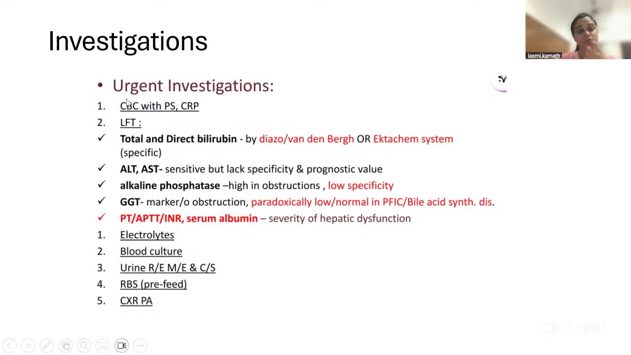
I know that there's already been a talk about LFTs, but just a very brief summary, ALT and A markers of damage to hpatices.
So they only go up when hpatices have undergone licis or necrosis, which means that the patient can have chronic liver disease but not have any raised ALT or A unless there's any active damage to their hypathosytes.
ALP and gamma GT um are markers of bilery epithelial cell damage. So anything that's causing damage to the bile ducts or somewhere in the bilary tree or an obstruction to the blossasis.
Um it's not completely exclusive though.
So you get some mild rays in either one with for example a predominantly damage there hpatocytes does cause a mild rise in the ALP and gamma GT and vice versa.
Um always remember that alkaline phosphotase is also present in the bone and placenta and gamma GT is the way of differentiating outer origin as opposed to bone or placentology origin raise ALP and then as I've mentioned albamin and clotting factors really are the markers of liver function as opposed to liver damage um where clotting derangement would be your first sign of of failure of hippatic synthetic function So start talking about jaundice.
Um and in simple essence it's raised bit of rubin.
Does anyone know roughly what figure bill rubin needs to get to before you can see it.
Yeah. So 30 to 50 is usually what's quoted. Um, and you can classify it in terms of what kind of bilerubin it is or where the pathology is. And we're going to talk about it in terms of where the pathology is. Is it not really involving the liver, a prehypatic jaundice, one due to liver damage, an intrahypatic jaundice, or a problem with bile excretion, uh, post-hippatic or obstructive jaundice.
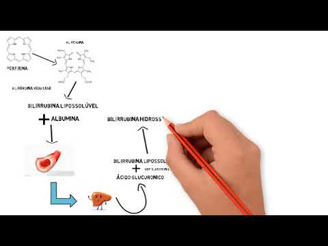
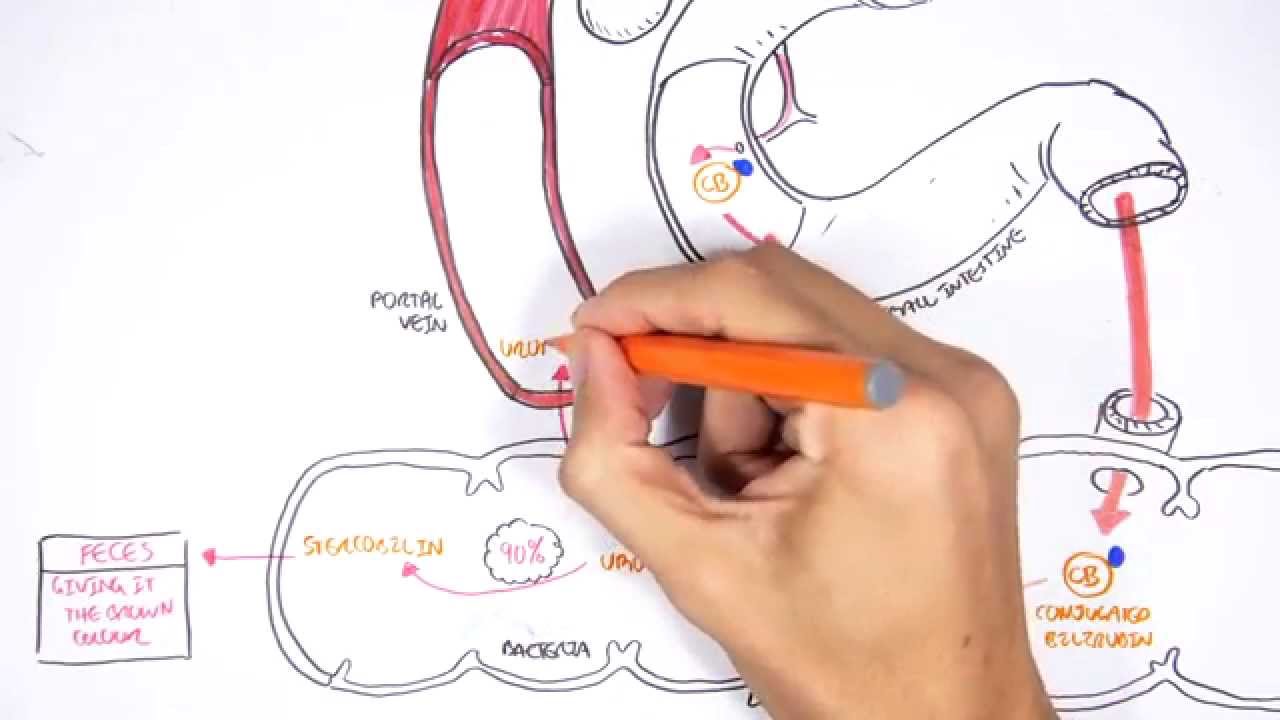
Um, so I've changed the the slide slightly. So just need you need need to summarize this uh production of bilarubin almost all of it comes from red blood cell breakdown which makes heem heem converted to berubin that berarubin is unconjugated it's just on its own and it's not soluble in water so as it circulates in the blood that berubin doesn't get pilted into the kidneys it gets taken to the liver it's acted on by an enzyme called UG GT which conjugates it. So it sticks on a glucaronide group onto the berubin and that makes it soluble in water.
Once it's soluble, it's then able to be excreted via the bile into the bowel and then from then on it's acted on by bacterial enzymes in the bowel. So that first conversions to something called eurobilinogen.
Um and the urbalogen is normally reabsorbed in part and then travels back by the bloodstream some of which gets left in the kidneys.
So urinary eurobilinogen is a normal finding and demonstrates the presence of bilerubin getting into the bowel.
It then gets converted to a brown pigment pigment called stircoinagen and that's what causes stool to be brown. So if you're not getting any berubin into the bowel, you wouldn't have eurobalinogen and your stool is pale because you've got no stircoin.
If you have a conjugated berubin in the blood, the berubin glucaronide that's water soluble so can be seen in the urine and makes the urine dark.
So just sort of there summarized. So if you got a conjugated hypogillaria then you get dark stools. If it's obstruct of dark urine, if it's obstructive, you also have an absence of urban in the urine and pale stools.
Prehipatic jaundice um is almost always due to either hemolysis or a condition called jar syndrome.
In most cases, the LFTs are normal. So if you're presented in your MCQs and it's a range of LFTs and just the bill of Rubin is up, it's reasonable to assume that it's a prehypatic jaundice.
What kind of tests would you do to differentiate the cause of a prehipatic jaundice?
What did he say? No. Here we go. I was going to say split the bill of rubin into conjugated and unconjugated. Yeah.
So you can you can prove it by asking for a direct and an indirect bill of rubin where a raised indirect bill of rubin is a raised unconjugated fraction but if we're thinking about a homoysis or jars what tests would we do?
So and someone say blood film. Yes.
So kums test and blood film. So Kum's test broadly is looking for antib um an antibbody mediated hemolysis.
Um and then a blood film you can look for evidence of himis.
Anyone know any other blood tests that you could do to look for evidence of himis?
You can do look for a lactate. So lactate dehydrogenase or raised lactate um but raised LDH particularly.
What does that show? So a raised LDH um is a feature of hemolysis and also you can get something called reduced hapttoglobin.
It's a protein that binds free hemoglobin. So hptoglobin goes down if you've got excess free hemoglobin.
The classical case that you'd get for condition called Zilbear syndrome is 50-year-old man has his uh has a hernia operation. It all goes fine. Um but the next day he's found to be he he appears jaundice. All his LFTs are normal apart from a raised bit of Reuben. He says this has happened before. Um he feels fine. and there's no abnormalities on examination and that's this case where in Jar syndrome patients when they come under some form of stress which can be surgery illness starvation their expression of UGT goes down which means despite the fact they're producing a normal amount of berubin they're not able to conjugate it and because it's an a raised unconjugated fraction their stools and their urine still appear normal and all their other LFTs normal.
What advice would you give to someone with Jar syndrome?
Do they have to be worried?
No. Yeah. So you can say there's nothing to worry about and tell tell doctors if it happens again because otherwise they might end up having all sorts of investigations to try and find out the cause.
Does anyone know the name of a another rare pediatric condition that sort of mimics your cri? Yeah. So one for bonus points pediatrics.
So it's prehypatic jaundice.
Intraypipatic jaundice is almost always conjugated hyperbillarenemia because the liver has capac will almost always conjugate all the berubin it has even if there's only 10% of the liver functioning. So in acute viral hepatitis it's usually a raised conjugated fraction but the liver is still excreting the bilarubin through the bile. Therefore stools still appear dark but with a raised conjugated fraction you get dark urine.
So for example a classic exam question would be uh man comes back from holiday in Egypt. He feels like he's got flu. He then notices his eyes are yellow, his bit of rubin's 100, his ALT is a thousand, his urine is dark, his stools are still dark.
think of some kind of acute viral hepatitis A or E.
But broadly um the causes of intropatic jaundice are sort of anything that can damage the liver both acutely or chronically um including paracetmal poisoning is an important one never to be missed. Um sort of acute on chronic would be decompensated cerosis which we'll talk about in a bit.
any of the any of the hepatitis viruses A to E all the metabolic diseases especially alcoholic and non-alcoholic fatty liver disease knackled um autoimmune hepatitis and the billery diseases now standing for what's PBCI yeah and primary sclerosin kangitis so despite the fact that Both of them predominantly affect the bile ducts. Um they both can lead to cerosis. Therefore they both could be considered to be a cause of intrahipatic jaundice and drugs as well. So drugs are quite an important one. Um can anyone name a drug that can cause liver damage?
Yeah, good one.
What do you say? Iso I can't say iso yeah is kind of almost all the almost all the all the TV drugs apart from if that be told um any others a very common paracetamol paracetimol of course there's an antibiotic that we give for cellulitis so flu clocks um patients on who are on highdose flu clocks should have their LFTs monitored weekly or sort of twice weekly because people do go into liver failure from flu clocks.
Postpipatic jaundice it's kind of a surgical thing usually um got something stopping the outflow of bile bile reflexes back and the conjugated bit rubin spills it out into the blood.
It's conjugated so it's water soluble.
So the your urine goes dark because you've blocked the flow into the bowel.
None gets into the bowel. No stircoin, therefore your stool is pale. No urinary eurobalinagen because none's being reabsorbed.
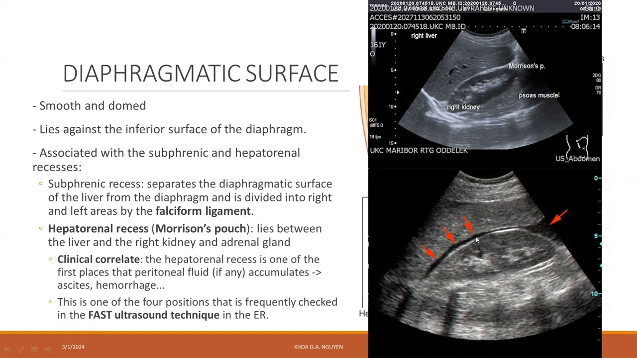
Key investigation is an ultrasound. So you do an ultrasound and what you're looking for is the dilated ducts.
So if you've got a dilated common bile duct, that suggests there's some obstruction. below the common bile duct that's causing back pressure. You've got dilated intrapatic ducts and a normal common bile duct that would suggest some obstruction further up. So the common hpatic duct best way to think about causes are like any obstruction in the lumen in the wall and outside. So gallstones is the most common of cause of an obstructive jaundice. Um, and we'll talk about them in a moment.
In the wall really, it's only strictest and cancer.
Um, so does anyone know anything about calangio carcinoma? It's really aggressive. It's really bad.
It is really, really bad. It's really sad. People get up when they're quite young. Um, does anyone know any conditions that predispose to gangiocarcinoma?
PSC. So it's kind of PSC patients tend to get clangio carcinomaomas.
Um and then the other one not to be missed is cancer of the head of the pancreas causing obstructive jaundice.
Any questions about jaundice rubin and sort of the principles behind it?
It's about recognizing the patterns of the raised liver enzymes and correlating it to the symptoms. Um, and exam questions can be phrased in any which way. So, for example, they could give you a set of LFTs and then ask what symptoms they're going to get. So, so cool stones um it it it's all about where it is and exactly where it is um which defines the pathology and the symptoms.
There are these poor jokes all the way through if you think they could carry on. Um and the the anatomy is quite important. So right left apatic ducts make common hpatic duct which is joined by the common the cystic duct to make the common bile duct um which joins the pancreatic duct at the ampular enters into the geodum of the sphincter body.
So if you have a stone in the cystic duct blocking the cystic duct completely, does the patient become jaist?
No. Great.
So just remember that cuz people always get confused. So in theory, if you've got a blocked gallbladder, you don't get jaundice just from that. The stone must be somewhere else. say anywhere along here or up to here to cause back pressure and and obstruction to bile flow.
If you've got a stones just sitting in the base of the gallbladder, not obstructing it, um, and people then the typical presentation is billery collic. That's where the gallbladder is contracting on the stones and they're getting pain from that, but the gallbladder itself isn't infected and it isn't particularly inflamed.
When you have a stone that blocks the outflow of the gallbladder, then the gallbladder becomes inflamed and they've got a stagnant pool of fluid and like anywhere in the body it gets infected. That's your acute cylitis.
And if you have the same thing, stagnant pool of bile but in the bile duct, that's collangitis.
Um, does anyone know what the the anatomical name for this area is?
Pouch. Good knowledge. Yeah, Hutman's pouch. So, if you bring that one up, you'll you'll really impress them.
Um, so comic patients are relatively well. They're the people who kind of come along saying, "Oh, I've got a bit of pain." They don't have a fever. They are not clinically paritinetic.
They might have ever so slightly raised inflammatory markers, but generally they're just a person with pain.
The typical story is the fat middle middle-aged lady who eats fish and chips gets right up quadrant pain. Um, but it can be more vague.
The one thing to note is that P it's described as being a collicky pain which you'd assume is intermittent comes and goes but it's very commonly actually quite constant might get a little bit worse and a bit better but it doesn't truly come and go unlike renal collic where patients can be rolling around and then better.
If you do an ultrasound, there should be no dilated ducts and the gallbladder will look relatively normal or it might look a bit old and fibrosed from chronic colicyitis but it won't be swollen and adematus unlike an acute coliccyitis where the stones blocking the outflow.
They've got an infected viscera so they probably have a fever. They've got raised white cell count, high CRP.
Um, they're generally unwell.
Their pain is a bit different. They might have started out with a bit of bitter collic pain, but it's more continuous. Um, what clinical sign is sort of textbook for acute choleiccyitis?
Okay. So, someone had to define Murphy's sign exactly.
pain stops inflammation on pressing in the right way. And isn't there pain?
Yeah, greater on the right hand side than the left. So the way to test is you go on the left in sort of the mid roughly mid-clavvicular line pressure just under the costal margin and then ask them to take a deep breath in and they might get a bit of pain there. But if you do the same in the right hand side, they get much greater pain. But as the the gallbladder descends and hits the hand, that's the theory. And examiners are often particular about the fact that it has to be greater on the right than on the left. Because if it was the same on both sides, what would you be thinking of?
Say peritonitis or pancreatitis, either one. So something else. Um it could still be chositis, but the they might well have a slightly raised ALT um as sort of run 50 60 70 and slightly raised gamma GT and ALP as well and the thing that you tend to read on the ultrasound reports is adeus gallbladder with stone impacted in neck or stone in cystic duct coitis um is again the classical description is shos triad um which is right upper quadrant pain and obstructive jaundice and rigors.
Does anyone know what Reynolds pentad is it if it extends to um have hypertension or ticardia or something like that. It's two words. Yeah. Well done. Um so Reynolds pentad is shock. This is just for fun. Like I wouldn't worry about it too much. Um with with shock and altered mental status. Obviously this is Sho's triad for right upper quadrant pain. Not to be confused with Shako's triad for multiple sclerosis which is like nestagmus relative afroillary defect and something else but there are two.
Basically, the patient is really unwell because they've got um ascending infection in their in the boduct. Um hugely raised LFTs. They can have bit of ribbons in the hundreds.
Um on the ultrasound you see a dilated common bile duct or a dilated pathic duct.
Um and the figure given is over 6 mm for the bile duct suggests dil um dilotation what's the so the the further investigations that they done then are either an MRCP not the examination but the um the uh a magnetic resonance calangia pancreattography or an ERCP what's the do you know why one would be done and not the other yeah so ERCP allows you to do the treatment. But why would you do an MRCP?
ERCP carries a higher risk.
Sure. So ERCP's firstly it's relatively invasive do an OGD and then split the spinter body and then go all the way up and there's a risk of pancreatitis. There's a risk of contrast reaction. Um there's the risk of the actual sedation and everything in itself.
Um, and also there's the fact that often you'll find in hospitals ERCPs are only done on a Tuesday in a in a DGH. So they might do an MRCP in the meantime to confirm the position.
What medical treatment is given to kangitis and colcistitis?
Antibiotics. Antibiotics. Which antibiotics? say yeah keen met because it's usually an anorobic infection but it's always reasonable in your exams to say antibiotics um it's always reasonable to say I would consider the hospital's local guidelines but for example I keporon and um metroniz would be appropriate uh and fluid resuscitation and analesia and sort of appropriate But really with colonitis you need to get the stone out. Acute kiccyitis usually they treat it medically and then when they're better take the gallbladder out. Is there any difference between sending kitis they're just two words? My understanding is that there two different words for the same thing because I can't understand how you have descending colanditis. So I just I' I've never heard it them being separated out.
Someone define kazi as my please Ian. What's the definition of kazan?
Okay.
It's the head of pancreas. It's got to be accurate. Okay. So, right. So, so the the the law goes that in the setting of an obstructive jaundice, if there is a palpable gallbladder, it is unlikely to be due to gallstones.
Why? Because it should be like Yeah. So a patient who has gallstones um has some degree of chronic coliccyitis like any form of chronic inflammation it results in fibrosis and contraction of the organ. So they have a small shriveled gallbladder which then in the setting of obstructive jaundice won't expand to become palpable.
So that would suggest that a patient who has gallstones, the gallstones won't be causing the obstructive jaundice because their gallbladder wouldn't be able to expand.
So you need to investigate for other causes like cancer of the head of the pancreas.
So it's often used to be sort of oh causes law suggests this is cancer the head of the pancreas but it's it could be other things like clangio carcinoma or um lymph node obstruction.
Okay.
Any questions about gallstones or jaw dis so far? How does um colander passing present? Um exactly like cancer the head of the pancreas.
Painless painless obstructive jaundice with weight loss.
um anorexia.
Then they do sort of a ultrasound scan dilated ducts might do an MRCP or an ERCP and the and the the classical description is a dominant strict dominant stricture in the bile ducts.
So the confusing thing is then that if a patient who has primary sclerosis and colangitis they commonly get these strictctures that develop in the barducts and then they have this one really big strictcture and the question is is that a cancer or is that just another benign stricture so that's a whole different question. So then they can try and take biopsies or stuff like that. Is it cancer off the b?
Yeah, a billery epithelial cell mundancy you often inoperable poorly responded poorly poor response to uh chemotherapy mean survival is less than six months the people I've seen with it are relatively young most of them have PSC panic in old people also more common in cerosis.
Uh I don't know if you can come out more about it probably probably enough to find any other questions.
Okay.
So uh is cerosis a diagnosis?
No. Cerosis is not a diagnosis. It's merely a pathology. It's like saying someone has lung fibrosis or they have um uh yeah myocarditis. It's not a a diagnosis. It's just how the liver looks under a microscope.
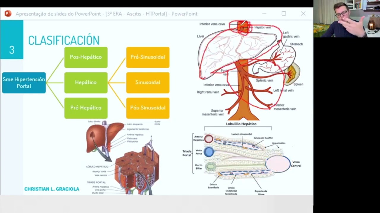
Um and the three features are that you've got disruption of the architecture, the normal lobular asin structure and with big bands of fibrous tissue going through it. And that means you're interrupting the normal portal blood flow in which is why you get portal hypertension. And then you've got these great big nodules that grow back.
So they've got a high rate of hpaticite turnover and that's what causes them to have an increased risk of hypathic cellular carcinoma.
Cerosis doesn't occur with acute liver damage. So if someone has an acute eskeemic hepatitis or one paracetamol overdose or acute thyroid hepatitis that doesn't cause cerosis. It's got to be something ongoing.
Um the causes of cerosis are broadly the same as the list I've put up before for causes of intropatic jaundice. Um except it's not including the acute causes. So I've taken out hepatitis A and E. Um, and I've taken out pies small overdose, but more or less the others drugs is extremely rare as a cause of cerosis. That shouldn't really be on there. I'm sorry.
So, the way I think of cerosis is that two problems uh which are um independent events. They're not mutually exclusive.
So you've got portal hypertension because you can't get the portal blood in and then due to l loss of normal hpaticy function you get liver failure or hpatocy failure but in most people with cerosis it's compensated in the same way that people with chronic kidney disease are not walking around with hugely high potassiums and clinically uremic. Um most people with cerosis have have got preserved synthetic function. So they're clotting as normal. They're not enkeapilopathic.
So it's common for most people with cerosis have features of portal hypertension but not hypathic failure until they have some kind of event that causes them to decompensate and then they develop a decompensated cerosis or acute and chronic liver failure.
But it's both of these together that cause your your features of chronic liver disease that you look for in examination. So, well, should we name some? Should we try and get to 15 pomethe?
Yeah, good one.
Baraces, Jupiter, hair loss. Yeah, lots of hairs.
We have to call it Nikia, but I except Luca Nikia. Yeah, good. We'll stop at 10. There's loads.
So, they're from a combination of both.
But portal hypertension per se, um the the problems really occurred due to your porto systemic anesmosis. So this is where the buildup of pressure in the portal system is then pushed blood through back into the systemic system to try and get out of the esophagus. They cause varies which I I'll talk about in a moment and back pressure along the splenic vein causes the spleen to enlarge. Um the clinical manifestation of that is hypersplenism where the platelets then get trapped and they get thrombocytoenia.
So low platelets in fact is a very good marker of um portal hypertension correlates quite well.
The anistimosis in the rectum cause hemorrhoids. Um they get capus because the umbilical the umbilical vein bless you recanalyzes and they get this cap medus. Um has anyone heard of portal gastropathy before?
So basically all over the stomach they can get these dilated veins.
Imagine the appearance of esophageal viruses but in the stomach extra um ascites occurs due to a combination of portal hypertension and liver failure. Um the mechanism is quite complicated but if you were asked to explain it you can just say the portal pressure backs up and there's poor reabsorption of fluid from the uh uh from the peritineal cavity which would normally be reabsorbed through into the portal circulation.
You normally do a um a peritineal tap to test it. Some patients have paracentesis. So where they take up off quite large volumes regularly.
Spironolactone is the diuretic you usually use to manage it in the first instance and then on top of it then they start adding in loop diuretics.
um SVP or spontaneous bacterial paritinitis is uh quite a dangerous condition where patients who have ascites and more rarely people who don't have ascites can get bad infection of the paratinium and they can become extremely unwell.
Oh dear.
Uh there are increased risk of portal vein thrombosis and has anyone heard of syndrome? Yeah. Yeah. Uh it's quite confusing but broadly they get renal failure due to their liver failure. If you treat their renal failure their li if you treat their liver failure their renal failure gets better in most cases of fatal syndrome.
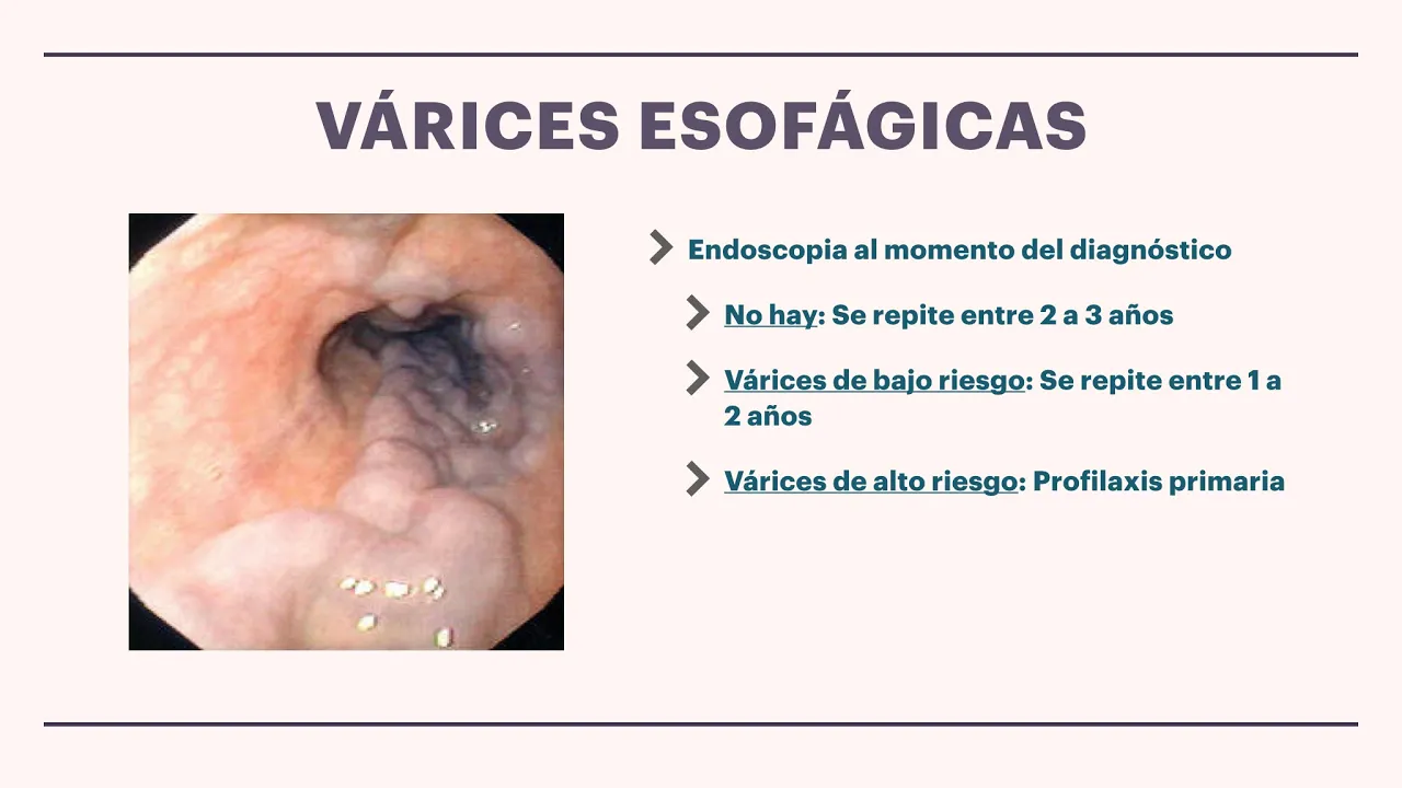
Faces sort of varicose veins at the base of the esophagus medically treated with beta blockers.
They're monitored with regular endoscopy to see if they're enlarging and then if they are then they can be banded. That's the best me method where they like the hemorrhoids tie a little band around them and they slowly drop off or scar therapy where they inject a scleros into um well fibros.
If they bleed then they get bad upper GI bleeds but I'm not going to go into the management of that today.
Yeah, beta blockers and regular OGDs.
Um so ascites just and is not always liver.
Um you can classify it like a plurusion into transidates and exidates. Exidates all your inflammatory things, transidates all your hydrostatic pressure things. So right-sided cardiac failure is quite an important cause of sites to remember in uh elderly middle-aged women. What do you need to think about for or consider the causes of ascites?
Yeah, ovarian cancer. Good. Um and then inflammatory and infective causes relatively rare.
What iatrogenic cause of a sites is there that you can think of.
What do you say? Shunt. Shunt. Yeah, good one. I had thought of that. So you can have a bit ventricular peritineal shunts in some patients with hydrocheilis and uh peritineal diialysis pouring 10 lers of fluid into their abdomen.
they're at increased risk of um they're increased risk of peritonitis.
So I realize we're going at quite the pace.
Please do stick your hand up or shout out if you've got any questions.
to compensated cerosis um the liver is still functioning and you haven't got any features of failure but you can still have portal hypertension and therefore still be at risk of varies and the regular OGDs and all this kind of thing.
So the three cardinal features of liver failure are encapilopathy coagulopathy and hypoglycemia.
Um but they tend to also come with worsening jaundice, worsening ascites, um infection which is actually the most common cause of death in liver failure and hypoalummia.
But obviously there's a whole host of reasons why they could have all these things. But if you're on the end of a telephone to the liver region, you're like, I'm worried my patients in acute liver failure.
So they're they're encapsulopathic and we're having to put them on dextrose to maintain their blood glucose and but their INR is maintained at 1.3. That's the kind of things they want to know. Do you have to have cerosis before is like cerosis like a stepping stone to failure or can you have a without cerosis?
Uh, yep. What's most common cause of liver failure in the absence of cerosis?
Yeah. So, decompensation of chronic liver disease would be in the setting of cerosis.
Um, but there are causes of acute liver failure. Most commonly paracetamol overdose after chronically chronic fat is cerosis.
Okay. Yeah.
Um, yeah, I can't really think of any other Yeah, I chronic liver disease implies cerosis.
Um, you can get funny things like non-cerotic portal hypertension though, which is interesting if you like portal hypertension.
Um, does that answer your question? Yes.
Uh, cryolopathy of liver disease is a It's quite a nice question for examiners to ask you. So why do people bleed with liver failure? The liver stops making clotting factors. You need bile to absorb fat. Vitamin K is a fats soluble vitamin and therefore you don't absorb any vitamin K. So that worsens your clotting derangement if you've got obstruction to the boflow. So people with obstructive jaundice should probably be on IM um vitamin K regularly.
um they've got low platelets and also the the toxins that build up in liver failure worsen their platelet dysfunction.
5 minutes.
Um so what one of my personal book bears hepatitis does not mean viral hepatitis it just means liver inflammation in general. Um, and you generally get this hepatitic on LFTs with raised ALT and AST.
Um, I'm raised bit of Reuben.
Reasonable tests to do are viral corology for the hepatitis viruses. What are what are the viruses are normally tested for?
CMV. Yeah, CMV and EBV.
And if you're testing for HEPS C, then often they'll test for HIV at the same it's reasonable to consent a patient to a test for HIV at the same time if you think that they've been exposed to the same risk factors.
Um need to test for auto antibodies. There are three three which are probably most important to test for which are antimicrobial AMA so anti mitochondrial antibbody SMA smooth muscle and anti- um anti- nuclear so ANA where ANA and anti smooth muscle are your autoimmune hepatitis antimitochondrial is very strongly associated with primary billary cerosis And then if it's an acute thing need to do a paracetamol level.
So quick way to remember hepatitis viruses A and E acute C stands for chronic B stands for both.
So A and E are only ever causing acute hepatitis and feal or fecal oral transmission.
C is only ever chronic and the most common method worldwide for transmission is iatrogenic.
So for example in in the Middle East when people go to the doctor the doctor has five needles and then if you're the sick person who needs a needle in the day then you get a previously used needle. So whereas in the UK the most um common method for transmission is introvenous drug use but it's got quite a high rate of chronicity unlike hepatitis B in the adult.
So in adults you're very unlikely to develop chronic hepatitis B whereas in children they are very likely to develop chronicity.
So, if you've got 40-year-old man with chronic hepatitis B and he says he once had sex with a prostitute when he was 35, that is unlikely to be the cause of his chronic hepatitis B.
It's much more likely that he had it.
Perinatal transmission from the mother.
So, just before we finish, I want to go through the minefield that is hepatitis B corology. um I've tried to sort of relatively simplify it down to two antigens.
So the surface antigen if that is present in measured in the blood HBS antigen um that demonstrates the person has some form of hepatitis B virus in their body at that time some kind of current infection.
If someone has the antibbody to the surface antigen, that means they don't have the antigen, they don't have hepatitis B in them, but they have been exposed at some stage. So they either have a previous infection that's been cleared or they've been immunized.
So when, for example, they measure our immunization level, they're measuring the antibbody level.
E is sort of a an antigen that comes out when hepatitis B replicates and it indicates current infection and replicating.
An anti-E antibbody means that you're mounting a response to an infection. So you'll only have that if you're um if you're stopping them replicate and you have been actually exposed.
So if someone's S positive, E positive, then they've got the virus and they're making more viruses.
If they're S positive but E antibbody positive then they've got the virus but because they're E antibbody positive they're not actively making anymore.
If you can imagine there's a spectrum between the two. So chronic hepatitis B patients have varying levels of E antigen E antibbody and then there's another one C and that's where it gets really confusing but if you just chronic inactive is S antigen positive E antibbody positive because this stops them replicating a previous infection is someone who has been exposed so they're S antibbody positive and E antigen E antigen E antibbody positive. So they've been exposed and they've stopped them replicating and they've had the virus and they've cleared it. Whereas if you've been immunized, you're only S antibbody positive because you've been given the S antigen immunized against it but have never actually had the virus.
sort of never mounted a response to hepatitis B replication.
In the textbooks they will go through more with C antibbody and antigen and dividing out IgM and IGG um which is nice but this is four which you can just sort of learn in the first instance.
Uh very quickly last thing PBC is only intropatic. It doesn't cause pillar strictures.
It's a classic it's a relatively classic autoimmune disease. So it's associated with thyroid disease. It's more common in women whereas PBC PSC primary sclerosin colangitis causes strictctures more common in men and is associated with inflammatory bowel disease.
So they sound quite similar but they're actually extremely different and both can cause sterosis but only PSC causes this uh show gallstones but um only PSC causes strictctures of the bile ducts.
Um I think we're more or less out of time.
Um just some metabolic disease nappled and chromatosis and subrenic absess and that's it.
Can I do nappled? So nappled is the so nappled is the most common cause of abnormal LFTs in the UK and the US. In the US, it will very soon be the most common cause of cerosis. So, it's pretty important.
Basically, it's the metabolic syndrome in the liver.
And um broadly, everyone with diabetes has some degree of napp. Nephold is the overarching term for fat in the liver, inflammation in response to fat in the liver and cerosis due to fat in the liver.
Um but broadly most people present at this stage when they have no features of napp left. that if you have a patient on the ward and you do their bloods and they have slightly abnormal LFTs and they've got a BMI of 40 and type 2 diabetes, it's probably nappable.
So it's important um metabolic disease really cool rally rare Wilson disease is too much copper um or you can't get rid of it from the pathocytes so they get liver damage and neurological damage and then they can have these acute events where they throw out loads of copper and they get renal failure and hemolysis.
Broadly it only happens to children.
Does anyone know a particular sign in the eyes that they say you get? Kaisen rings. Yeah. So um they can only be viewed correctly under a slit lamp. So uh if you're doing a hyp a a a liver examination or a GI examination and you you say to the the examiner, oh I can see kaichia rings. You sound pretty stupid. So you could say I could examine for Kaisopla rings if I had a slit lamp available.
So it's copper deposition in the edge of the cornea.
Alpha one trips deficiency is emphyma and cerosis.
Broadly people get problems with emphyma and not cerosis. They die from their lung problems not from their liver problems.
Hemocchromattosis is one of the most common autotosomal recessive conditions in Caucasians and it's due to excessive iron absorption not an inability to excrete iron because we all are completely unable to excrete iron. We can't actually get iron out the body and their features are that they get cerosis, cardiammyopathy and endocrine abnormalities.
And it's described as being the bronzed diabetes because they're meant to appear tanned and also get diabetes. But the diabetes is sort of mild type 2 diabetes, sometimes diet controlled um and often described as being slate gray appearance. So they kind of just look generally unwell rather than being kind of nice holiday to maicious type tan.
Um bonus question. Does anyone know what the typical osteoarthritis appearance for human chromattosis is?
Asymmetric panthropy.
Uh but you could get them with any kind of osteoarthritis but the the typical one is the first and the the second and the third MCP joints in the hand.
Yeah. So, oh, and then subrenic absess. Yeah.
Passing it bad. Very ill.
Any questions about anything to do with the liver or tree?
I appreciate there's a huge volume there. Um, but the most important bits, the bits went through first. So, LFTs, jaundice, gallstones,
Up Next

NAFLD and NASH: Diagnosis, Risks, and Management Strategies
@americanliver
12.3K views•2019-06-26

Graphic Medicine: Comics for Collaborative Healthcare Communication
@nationalpatientadvocate
189 views•2023-12-04

Neuroanatomy: Central and Peripheral Nervous System Divisions Explained
@AKLECTURES
136.2K views•2014-09-20

Stages of Labor and Vaginal Birth | Childbirth Animation
@nucleusmedicalmedia
52.1M views•2017-08-18
Related Study Plans & Knowledge Roadmaps
Structured learning paths in Medicine