












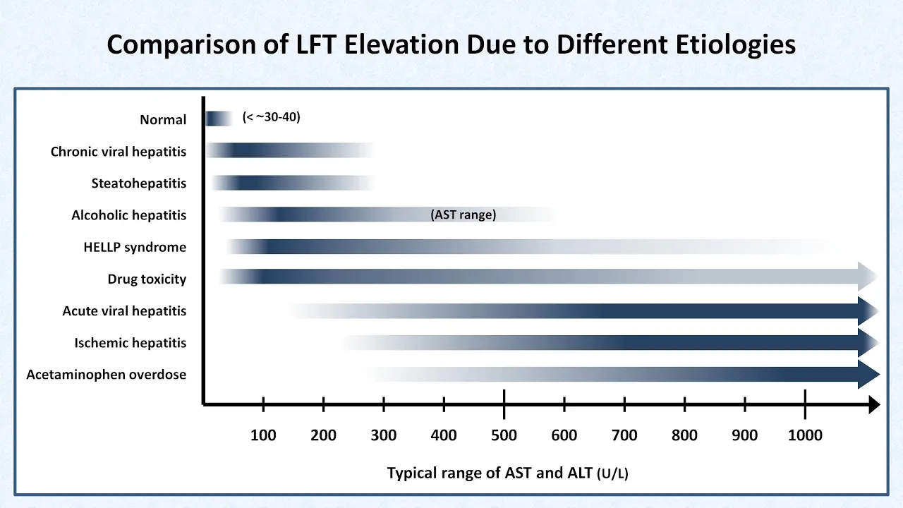



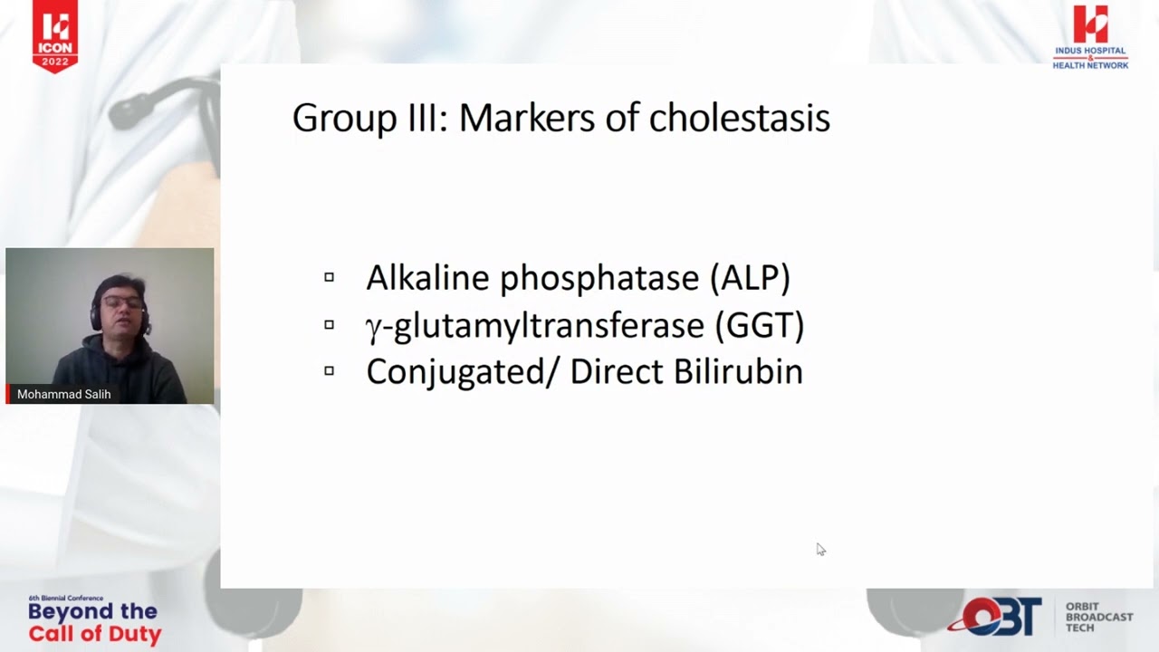

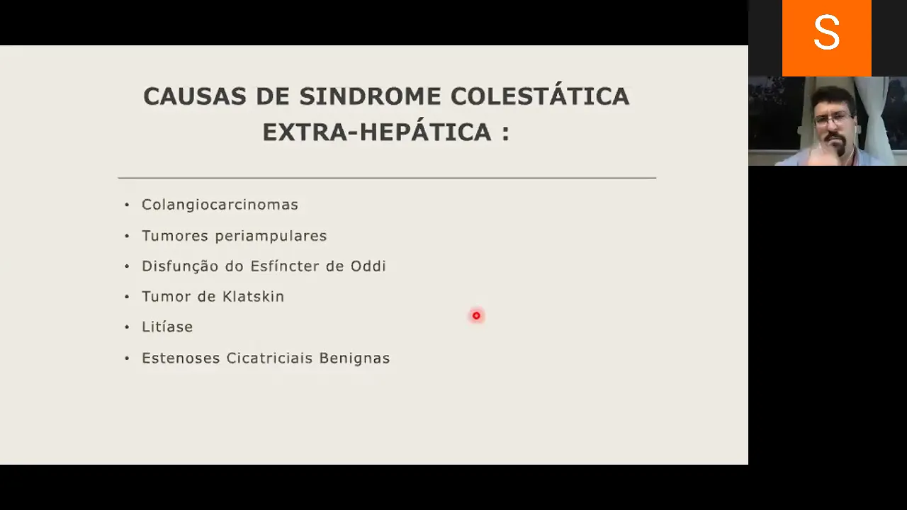





Liver Function Tests (LFTs) assess hepatic function and injury through specific enzyme and bilirubin markers: AST and ALT indicate hepatocellular injury (elevated in drug toxicity, viral hepatitis, ischemia, or metabolic diseases); ALP, GGT, and conjugated bilirubin indicate cholestasis (intrahepatic or extrahepatic obstruction); while unconjugated bilirubin elevation suggests hemolysis, Gilbert's syndrome, or rifampin effects. Diagnostic interpretation requires analyzing AST/ALT ratios, bilirubin fractions, and clinical context to distinguish between different mechanisms of liver damage.
Liver Function Tests (LFTs) Explained: Interpretation & Patterns | Clinical Medicine
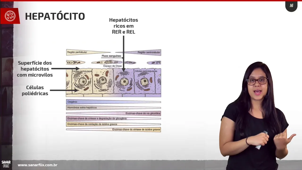
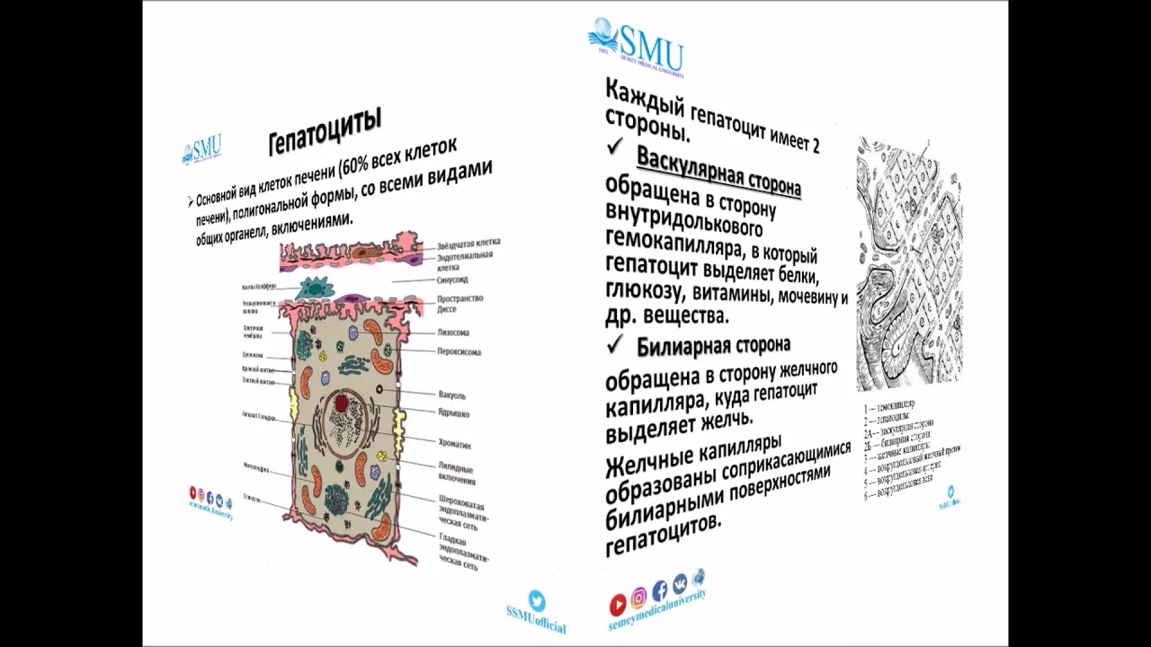
Added:Basic anatomy and histology of the liver, including the cellular structure of hepatocytes and the organization of the biliary tree.
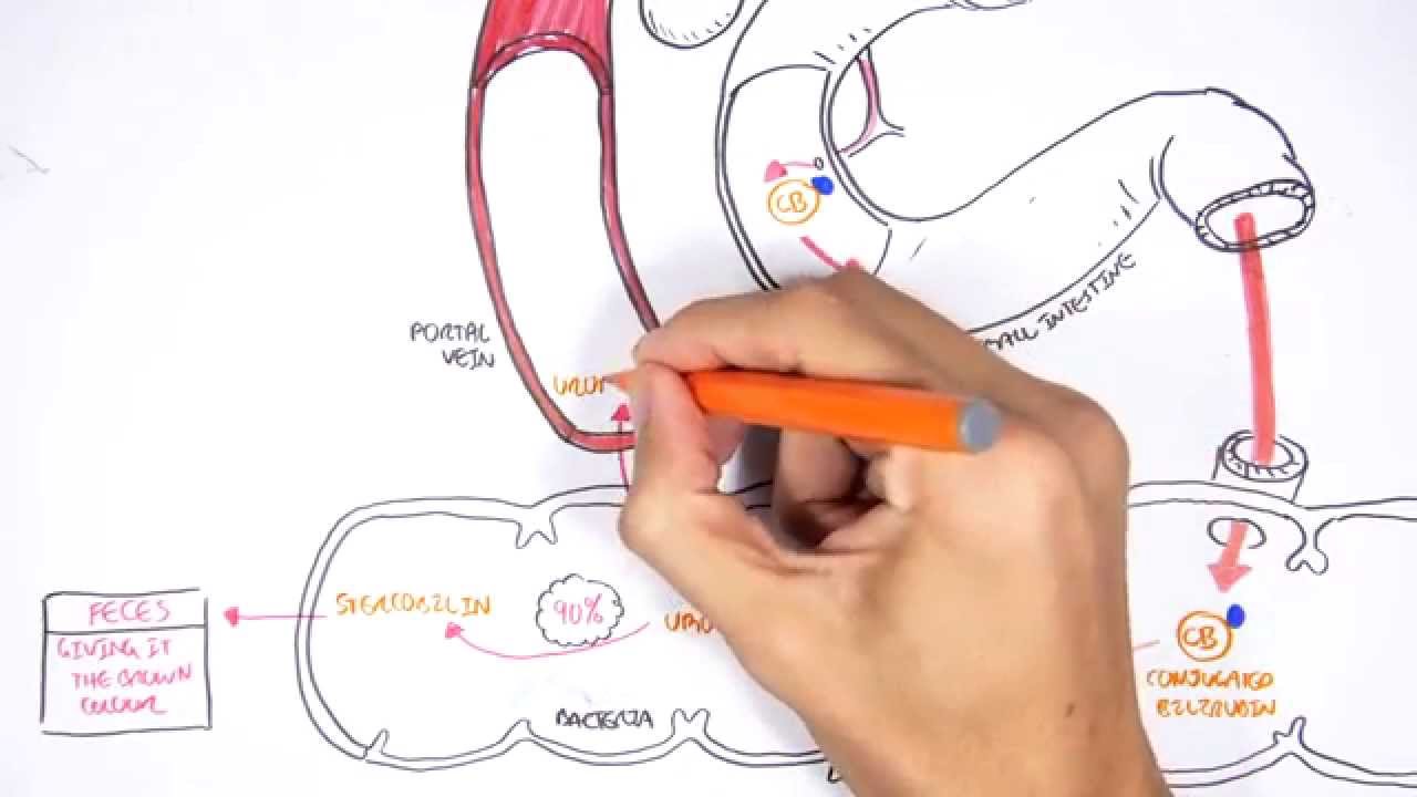
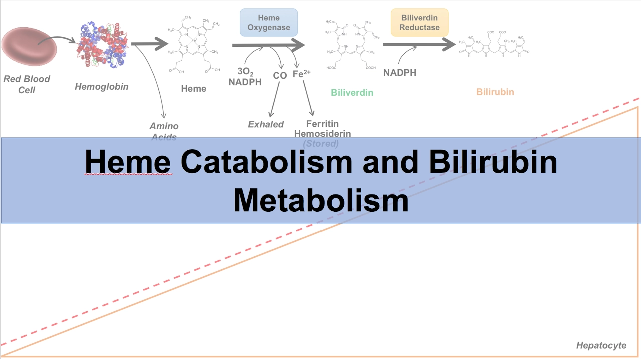
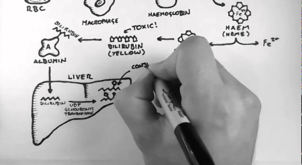
The biochemical pathway of bilirubin metabolism, including conjugation in the liver and excretion via bile.
The physiological role of the liver in synthetic function, specifically the synthesis of plasma proteins like albumin and clotting factors.
Fundamental understanding of cellular enzymes (specifically AST, ALT, ALP, and GGT) and what their release into the bloodstream signifies pathologically.
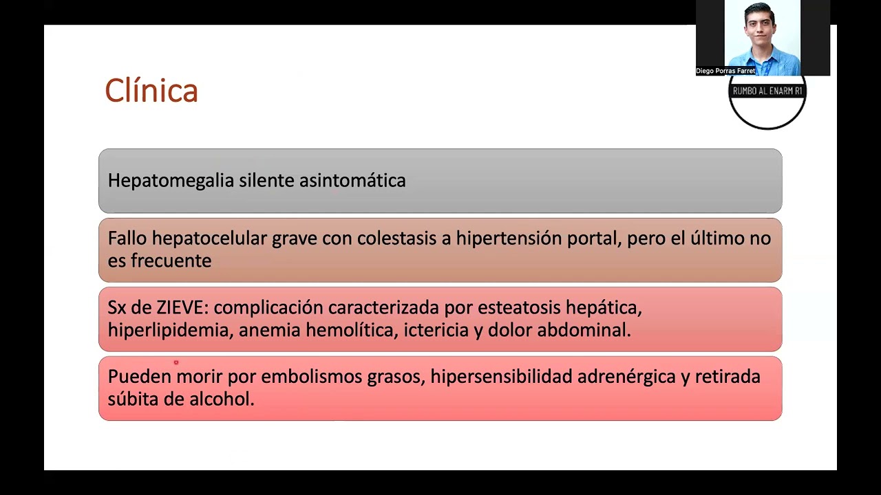
Differential diagnosis of specific liver diseases based on LFT patterns, such as alcoholic hepatitis, viral hepatitis, and drug-induced liver injury (DILI).
Advanced diagnostic workup for abnormal LFTs, including viral serologies, autoimmune markers, and iron overload panels.
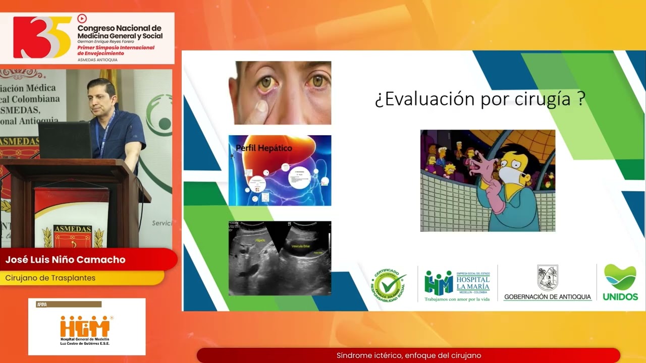
The role of hepatobiliary imaging (e.g., abdominal ultrasound, MRCP, ERCP) in investigating cholestatic LFT patterns.
Application of liver synthetic function markers in clinical scoring systems, such as the Child-Pugh classification and the MELD score for end-stage liver disease.
245.3K views7.8Klikes1:22:27Original Release: 2024-03-11
what's up Ninja nerds in this video today we're going to be talking about liver function test that's also known as lfts again this is a part of our clinical medicine section and I really hope that you guys are liking these videos I really hope they're making sense and they're really helping you to understand the topic really well and if they do please support us and some of the simple ways that you can do that is by hitting the like button commenting down the comment section and subscribing it really goes a long way also I really truly believe this if you guys go down the description box below click on the link that goes to our website become a member there's going to be many benefits to you that'll help you to understand these topics even better things that we have notes really good notes illustrations quiz questions and we're even developing exam prep courses so go check that out and really gain some understanding of these topics and master these topics all right let's talk about lfts so liver function tests is a really interesting term and sometimes I think it's a misnomer because when you look at lfts in the sense that you're trying to determine liver function it is really good it can give you kind of of a basic concept of basic hepatic function but also it's a misnomer because it also gives us a concept of liver injury and so when we talk about liver function tests I like to really call them like liver biochemical tests because it tells us about hepatic function and aob bilary injury so let's get a basic kind of introduction some small patho fiz and then we're going to go into some of these in more detail first thing is when we talk about a patient who comes in maybe they're coming in in jaist so they have a yellowish discoloration maybe they're coming in with rtopr quadron abdominal pain which we think that could be a hepatobilary injury maybe they're coming in with fulminate and cyop in these particular scenarios it's really good to have an understanding of hey I should probably send off some lfts for these patients and look to see do they have anything going on if you send off those LF te's there's three things that you want to be able to keep in your mind as you're working these patients up the first one is there evidence of a patoc cellular injury often times the way that we determine a patoc cellular injury is if I take this basic diagram here here's our aaty all right so here's the first part here is the hpat site this is just one component of the liver I'm looking at a bunch of these little hepatocytes here the next thing is we have an artery this is just representing the hepatic artery that supplies oxygenated blood to what to the hepatocytes so here I'm going to do this one in red here this is our hepatic artery right here okay now the other concept is that this one gives oxygenated blood this one removes any kind of particular deoxygenated blood and blood that's rich in relative nutrients that have been cleansed and cleansed of any toxins and so we call this one the TIC vein this is going to be removing blood from the liver and going to put that blood into the inferior vnea this one is going to be called the portal vein this one is taking a bunch of different blood from the git and from other organs Associated or accessory to the actual git and taking it to the liver rich in nutrients rich in toxins it's going to be cleansed and then the last one here is you have your bile duct or the bio canaliculi and this is really important because it actually helps to take things like bile which is rich in bile salts and Billy Rubin and cholesterol and take that down into the gut as we see here okay so these are the different components of our basic diagram we're we're going to use this a lot the concept I want you guys to understand is if there's injury to these Ayes they're damaged there's many different ways that we can damage these Ayes and when they are damaged one of the things that happens here is that they start to leak specific molecules or enzymes that are present inside of these cells and those molecules that are usually sitting here are going to be primarily a and ALT there is going to be other molecules in here such as Billy ruin but this is going to be the primary molecules that are indicative of direct injury or indirect injury to the ocytes so whenever have a patient gets these labs done the lfts and we see a a market elevation in the A and the alt this really tells me there's an injury to the osy and we'll talk about some of the reasons the ocytes get injured it could be directly from drugs and toxins Etc it could be from hepatic artery damage it could be from hepatic vein damage or it can be from biliary duct obstruction and we'll talk about those in more detail but if I see a massively elevated AAL I'm thinking of P cellular injury and we'll go through the differentials in the next topic if a patient then gets evaluated they're coming in again jaundice right precaution abdominal pain maybe incop AIC Etc then we should also get lfts and again evaluate is there another thing that's going on so same thing here hocy bile duct small intestine some bloodstream all right so what happens here is let's say that this patient has a problem and the particular problem here with chasis is there is a decreased movement of bile through the biliary system into the small intestine and there's two ways that this usually happens and we'll go into this in great detail one is we call this intrahepatic chasis and with intrahepatic chasis this means that there is not really a good movement of bile usually with inside of the liver so here's our Ayes here the biliary canaliculate within the or surrounding the aasy ites usually what happens is these hepatocytes make bile and they secrete this bile into the biliary system and then it's supposed to move through the biliary system and then from here into the small intestine to help to aid in digestion what happens here is that in these patients they don't excrete this bile and when they don't excrete this bile it builds up in the ocytes and then the pyes become agitated with that they become really affected by that and then what happens is these hepatocytes and biliary cells start secreting a couple different chemicals and they accumulate in a large amount and then what happens is some of these chemicals will back up into the actual circulatory system and these two chemicals that really are indicative of chasis is alkaline phosphate another one that we referred to here is what's called ggt and then the last one that's really also indicative of chasis is Billy Rubin and that's one of the components of the bile so again the concept behind having these elevations is that when the ocytes have difficulty excreting the bile the bile builds up inside of these hepatocytes or if there's injury to the biliary duct cells whenever there's injury to these cells they respond by increasing the expression of alkaline phosphate and Billy Rubin builds up in these cells and then moves into the the bloodstream and so when I see patients who have elevated Billy Rubin and elevated alkaline phosphate or ggt it really makes me think is there something going on in the liver cell or is there something outside of the liver that's obstructing the flow of bile and then it backs up backs up backs up into the bloodstream and this would be what we call extra hepatic kolasis and we'll go into some of the actual differentials associated with this one as well all right so two things I want you guys to think about whenever you see a patient who has elevated Billy Rubin Al fos ggt is think is there intrahepatic K stasis something where I'm not secreting B building up or I'm injuring the biliary duct cells and causing them to increase the expression of Al fos and ggt and as a result they build up in the bloodstream and then bile from the actual Ayes leaks into the actual bloodstream so you get increased Billy Rubin Al floss or ggt or is there an obstruction that's impairing the flow of bile and that's backing up into the blood the last concept that I want you guys to understand here is if we work a patient up and they don't show Market elevation a alt they don't show Market elevation in the AL fos The ggt and the Billy Rubin they only show maybe maybe a combination of a bunch of things and one of the things that we really want to understand is hyper bill rubinia this is really kind of the temperature the thermostat of what really liver function and liver injury looks like so there's a couple different ways that Billy ruin is kind of like moving through the actual bloodstream in the liver it's really important to understand it we have a basic idea here here we have like a maccrage and a maccrage is going to be one of the main cells that eats up red blood cells when it eats up red blood cells it secretes that molecule into the bloodstream and it's called Billy rubben and I'm going to put next to this Billy Rubin I'm going to put what we call so there's two different types of Billy Rubin one we call it unconjugated meaning it's not bound to glucuronate or it is conjugated it's bound to glucuronate this one is not so it's called unconjugated or indirect Billy so I'm going to put I Billy which is just a fancy way of saying this is unconjugated Billy Rubin this is released from the macrofagos from the macras it gets to the ocytes it gets taken into the ocytes from here there's an enzyme it's called U GT and urine gluc glucuronic transferase enzyme and it puts a glucuronate on when it puts the glucuronate on it's now converted into conjugated Billy Rubin which we're going to call so this is Billy rubben but it's a specialized Billy Rubin that we call Direct Billy Rubin or debilly it's a fancy word for conjugated Billy Rubin then that debilly is then going to be moved via transporter into the bile system and then from the bile system they'll come out into the small intestine or they aid digestion a couple different things will happen in this process that you have to be able to understand when patients have diseases we want to be able to understand which is the type of Billy Rubin that is elevated because you can have an elevation in both of these or one of them so there's three different types that we will discuss here one is if the bloodstream has elevated levels of indirect Billy Rubin we call that unconjugated hyper B Rubin emia so I want to know do they have unconjugated and I'm going to put Billy and I'll put an up Arrow to indicate that they have elevated levels of unconjugated Billy ruin do they have mixed do they have a decent degree of mixed Billy so in other words they have elevated both unconjugated and unconjugated and then lastly is it primarily elevation and only conjugated bility and we'll talk about what this particularly the differential is and what this can look like often times for these patients when you think about unconjugated it has to happen from the maccrage to this point all the way until we get to this point so any disease process has to occur above that enzyme or at the level of the enzyme anything from mixed can be a combination of disease that's occurring anywhere from here all the way to here and then conjugated is it's only occurring from this point after the ugt down to the small intestine so we'll talk about all those differentials but often times the primary thing is that these patients will present with an elevated Billy Rubin they can also present and that's what's going to help us here they can also present depending upon the type that they have they may present with a plus or minus we'll put here plus or minus an elevated an a alt or alos but the constant the the actual thing that you need to understand is is it depends upon which type and we'll go into that often times if you don't see any of these elevated and you just see unconjugated ailly it's homolysis and we'll go over each one of these I don't want to get too far ahead because we'll discuss these in detail but that's the big thing to think about does the patient have elevated as alt it's a p of cellular primarily do they have an increase in the alkaline phosphate ggt with Billy it's cholestasis do they have an increase in Billy and that's the primary one that we're trying to think of you want to know which type of Billy Rubin is elevated and that'll help us to think about the differential all right this is the concept of aatos cellular injury what about the test that we use for a patic function we don't have great tests for hepatic function we're better at being able to determine tests for hepatobilary injury but there is some test that we can utilize here one of the tests that we utilize for hepatic function is that the liver produces a particular molecule here and this molecule is called albumin and albumin is a plasma protein and what it helps to do is it helps to be able to regulate the oncotic or osmotic pressure in blood vessels so if you have let's say a disease and we'll talk about two particular diseases here one is you have a disease called curosis which is like a chronic liver disease it's like a slow kind of like simmering liver dysfunction over many years or you have an acute abrupt onset of decreased hepatic function in the scenario of acute liver failure you may really really see a lot of this decrease in hepatic function become super obvious so one of the things is that your albumin levels will drop if albumin drops then we don't maintain as much of this osmotic or oncotic pressure in the bloodstream so this is going to be impaired there's a decrease in the osmotic pressure if there's a decrease in the osmotic pressure as an effect of this so we'll put here on the side a decrease in osmotic or oncotic pressure it's the same dang thing the process here is that fluid is going to leak so it's going to cause capillary leakage and whenever fluid leaks out of the capillaries it leaks into particular types of interstitial spaces or cavities so what are some of these well one is it'll leak into the plura and it'll leak into the actual parium and so they'll develop potentially what's this what's this called whenever you have lots of fluid building up in the actual parum aites what's it called when you have fluid building up within the plural cavity plural affusions so often times these patients can develop what we call plural affusions and they can also develop something called aites so watch out for a patient who has decent amounts of plural affusions as a result of the significantly decreased alumin as well as watch out for aites and this would be aites that has a sag that's usually less than 1.1 because it's more of an exudative type the other thing is that fluid can leak into the interstitial spaces in the legs and these patients can also develop some pretty good edema so look for any kind of like generalized pedal or pitting type of Edema so we'll put pitting edema this is a big thing to watch out for all right so that's one of the big things to watch out for without buum is a lot of more ademus types of appearances the other thing that we really used to kind of give us an idea of hepatic function is that the liver also produces a lot of what's called uh anti-coagulants and procoagulant so it'll produce a bunch of different molecules here and if in this scenario we don't produce albumin because the liver is dysfunctional right so it's damaged because of acute or chronic deliver disease so acute is usually just a profound AOC cellular injury or a profound types of damage to liver without ability to compensate and regenerate where sorosis it's it's repeated injury with fibrosis and nodular degeneration either way we're not going to produce these clotting proteins what are some of these clotting proteins that are often times produced by the liver in normal healthy individuals well there's what we call Factor two Factor 7 which is a really big one factor nine Factor 10 and then another two which are are three technically if you want to think about these other ones we call this one anti-thrombin 3 which is this guy and then Pro protein C and protein s there's lots of these but the big ones that I think are really really important here are going to be these ones right here because these are what we call procoagulants these are what we call anti-coagulants in liver disease where you lose the ability to produce these things you're going to have a decreased number of these and a decreased number of these but if something happens to these patients that tips them out of this abnormality they can definitely have less of these especially the procoagulants and this is the problem when you have less procoagulant these are things that wants to induce clots what do you think is going to happen these patients will now not be able to form clots so they have an increased risk of bleeding so they're going to have an increased risk of bleeding or some type of disease that we call where there's inappropriate ability to form proper clots or in that scenario what do we call that we call that coagulopathy and the ways that we look at this is two particular ways one is that if you have a decrease in the procoagulant we see this Pro potential process bleeding and we'll talk about what that particularly looks like but the other concept here is when you have less of these procoagulants it Alters a particular lab value and one of the particular lab values that's really dependent upon Factor 7 is called the PTINR and what we will notice in patients with really profound liver failure is that their PTINR will start to go up at least to greater than 1.5 and that's usually indicative that they're not able to properly clot so that's another way that we can look for this type of profound decrease in hepatic function or we can look at it clinically where they develop bleeds in the brain IES they develop bleeds from the nose or from the mouth or on the skin like peal lesions so mucocutaneous bleedings or intracranial hemorrhages or they bleed from the git such as hematemesis or maybe a lower GI bleed these are big things to think about and so something else that actually worsens that coagulopathy and increases the risk of bleeding is the liver also produces a hormone called TPO we call this thrombo potin and when Throop potin is not able to be produced it's supposed to tell the bone marrow to make platelets you won't make the platelets what does that call when you have decreasing number of platelets thrombocytopenia so these patients can have what's called thrombo cytopenia relatively chronically and that definitely propagates the risk of coagulopathy it won't affect the INR but it will cause them to maybe be a little bit more coagulopathic the last thing we'll talk about this more in acute liver failure and curosis we just don't have tests for these that are really really great um one thing that we can also look at to a certain degree is that these patients may lose the ability to clear ammonia and so ammonia levels May be potentially elevated in patients with underlying hepatic dysfunction and the other thing is that Billy ruin is a marker of liver injury but it's also a marker of liver function that's weird right it can be a measure of both and so when patients also have an inability to conjugate Billy Rubin and excrete Billy Rubin they may potentially have an increase in Billy that's maybe not even related to the liver injury itself it could just be related to the inability to conjugate or SC Billy Ruben and these are also different tests but often times the B big ways that we kind of do this on the exam is by looking at PTINR risk of coagulopathy and low albumin there used to be other tests that we would do with these such as looking at ammonia looking at Billy Rubin levels with certain types of weird things we don't often do that anymore because they're kind of scary they used to do things like check the Billy Rubin levels after you give a patient refampin if they have liver disease or there used to be one lady who used to say let's give ammonia to to people and if they have good liver function they'll clear it if they don't they won't clear the ammonia and they'll develop incopy we can't do that to patients that's not ethical so we don't really do those things anymore but that gives us an idea now of how we should look at these tests the big ones that are really going to be tested on your exams is going to be this one so we're going to go into each one of these into great detail let's do that now all right my friends so now a patient comes in they get their lfts done when you get the lfts back you see oh their a and their alt is really elevated I think it's important to have an understanding of is this an acute enzyme elevation is it a chronic enzyme elevation so does it look like if you have a comparison to their prior lfts what does it look like does it look like it's about what it's usually at or did it look like it really jumped up acutely into some pretty high numbers and when I consider like an acute like massive elevation I'm looking for really at least greater than a thousand so really to call this like an acute and significant elevation I'm looking for levels that are greater than a thousand of the A and the alt and that's pretty significant when I see acute and massive elevations in the lfts the St and the LT there's a couple differentials that will come to my mind that these are the only real things that can cause massive elevation the first one is I like to think about drugs so we call a Dilly so a drug induced liver injury and there's really only one of these drugs that has been really commonly known to cause pretty significant hepatotoxicity have given in large amounts at least way more than four grams per day and that's usually Tylenol so that's one thing is I'm going to look at the patient's history maybe I'll check a Tylenol level and if they're really taking large amounts of Tylenol and their level suggest that that could really be indicative of a Dilly that caused this greater than a th bump the other one that I think about is vascular so vascular causes which will go into pretty significantly this is usually going to be a really really big one and I'd say out of the vascular causes two are probably going to be the most significant and that's usually arterial and the other one would be like a bud kiari syndrome so vascular causes and I'd say the biggest one out of all of these is going to be es schic hepatitis and we'll talk a little bit about that one a little bit later the other one that really causes pretty significant elevations is viral so usually viral infections are going to be a big one um and there's really a couple I'd say he hepatitis A is relatively rare Hepatitis E somewhat common especially in pregnant patients Hepatitis B virus especially with co-infection of D has been shown to be a pretty common one that can cause both acute and chronic another one is going to be autoimmune so autoimmune disease can kind of like be a slow and Kettering one as well with chronic elevations but if you get a pretty good autoimmune kind of attack your your patients can have pretty significant Mass massive elevations in their enzymes and the last one I really don't like to kind of avoid just because it really can cause these problems is when a patient develops an accute biliary obstruction usually like a kocal aasis these can really cause some specifically um decent elevations in the As and the alt so these are the things that I want you to think about if a patient comes in and they definitely have increasing ests and alts think is it massive if it is you already have like a differential to go through and we'll talk about these a little bit more if it's not that and it's a little bit like lower and I like to use like usually maybe it's chronic or it's very small elevations like less than 500 would really kind of make me think that it's not necessarily one of these things the things I like to think about in this particular scenario could also be Dilly so this would usually be more particularly alcohol uh related and we'll talk a little bit later cuz alcohol has really been linked to this like particular ratio um and one of the things that we often say a lot is that alcohol that ratio is usually where the a to alt ratio is usually greater than two so the ratio there I think is helpful as long as it's in the lower numbers less than 500 as you get up to the higher one I don't really feel like this ratio consistently kind of works but this is something to definitely think about on your board's exam the other one would be metabolic causes so metabolic diseases I know that a lot of the time PE people will think that that if a patient comes in and they have like these massive elevations that it could be Wilson's disease or hemocromatosis as much as we want to will that into existence I don't think that's the case you'll be looking at right case reports often times Wilson's disease won't generally present as much acutely and neither will hemocromatosis and the other one that generally won't present with these massive ones is going to be uh non-alcoholic fatty liver disease so those three particular ones I would really be thinking about in the non-alcoholic fatty liver disease patient I would also be thinking about this in the Wilson's patient and I would also be thinking about this in the hemocromatosis patient okay but these are the ones that I would definitely potentially consider if I'm looking for a metabolic disease obviously this one obesity hyper lipidemia uh hypertension other kinds of factors of obes U metabolic syndrome this one Kaiser flesher Rings CNS dysfunction this one hyperpigmentation pancreatic dysfunction with diabetes and restrictive cardiomyopathy are common the other one that I think is really important is going to be like viruses but it's often time a very particular virus and this is usually hepatitis C virus sometimes Hepatitis B virus as well but hepatitis C virus is the really the big one that I want you to associate with more of that chronic types of liver disease and then the last one is pretty common and this is therosis therosis is obviously a very chronic dysfunction of the liver over time so this is also because as you cause damage to the liver as you impede more of the liver function function by causing exessive fibrosis and aul degeneration you lose cells that have the capability of releasing as and ALT so levels of levels will actually slowly dropped almost being normal over time a pseudonormalization so let that help you to guide what your differential is right off the get-go but then once you get these levels of an elevated as and ALT develop a differential that it could either be direct prink injury vascular dysfunction or finally could it be a biliary obstruction so the first first thing is what if it's a vascular dysfunction there's a couple different things here so here we have the TIC arter remember I told you guys remember this diagram this one delivers oxygen rich blood to what to the liver well what if there is a decreased delivery or no delivery of oxygen to these ocytes that would be a problem and then as a result these cells will start becoming injured as they get injured they'll pump out what increas amounts of a and ALT in this particular situation we already kind of talked a little bit about this this is a vascular cause and Vascular causes such as arterial related this will cause massive elevations this is usually what we refer to as es schic hepatitis and often times the most common cause of a patient developing aeic epati is going to be profound hypotension so I really want want you to look at this when a patient has really really low blood pressure and this could be seen in a couple different types of shocks you could see this in cardiogenic shock hypemic shock septic shock so look for a patient who is in overt shock and this is caused decreased perfusion to the tissue decreased oxygen delivery and a Pell injury so that'd be one type of vascular dysfunction so that' be the AR Tre related type what if it's a Venus related disease so the Venus system is supposed to take blood from these ocytes from the sinusoids and eventually return this to the inferior vnea what if though in this scenario there is an obstruction or high pressure high back pressure that's impeding the outflow this will lead to what we call Venus congestion and that Venus congestion if blood is not able to leave the liver it causes higher liver pressures higher cyos soidal pressures and those pressures will definitely start leading to injury to the liver because it's not able to drain properly there's a couple different things I want you to think about here one is what if the back pressure is really high they have what's called an elevated Central Venus pressure that would be in the scenario of what we call right heart failure so look at the patients's echo look to see if they have a history of right heart failure and the other one is a clot and this is usually what we call Bud kiari syndrome usually you see this in a patient with hypercoagulability you see this in a patient with malignancy or you see this in a patient with poly cymia but either way blood is not getting out because there's a clot or there's high pressure that's preventing it from leaving the next one is going to be pinkal injury all right so this is a really really big one and this is where there is direct injury to the liver cell itself and as you damage these liver cells you'll see these pumping up levels of a and ALT there's many many causes of these we're going to need to write a couple of these down so the first one and I think by far the biggest thing that you want to be considering and a patient with massive elevations is did they take any drugs it's the first one that I think is really really important so did they pop any Tylenol pills in large amounts all right that's the first one or do they have have a history of chronic alcohol use there's a lot of different things that we can try to utilize to our advantage with this with drug induced you can check a Tylenol level if they're taking at least levels greater than four grams per day this is definitely going to cause this type of problem the other one is alcohol alcohol use excessive alcohol use can cause elevated ggt levels but also affect that ratio that we talked about the as to alt ratio greater than two so think about elevated ggt history of ethanol use and think about the as to alt ratio the other one that I think is really important that really caused this is metabolic diseases all right especially more of the chronic kind of like slow and Kettering elevation and that types of causes there's many of these I don't want you to get too caught up about all of these we talk about them a lot more in the inherited liver diseases but this is going to be things like hemocromatosis often times with this one this will cause more of the slow kind of like slow chronic elevation Wilson's disease slow chronic elevation alpha 1 anti-rip and deficiency slow elevation and the last one is non-alcoholic fatty liver disease that'll cause a slow elevation with metabolic diseases the things that will make you think about these is obviously they won't be massively elevated but you're going to have a bunch of different tests for these we'll talk about them a lot more but this one's related to elevated iron elevated copper decreasing levels of alpha 1 and this is usually associated with metabolic syndrome so we'll write down each one of these think about elevated iron levels elevated copper levels and metabolic syndrome in other words the patient with obesity kind of like slowly elevated uh glucose levels because of what we call insulin resistance hypertension dyslipidemia there's one other thing often times with a non-alcoholic fatty liver disease what happens is their ratio also changes a little bit too and so sometimes we'll see kind of an opposite you'll flip the ratio with alcohol in comparison to non-alcoholic fatty liver disease so you'll start seeing that their alt in comparison to their a will actually be flipped a little bit and so that's an important one to remember all right so these are the big things to remember here the last um kind of thing that I want you guys to remember besides metabolic diseases is autoimmune so autoimmune is this kind of like really interesting disease it doesn't often cause like super obvious presentations that you would think uh so in patients with autoimmune hepatitis maybe you'll get lucky and they'll have some other autoimmune diseases maybe they'll have some joint diseases and other things like that but often times you won't see anything specific and so often times when a patient has a massive elevation in their enzymes you think autoimmune and you want to test for a couple different antibodies that have been linked to directly injuring pra a couple of those are going to be Ana anti-smooth muscle antibody IGG and if you feel a little extra more particularly in the younger patient population you can even consider an anti lkm1 uh liver kidney microsomal one antibody but often times these are the big things that at least this one is the highest yield one the anti-s smooth muscle antibody that you really want to think about so these are things to think about in your differential drugs metabolic diseases usually this will cause other features like pancreatic injury so diabetes restrictive cardiomyopathy this one Kaiser flesher Rings CNS dysfunction this one osma this one usually you're seeing Pro prominent features of obesity the last concept is bilary obstruction when a patient develops an a bilary obstruction usually the most common cause is they get a stone and when you get a stone inside of these like biliary ducts so here we have like a bile duct and in this bile duct there's a stone that stone will cause a massive back pressure to kind of be exerted onto the liver on top of that things like bile will leak into these and bile acids and bile salts love to cause direct injury to the ocytes so there's a lot of oxidative stress there and that will cause these massive elevations in as and ALT the big thing to remember for this one is by far the most common cause for this is going to be kocal lithiasis so I would really really look for this one in a patient with KOCO lithiasis and then maybe a patient with really profound elevated levels with an infection appearing so something like ascending colitis so we call this ascending colitis all right one other thing with biliary obstruction that's really interesting all of these that we talked about will cause massive elevations in ASD and ALT usually alone with biliary obstruction the big thing to remember is this one will also cause a market because it's a chasis effect you'll get Market elevation and alkaline phosphate ggt and you'll get elevation in Billy Rubin within the bloodstream you can get this with some of these other ones but not to the same like degree that you'll get with aary obstruction so really think about that in the differential when you see massive elevations in Al flos ggt and Billy Rubin in a combination with elevations in as and ALT all right now let's go into the diagnostic approach of this so we have an idea now of the pathophysiology really of this we kind of have the causes we broke down a lot of these and I think now we can get generate a pretty good diagnostic approach elevation and asdl okay I remember off the R value if it's really elevated and I don't really have any significant elevation on my alos ggt it's probably a PO cellular I can say the RV value would be in this scenario greater than five if it's a Pell then I got to start thinking of the causes if it's a Dilly then there's two types Tylenol toxicity so I'd have an elevated Tylenol level and on top of that with Tylenol toxicity what kind of elevation do they have massive elevations greater than a thousand that would suggest the Tylenol toxicity if it's like less than 500 it's not super high and their as to alt ratio is greater than two that suggests more of that alcoholic liver disease patient vascular causes are always going to be pretty high you're at least going to have them greater than 500 maybe even greater than a thousand because of that again think about the scenario itself if I get the liver ultrasound with Doppler flow I'll be able to see a clot in the aaic veins and if that is the case it's likely buari if I get an echo and I see that they have a very blown out dilated right ventricle with a poor systolic activity then I can say it's probably right heart failure with high central venous pressures and then lastly if they have a history of recent hypotension or profound shock with lactic acidosis multi-stem organ dysfunction and on top of that a massive elevation in lfts against probably I say it's es schic hepatitis the next thing is if I have these hepatotropic viruses so a e and even some degree B if it's massive elevations I could say that it's probably those acute uh hepatitis like A&E and if it's less than 500 I can probably go to the length of saying that it could be Hepatitis B and C again Hepatitis A and E are going to be more fecal oral transmission b c are going to be more bloodborne or sexual transmission you can see the other types of viruses such as um herpes simplex and CMV but it's usually more in the imuno compromised state but again this would fit in that category of viral hepatitis and a patient who has a massive enzyme elevation especially in the thousands I would always send off an Ana an anti smooth muscle antibody and a soluble IG and if that comes back positive you have a pretty good confidence that they could have autoimmune hepatitis but they may need a biopsy to confirm especially if you start them on steroids the next thing is I'm kind of running into my metabolic disorders so this is going to be those metabolic diseases so first thing I would want to know is do they have non-alcoholic fat liver disease is there obesity is there hyp lipidemia is there hypoglycemia or diabetes if that is the case it's probably non alcoholic F liver disease and especially look at that alt a ratio alt should be greater than a do they have any evidence of uh empyema as well as a reduction in their alpha 1 antirion levels that could be alpha 1 Anson deficiency is there transfer saturation and fertin levels high that tells me that iron is very high in the blood bound to transfer and iron is very high in the cells bound to fertin so these levels are going to be high and then if I really think that it's a primary cause of hemocromatosis check the HF Gene it should be positive plus look for restrictive cardiomyopathy hyperpigmentation and even potentially diabetic like diabe enic like states that would suggest hemocromatosis lastly does the patient have an increase in urinary copper so much copper in their bloodstream spilling over into the urine or a lot of copper that's binding up ceruloplasmin and reducing the free levels of ceruloplasmin if so that suggests Wilson's disease especially if they have Kaiser flesher rings and movement disorders that are present this is a way of being able to diagnostically approach this in a more simplistic way all right my friends so now the patient has come in they're presenting jaundice right right usually if you have a patient who comes in and they kind of present a little bit weird and they seem like they maybe have some R uper quadrant abdominal pain maybe they have some iny opathy or they have jaundice you check those lfts but I think really more particularly in the setting of jaundice you definitely want to be checking the lfts and be thinking is their chasis so so you get the lfts the lfts definitely show that these particular things are elevated in the bloodstream they have an increase in Billy Rubin they have an increase in alkaline phosphotase and they have an increase in ggt if these are elevated it definitely super suggests that this is a chasis type of problem and so then you have to start thinking okay what was that differential that Zach said prior well it's either intraop patic problems with movement of bof flow or production of Bio or it's an obstruction extra hepatic so outside of the liver usually involving the common bile duct so let's say that there is an elevation in these particular molecules we then have to start kind of dissecting through this and think thinking what would cause intrahepatic colestasis and there's two things one is their hepatocytes are supposed to produce bile right that's their that's one of their big jobs and so it should take and produce bile and put bile from the Ayes into the biliary ducts right intraa but let's say that there's certain types of problems diseases that impede this ability to secrete bile if you can't secrete bile particularly a couple things build up in the cells and one of them is these like bile acids or just generally lots of bile builds up as bile builds up this can cause aat toxicity it increases oxidative stress and so it will start causing injury to the ocytes and so you may see a little bit of an elevation in the a a little bit of elevation in the alt that's one thing but here's the problem when there's lots of bile because you're having difficulty being able to do this particular process what this does is for some reason it signals the aaty to start increasing the expression of another molecule that we put out here into the bile system you know what that molecules are Al fos and ggt it's a weird mechanism we don't have a really great understanding of it but that's one particular thing that'll happen is you'll increase the expression of alkaline phosphatase and increase the expression of ggt so it'll kind of really put a lot of stress on these hepatic cells and the biliary cells and it'll stimulate this increased expression if we have more alos and more ggt being expressed in the biliary tree these will eventually leak into the bloodstream and so now some of these molecules will start trickling their way out and trickling their way into the bloodstream and that will cause the elevation the alkos and the ggt is it's a response to inability to excrete bile into the bile system causes a increased expression of alos and ggt that causes them to leak into the blood but how's the bile particularly the Billy Rubin component getting out well when bile is built up one of those components is Billy Rubin and Billy Rubin is a really important component here and so I told you that some of these ocytes may get injured and if the Ayes may get injured they may leak specific substances out here into the bloodstream and one of those particular components is called Billy ruin so we now see how inability to excrete B can lead to a transient rise in alkos in gdt and the release of Billy ruin from the ocytes into the bloodstream what's causing this particular problem is the question there's a couple things one is what we call intra we call this chasis of pregnancy actually we call this K stasis of pregnancy this is a huge huge one cuz in patients who are in like their close to third trimester or going around that they somehow create this weird effect where usually there's high levels of estrogen and high levels of estrogen have really been linked for some reason to inhibiting this particular process so high levels of estrogen are definitely linked to the inability to excrete particular bile components into the bile system so that's one thing that can definitely do this that's particularly a problem another one is medications and I bet you can't guess what are some of these medications that have really been linked there's so many but one I think that's really really important to make it easier on you is what's the jugs that really increase estrogen oral contraceptives and so oral contraceptives have been really really heavily linked to this as well to increasing the estrogen content and therefore inhibiting these Transporters from excreting bile those are two big things that really are impeding this process there's many other medications like anti-epileptics like phenin and carbamazapine and a bunch of other stuff but I don't want you to focus too much on those really think about these two the other things is that there's something that's actually causing either fibrosis or inflammation of the intrahepatic ducts so what if these intrahepatic ducts are really really inflamed or there's fibrosis there right that could be potential or infiltrative diseases there so there's two things that I really want you to think about here this is a really really big one one is what's that disease called introp patic inflammation of the biliary Ducks only those PBC so I really don't want you to forget about this one called PBC primary biliary colangitis the big thing to remember associated with this one is the anti mitochondrial antibody that is particularly induced and related to this one it's that autoimmune type of process there's one more could be bunch of different diseases I don't want you to get too crazy here but infiltrative diseases are a big one as well this could be malignancy this could be things like sarcoidosis Etc don't get too crazy really think about this one as a really really high yield one and really think about this one okay the reason why is these are really great because this gives you a good historical feature on the exam and this one's probably one of the big ones that has an exam related antibody that's associated with it okay either way in these particular scenarios these are a lot easier because now they're impeding the ability of alkaline phosphate ggt and what's another molecule that's actually present inside the billiary tree if this is working Billy Ruben I'm going to put Billy Billy's also going to be here as well and if you have something that's impeding the flow in the liver that's going to lead to these guys backing up and going back into the bloodstream so PBC infilt diseases will impede intrahepatic flow chasis and medications will impede the secretion of bile that's the big component there all right now we come to the one that's like super easy and very very very common what if I have something that I'm producing bow well it's flowing through the intop patic ducts well but then when it gets out here and it's running through the extratic ducts like the common bile the common aaic duct something is obstructing the movement of of this flow here now we're outside of the liver what are some potential diseases that really obstruct the flow and prevent this bile from moving forward causing ggt Al fos and Billy to not be able to move forward they end up back flowing into the blood so what are some of these things that could actually obstruct the flow what if there's a stone I think that'd be a really really big one here so what's that called when you have a stone blocking up the common B duct it's kocal aasis so if a patient has kocal lithiasis that's a really big one or if they have an infection of the bilary fluid proximum to that like ascending colitis that could be some big ones so really be thinking about a patient with a profound radon abdominal pain and profound jaundice and increase in these enzymes definitely want to think about this one the other one that's really really important here is going to be primary sclerosing colangitis so primary sclerosing colangitis this is interesting because it's not aone so this one's a stone so this one's a stone this one is usually some type of stricture so it's usually like inflammation chronic inflammation of the intra and extratic bilary ducts that causes strictures and really impedes flow this one's really really interesting it's kind of difficult to diagnose but we'll talk about some diagnostic tricks a little bit later the last one is if it's not a stricture it's not a stone it's often times a tumor and so usually a pancreatic carcinome should definitely be on the differential here as well especially in a patient with chronic pain right they have hypercoagulability of some sort and they have jaundice you definitely want to be thinking about this one and the concept behind this tumor is it's usually at the head of the pancreas and you guys got to remember the bile duct it it actually comes down and then it joins with the main pancreatic duct usually the head of the pancreas so if there's a big tumor that's compressing right there you're not going to be able to get bile out and into the duodenum so this will be impaired usually at the most distal level and that'll cause the bile back flow the big thing to think about here is that if you think about an extra patic holy stasis for all of these there's something that's obstructing the flow and so if you obstruct flow let's say like right here right here I have I'm just going to use this as an example a stone everything or it could be an any of these situations everything proximal to this will start building up pressure and as you build up pressure you pump open those kind of like bile ducks a little bit so with extratic chasis the dead giveaway is when you do exams like you do what's called right uper quadrant ultrasounds is you look for what's called ductal dilation if you see ductal dilation it usually suggests more of an extra patic obstruction of some sort if you don't see ductal dilation it likely suggests an intraa s and you got to go thinking about these particular scenarios so often times that's what we're going to talk about in the diagnostic approach next all right that's a lot how do I really put all of this together into a systematic approach first thing is again I'm going to get my lfts is the alkaline phosphatase really high is the ggt really high okay cool it suggests more of a coatic process if I have a high Bly ruin that even adds to it all right cool my R value in this particular scenario is probably less than two so what am I going to do next well the next thing that I'm going to do is I'm going to go ahead and say okay let me get a right upper quadrant ultrasound you're like wait what why the reason why is I want to say if there's an extra hepatic obstruction if there's a hepatic obstruction or some type of obstruction of the biliary duct outside of the liver then what happens is all the kind of like part of the B duct proximal to that obstruction will be super dilated so I want to know is there ductal dilation or not if there is not then it's probably not extratic obstruction it's probably interoptic holy stasis so I think about a couple things one is that weird thing I told you which could be viral hepatitis send off all your hepatotropic viruses and if they come back positive and they have this type of presentation and maybe even have a small elevation as and ALT it's probably a viral hepatitis the other thing is send off the AMA the antim mitochondrial antibodies because if those come back positive it could be primary biliary colangitis if this is negative and this is negative then and only then will you make the slight assumption that it could be medication or hormone related so it could be chases of pregnancy or it could be drug induced like oral contraceptives antibiotics anti-convulsants however if you remove these particular triggers or the patient gives birth and then afterwards they do not improve you should then start thinking about infiltrative diseases and that's in when and only when you would consider a lever biopsy to rule out that they don't have sarcoidosis amaly dois or malignancy you would not go to this any other way you get the Rau ultrasound though and it shows ductal dilation boom we got a pretty good con uh at least a consensus that there's an extratic biliary obstruction and then I start thinking okay what could tell me the cause I could get an MRCP which is like a magnetic resonance cango pancreatography lights up the entire area of the biliary tree or an ercp but this is pretty invasive and I wouldn't really do this unless I'm going to actually like do something that I'm treating it for so MRCP is often times the first line when I do this I get a good look at the entire biliary tract and I can see a stone which would suggest chalasis I could see strictures which would suggest primary sclerosing colangitis you see all this beaded like a picture here this is super characteristic of primary scorose and colangitis and then lastly I would even be able to see a pancreatic Mass especially on MRCP or clango carcinoma like a biliary mass that's obstructing the biliary tree and that suggest a malignant process and that's how it go about figuring out the catic injury all right my friends so off to the last one I think again if a patient comes in they have jaundice they have some right upper quadrant abdominal pain I think those are always good reasons to get some lfts again a alt predominant elevation aat cellular injury Al fost GT Billy Rubin predominant elevation chasis what about the patient who presents with some elevated Billy ruin some some some very elevated Billy Rubin and that Billy Rubin that's elevated the different types of Billy Rubin that we'll go into a little bit of detail we already have an understanding of this based on the physiology of Billy ruin metabolism but if unconjugated Billy Rubin is really elevated what could that tell us if mixed Billy ruin is elevated what could that tell us and then finally if it's just conjugated Billy ruin that's elevated primarily what could that tell us again these are all part of how we should analyze lfts so first scenario unconjugated Billy Rubin is really high so when I look at the bloodstream I'm going to see a very profound or to some degree profound elevation and unconjugated Billy um again understand that unconjugated Billy if it's elevated it's again the same terminology per se as indirect Billy Rubin all right so we have to ask ourselves the question what is the reason that we're having so much unconjugated Billy within the bloodstream and the first one I already kind of gave you a little bit of a start off one is homolysis so homolysis is a very profound cause of a patient developing uh particularly elevated levels of unconjugated or indirect Billy Rubin so homolysis there's so many different types we'll go through that more in the hematology lectures but if a patient has homolysis CLE cell or G6 pdh or autoimmune hemolytic anemia or whatever it may be they're busting open these red blood cells you have to know that red blood cells contain hemoglobin hog globin contains heem and heem is the general structure that when it gets broken down makes unconjugated Billy Rubin so we're breaking up more red blood cells than usual you're going to generate an increase in unconjugated ability ruin I think one of the big things to understand though is that when patients have homolysis there's other things that can make us see that that could be the cause so if we see an UNC conjugate ability that's really elevated what could make us think it's homolysis well the big thing with homolysis is you want to look for specific things like for example if I break down red blood cells what would happen to my red blood cell count it would start going down so I would see a decrease in the number of red blood cells what would happen to the molecule that binds hemoglobin well there's a molecule called haptoglobin and if it's in the free form that's good you'll be able to count that level but if it's bound to hemoglobin you won't be able to count that and so when haptoglobin is bound to hemoglobin the haptoglobin levels go down so we'll notice that the apoglobin levels will start to drop now the other level that really indicates homolysis is LDH when these cells are popping open they release LDH into the bloodstream and so when LDH is released it indicates lots of homolysis so the more homolysis the more elevated the LDH levels and then lastly and whenever those red blood cells they get popped open we can really see that really well when we look under peripher blood smear and we call those schistocytes and so look for these if you see some presence of schistocytes on the peripheral blood smear in combination with the above findings suggest homolysis all right if no homolysis findings is present in the setting of unconjugated hyper billar rubinia then go to the next step The Next Step here is unconjugated ability Rubin will bind out buum and will get taken into the liver cells via transporter protein so now we're going to have lots of unconjugated I'm going to put unconjugated Billy here if there's lots of unconjugated Billy what happens is you have to remember there's this transporter helps to bring the unconjugated Billy Rubin into the actual hocy what if this transporter is in some way inhibited or dysfunctional and it can't bring the unconjugated Billy Rubin into the aaty what will happen to the level in the blood it'll go up and so there's an actual drug that's been been commonly associated with this and it's called rampin so refampin will cause transport dysfunction so refampin will actually work here on this transporter and inhibit it so think about a patient who is taking tuberculosis drugs as a potential cause if you find that in the history and the homolysis labs are negative you can potentially consider that one all right another thing unconjugated ability ruin is supposed to get conjugated and when it gets conjugated it gets conjugated via this enzyme here this enzyme is called urine glucuronosyl transferase that's these little blue guys here what they're supposed to do is they take glucuronate add it onto the unconjugated Billy Rubin and then turn it thus far into conjugated Billy ruin I'm going to put it as conjugated Billy again realizing that this is also referred to as indirect Billy and this is also referred to as direct Billy if this enzyme is working it'll stimulate this step but what if in certain patients they have a decrease in this enzyme if they have a decrease in this enzyme this step will be inhibited will you be able to take all of this unconjugated Billy ribbon and turn it into conjugated Billy ribbon no so there'll be a drop there'll be a decrease in the conjugated Billy ruon and you'll continue to build this puppy up thing about unconjugated Billy ribon is it doesn't have a chain on its length right so for example if UNC conjugate comes into the cell and you chain it with a glucer on it can't leave the cell but if it's not chained it can leak right out of the cell into the bloodstream so what's going to happen to the unconjugated ability Rubin levels in the blood they're going to go up so you think about three particular causes one homolysis two refampin three decrease effectiveness of the ugt this one we call Jil Bear's disease or Gilbert's Disease however you want to pronounce it but it's a decrease in ugt enzyme function oft times this can be completely asymptomatic the big thing is that when a patient develops stress so some type of physiological stress let's say really can decrease the activity so I'm putting stress but this can beat anything it's not just the emotional stress it's more physical stress on the body sepsis critical illness whatever it may be often times when these patients get really sick that enzyme goes down that enzyme goes down even more you build up more of the Billy so often times when patients get sick and they have Jil Bear's disease they present with profound jaundice okay next thing here I think this is helpful because we have the causes of how we're going to work this patient up the next concept is how does all of this help us maybe more of the diagnostics for the clinical exam which is interesting unconjugated I sorry conjugated Billy has a transporter that leaks it out of the aasy into the bile duct from the bile duct it's supposed to leak this puppy out here into the intestines here in the intestines we're going to have that conjugate ability the the conjugate I'm going to put this is C Billy conjugated Billy ruin will eventually get metabolized when it gets metabolize there's a bunch of bacteria that's naturally present inside of our uh GI Flora part of our intestinal Flora they're going to metabolize the conjugated Billy Ruben into what we call Euro Bogen and then the neurogen will further get broken down into what's called stco billin and stco billon is what gives the pigmentation to our stools so this is this process Cate kind of catalyzed by enzymes and this one's just naturally this process here some of this urobilinogen not only goes into stco billin but some of it gets reabsorbed and gets taken back via What's called the portal vein so I'm just going to write this down so you guys know this vein this is called the portal vein and then from here it generally kind of comes back into the actual bloodstream and into the liver but some of this will get into the bloodstream so it'll come back go here to the bloodstream but I'm just going to represent it kind of coming off here eventually what happens is this urobilinogen when it goes to the actual kidneys it gives some degree of color to the urine now in this scenario there's less conjugate ability Rubin there's still going to be some eurobin it's just going to be a little bit less this urobilinogen will then go to the kidneys and give a color to the kidneys it gives it that yellowish discoloration if you will and if your a binen is somewhat normal-ish then the urine should be appropriately yellow it won't be dark so I'm just going to put here just really important here no dark urine and there's one other thing there's not going to be dark urine Circle billain there's still going to be a decent amount of circle billon being produced if there's less Circle billon really really important here your poop will become pale you're not going to have pale stools so if you don't have pale stools and you don't have dark urine in this patient think about unconjugated or mixed hyperbar rubenia then check do they have homolysis they don't think about refampin are they taking it no think about Jill Bear's disease in the scenario of stress then again consider no dark urine no pale stools okay it's not any kind of conjugated hyperbar rmia it's likely unconjugated this is the big thing for this one all right we go to the next scenario we have another patient that comes to us similar kind of process here all right so let's go through this in the same way this patient has again how do we do this part here when you break down naturally this happens normally when you break down red blood cells you produce a molecule called unconjugated Billy now you're not producing a excessive amount than you usually do in this scenario because there's no homolysis unconjugated Billy is still getting taken into the liver cells because there's no increase uh there's no kind of like transport dysfunction here when it gets taken into the liver cells you're going to have a normal amount of unconjugated Billy you're going to have a normal intact ugt enzyme if this enzyme is intact and it is properly functioning then it will lead to the stimulation of this getting converted into what's called conjugated Billy Rubin here's the difference conjugated Billy will then also be excreted out into the billiary duct now you're trying to kind of come up with an interesting concept here this patient has mixed hyperb rubinia well how okay well the process here is what if I take this liver cell and it's got normal red blood cell breakdown normal transport function normal ugt normal excretion ability but what I do is I just bust open this cell and in this busted I when I bust open this cell I'm going to release a normal amount a normal amount of conjugate ability and a normal amount of unconjugated ability but they're going to leak into the bloodstream when these leak out into your bloodstream what you'll start seeing is is you'll start seeing over time more and more and more unconjugated Billy and conjugated Bly in the bloodstream then you should in the cell it's normal but out in the bloodstream you'll notice that this will start causing an increase in the unconjugated and an increase in the conjugate I'm going to kind of abbreviate these again unconjugated indirect Billy conjugated direct Billy the increase in both of these is from hpat cellular injury another words I've damaged these hepatocytes so if I have a patient who has hpat cellular injury I'm going write that down here hpat cellular injury this will then stimulate an increase in what's called total Billy ruin an increase in the total Billy rubben what are causes of AOC cellular injury we already talked about this this should be in your fresh in your brain drug induced Tylenol alcohol autoimmune hepatitis could be vascular causes such as esic hepatitis Bud Ki syndrome right heart failure it could also be to metabolic diseases Wilson's disease hemocromatosis non-alcoholic fatty liver disease alpha 1 anti tripson deficiency these are big things to be thinking about patient with curosis acute liver failure all of these scenarios are hepatocellular injury and they'll cause a rise in both of these the other concept is is that if you have a patient with aatac cellular injury what other things will will be elevated oh shoot Zach if if that's happening here then I'll also notice that I'll have an increase in the things that are also present in these cells such as a and ALT so when a patient who has an increase in their a and their Alt with a combination of an increase in total Billy Rubin mixed that really suggest hpat cellular injury and then think of the cause all right cool all right so now conjugated Billy Rubin in this scenario is normally excreted so it's normally released into the billiary ducts when the conjug Bly ruin is released into the billiary duck normally it'll then move out here and get into the small intestine when it's into the small intestine what do we say happens again you have conjugated Billy will get converted into what's called Euro binen all right so you're still going to have these guys and then what happens is we know that eurobin is going to be formed by breaking down conjugate ability from bacteria this can then get recycled if it is recycled some of this urobilinogen will then go to the kidneys when it's taken to the kidneys it'll then help us to make urine appear more yellow not dark so you're not going to really have dark urine here and you're still having eurogen eventually being converted into stoin and so so therefore if you're still making stco Bell in here you're not going to have these pale stools as well so no pale stools and no dark urine that is also helpful all right now in some scenarios some of the textbooks say that the stools may be like slightly pale and the urine may be slight it's not very helpful I think the best scenario to think about whenever you have dark urine or pale stools is this last scenario that we will talk about all right cool so a patient comes in they present with right quadrant pain maybe some jaundice you get some lfts you see elevation in these and you see elevation in these you think about a p cellular injury patient comes in with jaundice no increase in as no increase in alt and it's primarily unconjugated you think about homolysis refampin and Jill Bears last scenario you have a patient come in with right up guadron abdominal pain and some jaundice you get their levels and their lft show that they have a profoundly elevated conjugated Billy Rubin that's really important so we're going to go through this pathway it's going to be literally ingrained into your brain so you have your red blood cells they're normally broken down not excessively more than usual but they're broken down they give way to what's called unconjugated Billy whenever you make this unconjugated Billy you're not making it more than usual you're not making an excessive amount from this homolysis this is still going to be taken up by the liver normally it's then going to be metabolized by these normally functioning ugt enzymes these are intact so you're going to have this occurring this occurring and this occurring you're still going to make conjugated Billy but here's the difference my friends the conjugate Bly is made but maybe there's a problem with being able to excrete it there's transport dysfunction so this part right here is inhibited oh if that's inhibited I'm not going to be able to get conjugated Billy Rubin into these biliary ducks eventually it'll build up in the cell and eventually some of these will start leaking into the bloodstream so that's one thing so one thing I'm going to put out over here is that this conjugated Billy will start Rising slowly into the bloodstream okay that's going to start Rising slowly in the bloodstream what's a disease or at least two diseases that cause transport of the conjugated Billy Rubin out of the actual aasy into the bod doct let's write these down one would be we call this very interesting so this is going to be a transport dysfunction these are super rare like crazy R right they love to ask these though sometimes on your exams one is called duban Johnson syndrome and the other one is called um what's called rotor syndrome so you have two types one is called duban Johnson syndrome and the other one is called rotor syndrome and these the whole purpose here is that this is causing this particular process to occur you're not excreting conjugate ability ruin okay that's a problem because if I don't excrete the conjugate ability Rubin it builds up in the cells if it builds up in the cells eventually it will leak into the bloodstream all right that's cause what's another one all right what if inside the liver there is a problem where I'm having also difficulty excreting bile so you know bile is also supposed to be excreted here well there's a disease we talked about this before in colestasis it's called intra hepatic chasis do you guys remember that so it's kind of like a process where you're supposed to be let's use this kind of like transporter like right well I say right right here I have like a transporter of some sort and it's supposed to help me to push some of this bile contents out here or the liver so let's say here's a bile duct I'll use like a dotted line let's use this here's a dotted line and this dotted line indicates everything above is intra hepatic everything below this dotted line is extra hepatic so if this is intrahepatic and there's some type of dysfunction with either excretion or there's some kind of like inflammation or strictures or infiltrative disease it's going to impede the bof flow into the extra hepatic ducts what are these called it's entatic holy stasis we talked about these you guys remember just a couple of them the most important ones please don't go crazy just the most important ones one was PBC right the AMA related and the other one was CH stasis of pregnancy and then we said that it could also be related to oral contraceptives last one is what if I have something that's extra patic so I'm supposed to be allowing flow to move this way eventually at the pancreatic duct and then I cause obstruction right at this level it's a stone it's a stricture it's a tumor that's going to be extra hepatic hoasis so if a patient has extra hepatic chasis this is going to be affecting the common biliary duct generally this is going to be again what were those three most common causes I told you guys about it's a stone such as kocal lithiasis or it's going to be a stricture or it's a tumor so this would be Ki doco lithiasis it'll be primary scorose and colangitis or a pancreatic tumor where usually at the head of the pancreas so this is the concept here if I'm having inability to excrete it transport dysfunction inability to excrete and inability to move it for because of an intraop patic obstruction intraa hoasis or an obstruction from a stone tumor or stricture extra hepatic of the Comm B duct that's going to impede the movement so I'm just basically not moving conjugate ability reubin from the level of the aaty all the way down to the biliary system in this particular scenario what's some of the other molecules though that are in the biliary system that'll help us that'll also leak out into the bloodstream you guys remember please tell me you remember these molecules were alkaline phosphatase and ggt so if a patient has a profound elevated ggt a profound elevated alkaline phosphatase in the setting of intrahepatic chasis and extrahepatic chasis that also really helps the diagnosis usually this is the only one that will not have the alkaline phosphate and the ggt I'll put like a little like symbol here to remember that no increase in alos and ggt so again no change in alos and ggt these ones they will have increases in alos in ggt now keep understanding this topic here don't excrete it can't move it along what's else going to happen here am I going to get any conjugate ability ruin out here into the actual intestines no there's going to be a really really low number of conjugate ability another concept is if there's less conjugate ability can I make what's called Euro binen no cuz I don't have enough of this this guy so this process is also going to be impeded that means there's less urobilinogen to travel via the ocytes and eventually get into the bloodstream and eventually go to the kidneys so now from here there's going to be a decrease in urobilinogen if there is a decrease in the urobilinogen that's a problematic feature because it's supposed to give some of that kind of like more of the yellowish coloration to the urine here's the other concept there's a lot of conjugated Billy lots of conjugated Billy Rubin when actually released into the urine if you got lots and lots of conjugate ability it makes the urine much more darker appearing so now if you have an increase in conjugated Billy and a decrease in eurobin guess what's going to happen to their urine they will have a very dark appearing urine this is the one that you will see this in you won't see this really darker in the other ones same concept less your binen what does your Bogen eventually get metabolized into in the poop stco billon are you going to make as much sterco Bellon if you don't have your binen no what's going to happen to the stools you're not going to have that kind of kind of pigmentation to the stools and so you will then form and stimulate the formation of pale stools so less binen means less stco billin less pigmentation to the actual stool and you'll end up with pale stools so if a patient comes in and they get their lfts and it shows increases in conjugated Billy Rubin with no change in alos and ggt transport dysfunction increas in alos increase in ggt increas in conjugated Billy Rubin with dark and pale stools think about introp patic and extratic chasis okay now that we've gone through all of this let's cover the diagnostic approach let's put a lot of this to practice because it is a lot first thing there's an increase in total Billy Rubin so in other words there's an increase in unconjugated and conjugate a billy Rubin the next thing that you'd want to do is fractionate out your Billy Rubin and figure out which one is elevated is it conjugated or unconjugated so let's say that you do that and you find that your indirect Billy Rubin or your unconjugated Billy Rubin is elevated what does that make you think about okay that was homolysis that was refampin or it could be ugt deficiency like cricin AAR or um Jill Bear syndrome so let me go through this first thing if I have this is I should check what did Zach say oh homolysis Labs if the homolysis labs are negative meaning that I'm going to have a normal hemoglobin I'll have a normal LDH a normal haptoglobin and no like abnormal reticul sites then I'm going to say it's probably Gil bear or Crigler Nar it also could be another potential pop process such as refampin because again that had nothing to do with homolysis if the homolysis labs are positive there's a drop in hemoglobin a drop in hematocrite there's decreased haptoglobin there's increased LDH and increased reticulocytes it's probably hemolytic anemia and I'd have to further work that up which we'll talk about in our anemia section in hematology if there is an increase in unconjugated and conjugated Bly Rubin this is a mixed hyper Bly rubinia so this is where it starts getting a little bit more interesting because it's probably a hepat cellular injury so if there's an increase in a and ALT it's definitely hepatocellular and I'm going to work these patients up the same way I would work up any other aatos cellular injury the next one is if the conjugated bil Rubin is the only one that's significantly elevated then it's probably a conjugated hyper B rubinia so then I got started thinking okay is that like a maybe that transporter dysfunction is it intraa chasis or is it a biliary obstruction that's out outside it's in the common bile duct okay well first thing Zach also said is that alos if it's elevated and if ggt is elevated it's definitely coatic interoptic or extratic obstruction wait how did Zach say that I determine the difference between intop patic chasis and extrap patic wasn't it the right upper quadrant ultrasound and if that was present and I showed me that there was ductal dilation that has to be extratic obstruction things like a stone a stricture or Mass if it was negative that's probably intrahepatic chasis and then I should check the viral Hep FAL serology to see if it's viral hepatitis I should check an AMA to see if it's PBC and if all of those are negative think about chasis of pregnancy drugs and if they don't improve get a biopsy to real out infiltrative diseases right but if the a the alt but more importantly the alkaline phosphatase is normal and it's just an increase in the conjugate ability ruin I should start thinking about those weird transporter dis disord such as duban Johnson syndrome or rotor syndrome and that's how we talk about hyperbar rubinia and my friends that covers lfts I hope it made sense I hope that you guys enjoyed it and as always until next time [Music]
Up Next

6:25
Elevated ALT Causes: 7 Common Reasons for High Liver Enzymes
@KenDBerryMD
259.4K views•2022-06-21

48:47
Graphic Medicine: Comics for Collaborative Healthcare Communication
@nationalpatientadvocate
189 views•2023-12-04

56:38
Hypothalamus Anatomy and Function: Diencephalon and Nuclei Explained
@NinjaNerdOfficial
676K views•2020-11-10

3:05
Stages of Labor and Vaginal Birth | Childbirth Animation
@nucleusmedicalmedia
52.1M views•2017-08-18
Related Study Plans & Knowledge Roadmaps
Structured learning paths in Medicine
medicine
Abnormal LFTs: Diagnostic Workup & Pathology
01Liver Anatomy and Basic Physiology
02Introduction to Liver Function Tests (LFTs)
03Hepatocellular vs. Cholestatic Patterns
+ 3 more learning modules
12h6 modules
Explore Roadmap
medicine
Analyzing Blood Smears: RBC, WBC & Platelets
01Introduction to Blood Components & Smear Preparation
02Normal Morphology of Blood Cells
03Erythrocyte Pathologies & Morphological Variants
+ 2 more learning modules
12h5 modules
Explore Roadmap
medicine
Mechanical Ventilation: Waveforms & Alarms
01Anatomy and Physiology of Breathing
02Introduction to Mechanical Ventilation & Core Modes
03Ventilator Settings and Waveform Interpretation
+ 2 more learning modules
12h5 modules
Explore Roadmap
medicine
Neuro Exam: Localization & Stroke Syndromes
01Foundations of Neuroanatomy and Vascular Supply
02The Neurological Examination: Step-by-Step
03Principles of Neurological Localization
+ 3 more learning modules
18h6 modules
Explore Roadmap