This video demonstrates the fabrication of microfluidic organ-on-chip devices with integrated electrodes for direct quantification of transendothelial electrical resistance (TEER), using a blood-brain barrier model. The key fabrication steps include mixing PDMS base (27g) with curing agent (2.7g), degassing the mixture for 45 minutes, pouring onto a silicon mold, curing at 60°C for 4 hours, and cutting into top and bottom chip parts. This technique enables direct measurement of barrier function in organ-on-chip systems, facilitating research in blood-brain barrier function, drug discovery, and personalized medicine.
Organ-on-Chip Fabrication with Integrated Electrodes for TEER | Protocol Preview
Added:Deep Dive
Fundamental principles of microfluidics and soft lithography (such as PDMS molding and bonding) used in organ-on-chip fabrication.
Organ Chip Engineering featuring Dr. Milica Radisic | The Stem Cell Podcast
334
Polydimethylsiloxane (PDMS) revolutionized microfluidics and organ-on-a-chip technology in the 1990s due to its soft lithography compatibility, gas permeability, and ease of prototyping. However, PDMS has limitations for widespread application: it absorbs small hydrophobic molecules (interfering with drug testing), is expensive at scale, and requires hours of curing time per device. For mainstream adoption, the field is moving toward multimaterial processing combining injection-molded plastics (for structural components) with embedded hydrogels or elastic polymers (for cell contact surfaces). The key principle is matching substrate stiffness to target tissue (from ~1 kPa for brain to ~100 kPa for muscle/bone). Future directions include biodegradable, recyclable materials and sustainable polymer sources to address environmental concerns associated with current plastic-based devices.
Click to watch →

PDMS Microfluidic Device Fabrication: SU-8 Soft Lithography Tutorial
66.9K
This video demonstrates the complete process of fabricating PDMS microfluidic devices through soft lithography, which involves creating a master mold by patterning SU-8 photoresist on a silicon wafer using UV lithography, then casting PDMS (prepolymer:curing agent ratio of 10:1) onto the mold, curing it, punching inlet/outlet holes, treating the PDMS surface with oxygen plasma to make it hydrophilic, and finally bonding it to a glass substrate to create a sealed microfluidic device.
Click to watch →
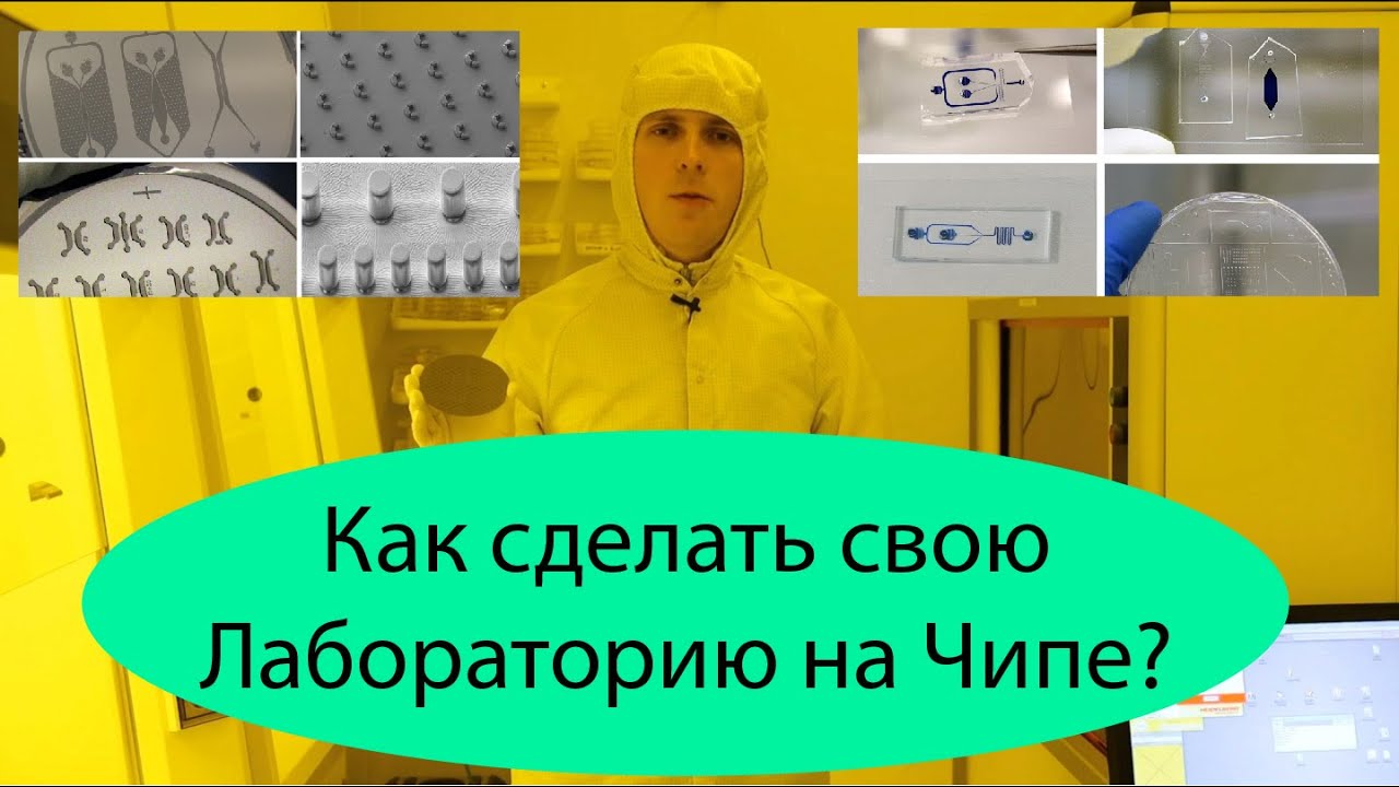
Мягкая литография
7.4K
Soft lithography is a microfabrication technique that uses flexible polydimethylsiloxane (PDMS) to create microfluidic devices, where a master form fabricated on a silicon substrate using photolithography serves as a mold to produce multiple identical devices through PDMS replication, enabling rapid prototyping and cost-effective production of microfluidic systems for biomedical research and applications.
Click to watch →

PDMS Replica Molding and Bonding
21.1K
This video demonstrates the complete process of creating PDMS (Polydimethylsiloxane) replicas through replica molding and bonding. The process involves mixing PDMS base with curing agent in a 10:1 weight ratio, thoroughly mixing for one minute, degassing the mixture under vacuum to remove bubbles, casting the degassed PDMS into a 3D-printed mold, performing a second degassing after filling, curing at 65°C for two hours, demolding using a spatula technique, creating inlet/outlet holes with a punch tool, cleaning surfaces with scotch tape, applying corona treatment to oxidize surfaces for bonding, and finally bonding PDMS layers together on a 95°C hot plate to create sealed microfluidic devices.
Click to watch →
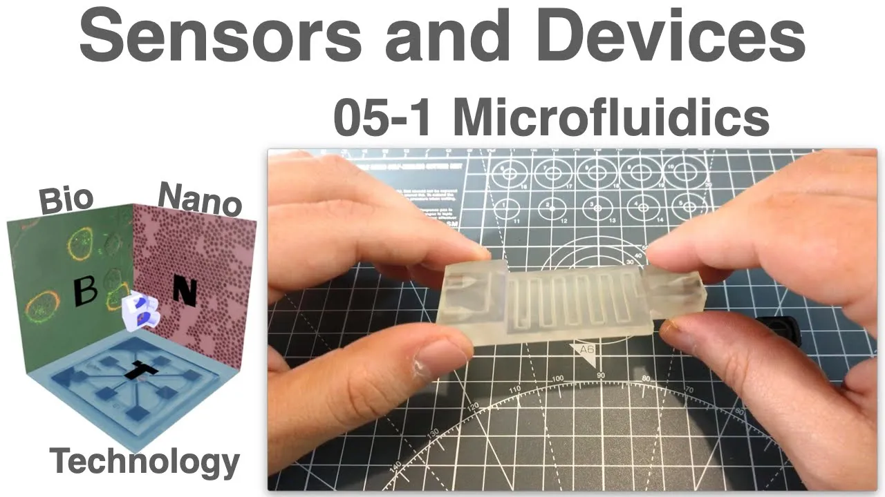
Microfluidics Fabrication Methods: Sensors & Devices Lecture
29.3K
Microfluidics offers significant advantages for sensors including reduced reagent volumes and enhanced sensitivity through thin channels. The field spans physics, chemistry, bioengineering, and chemical engineering. Glass microfluidics provide extreme solvent stability and miniaturization but require external manufacturers with long lead times and high costs, plus they are fragile. The replica molding method using PDMS revolutionized microfluidics by enabling lab-based fabrication. PDMS was selected for its refractive index matching glass, similar chemistry for surface functionalization, and transparency. The process involves photolithography to create masters, PDMS casting and curing, and plasma-mediated covalent bonding to glass, allowing direct microscopic observation through the glass substrate.
Click to watch →
Deep Dive
The biological significance of cellular barriers (endothelial and epithelial) and the role of tight junctions in tissue permeability.

Gap junction & Tight Junction (Lecture-11) | Cell Signaling | GATE Life Science 2024
610
Tight junctions are specialized cell-cell junctions that seal the space between adjacent cells, preventing any molecular passage including ions and small molecules. They are crucial for maintaining cellular compartmentalization and do not participate in electrical or chemical synapses. Tight junctions are predominantly found in epithelial tissues such as the intestinal and urinary bladder epithelium, where they prevent food particles from leaking out of the intestine and urine from seeping out of the urinary bladder. The structural basis of tight junctions involves three key protein families: occludin, claudins, and junctional adhesion molecules, which are linked to the actin cytoskeleton through zonula occludens proteins.
Click to watch →

Cell junctions Part I OCCLUDING JUNCTIONS
16K
Tight junctions create barriers between cells with two functions: functional barrier (prevents diffusion, requiring active transport for osmotic balance) and protective barrier (ensures endocytosis at apical surface, exocytosis at basal surface). Epithelial cells have apical (absorptive), basal (secretory), and lateral surfaces. Tight junctions are found in tight epithelia (kidney distal tubule, collecting duct, lung, gut) but absent in leaky epithelia (proximal tubule). This directional control maintains tissue integrity and proper cellular organization.
Click to watch →

Tight junctions, desmosomes and gap junctions
175.8K
Tight junctions are specialized structures found in epithelial tissues that serve two critical functions. First, they limit the paracellular passage of molecules and ions through the space between cells, meaning most substances must enter cells via diffusion or active transport to pass through the tissue. This creates tighter control over what substances can cross the epithelial barrier. Second, tight junctions prevent the lateral movement of integral membrane proteins between the apical and basolateral surfaces of cells. This compartmentalization preserves the distinct functional specializations of each surface, such as receptor-mediated endocytosis occurring exclusively at the apical surface and exocytosis occurring at the basolateral surface.
Click to watch →

The REAL Cause of Clogged Arteries | Dr. Malcolm Kendrick
262.4K
Endothelial cells lining blood vessels are connected by tight junctions that form an impenetrable barrier controlling what substances pass between cells. These junctions are so tight that even small molecules cannot pass through without specific cellular mechanisms. This explains why substances cannot simply diffuse across endothelial barriers based on concentration gradients alone.
Click to watch →
CSIR-UGC-NET 2026 | Cell Signalling & Communication | L11 Tight & Gap Junction | Dr. Ravina Rai |
1.7K
Tight junctions have a dual role in epithelial tissues. They function as barriers that prevent the passage of ions and molecules through the intercellular space. At the same time, they regulate selective permeability by allowing the passage of certain molecules through claudin channels. Tight junctions also support transcellular transport by maintaining cell polarity and ensuring that transport proteins are located in the correct positions. This dual function is essential for maintaining tissue integrity and controlling the movement of substances across epithelial barriers. Tight epithelia have low permeability (kidney collecting duct), while loose epithelia have high permeability (small intestine).
Click to watch →
Deep Dive
Basic electrical circuit theory, specifically Ohm's law, electrical resistance, and impedance in the context of biological media.
Ohm's Law for Neurodiagnostics (Quiz for one ASET-CEU)
4.5K
Ohm's Law (V = I × Z) defines the relationship between voltage, current, and impedance in electrical circuits, and in neurodiagnostics, impedance meters (using AC voltage) are essential for measuring tissue impedance because biological tissues have capacitive and inductive properties that affect different frequencies differently, unlike ohmmeters (using DC voltage) which only measure resistive components and cannot accurately assess tissue properties.
Click to watch →

Seminario DBSS: BIA & Análisis Vectorial de Impedancia Bioeléctrica (BIVA) | ¿Ángulo de Fase?
599
Electrolytes are substances that conduct electricity when dissolved in water, typically metal ions like sodium, potassium, and calcium. The human body contains electrolytes that allow electrical current to flow, which is the basis for bioimpedance analysis. Electrical circuits require closed loops for current flow, with components like batteries (voltage sources), conductors, resistors, and capacitors. Voltage is the energy needed to move electrons, current is the amount of charge passing through a cross-section, and resistance is the opposition to current flow. Ohm's Law (V = I × R) relates these three quantities. The human body exhibits electrical resistance that varies based on body composition: fat tissue has high resistance due to low water and electrolyte content, while lean muscle has lower resistance.
Click to watch →

RRB ALP 2026 | RRB ALP Previous Year Question Paper | RRB ALP Science Previous Year Question
783
Ohm's law states V = IR, where current is proportional to voltage and inversely proportional to resistance. For a 2 ohm resistor with 2 volts, the current is 1 ampere. Hemoglobin in animals transports oxygen and contains iron (red color), while chlorophyll in plants enables photosynthesis and contains magnesium (green color). Both serve as essential pigments enabling critical life functions in their respective organisms.
Click to watch →

M2 P3 1
100
Electrical impedance (Z) combines resistance (R) and reactance (X) to describe how AC circuits oppose current flow, where resistance dissipates power as heat and reactance (inductive or capacitive) affects the timing relationship between voltage and current; Ohm's Law (V = IR) defines resistance as the ratio of voltage to current, with resistance calculated from material properties as R = ρL/A, and conductance (G) being the inverse of resistance measured in Siemens.
Click to watch →

Game Changer GK Series For SSC MTS 2025 | Lecture - 7 | GK Series For MTS 2025 Based on New Vendor
7.7K
This segment covers two fundamental scientific principles. Plant hormones (phytohormones) regulate growth and development: Auxin promotes apical growth in main stems, Gibberellins promote stem elongation, Cytokinins delay leaf senescence, Ethylene is the only gaseous plant hormone promoting fruit ripening, and Abscisic Acid acts as a growth inhibitor. Ohm's Law states that at constant temperature, current is directly proportional to potential difference (V = IR). When potential difference doubles while resistance remains constant, current also doubles. These principles form foundational knowledge in biology and physics, essential for understanding plant physiology and electrical circuits.
Click to watch →
Deep Dive
Standard cell culture techniques, including maintaining sterile environments and seeding cells within microfluidic channels.
Aseptic Technique Training – Essential Cell Culture Video
457.2K
Successful cell culture requires proper laboratory setup with essential equipment including a cell culture hood, incubator, water bath, centrifuge, refrigerator/freezer, hemocytometer, and microscope; maintaining a sterile environment involves opening all materials within the hood, wiping surfaces with 70% ethanol, organizing items systematically (pipettes in front, reagents at back, waste containers in corners), and avoiding breathing or talking into the hood; mammalian cells should be cultured at 37°C with 5-7% CO2 and high humidity, with flasks evenly spaced for proper gas exchange; reagents should be warmed to 37°C for 10-20 minutes but not left excessive time to prevent degradation; daily microscopic examination is essential for monitoring cell health and detecting contamination.
Click to watch →

Mod-01 Lec-46 Biomicrofluidics
1.8K
Microfluidic cell culture systems offer transformative advantages over conventional methods: precise control of cell numbers and density, accurate placement in complex geometries, improved assay accuracy and throughput by orders of magnitude, spatiotemporal control over biomolecule concentrations enabling diffusion gradients, low reagent consumption, automated long-term culture without manual intervention, and delivery of both chemical and mechanical signals. Implementation involves soft lithography for channel fabrication, cell seeding, and microscopy-based analysis. Fluid flow is essential for nutrient delivery and oxygen supply, but creates mechanical stresses requiring optimization of flow rates and static incubation periods. Subcellular assays can study intracellular transport phenomena. Applications include targeted drug delivery through complex networks and sperm selection using laminar flow principles.
Click to watch →

WEBINAR | Flow and medium recirculation in microfluidic cell culture, by Lisa Muiznieks, PhD
2.4K
Microfluidic cell culture enhances physiological relevance by adding controlled fluid flow that mimics natural conditions, enabling constant nutrient exchange, dynamic gas exchange, waste removal, and mechanical stimuli like shear stress; medium recirculation using pressure-driven flow controllers with MUX valves allows continuous, unidirectional flow through microfluidic chips, enabling extended cell culture experiments, incorporation of circulating cells, and multi-organ chip integration for more accurate drug testing and mechanobiology studies.
Click to watch →
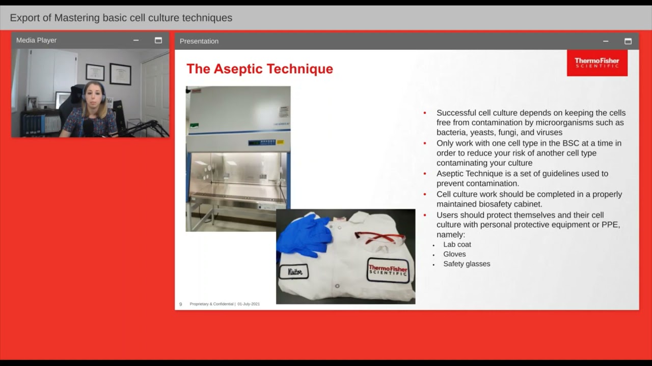
Mastering basic cell culture techniques
21.6K
Cell culture is the process of maintaining plant and animal cells alive and dividing in vitro, requiring specific tools including complete cell culture media (basal media plus supplements like serum and glutamine), appropriate culture vessels, and an incubator. Key techniques include subculturing (passaging) cells at optimal confluency (70-80% for adherent cells), practicing strict aseptic technique to prevent contamination, and regularly monitoring cell health through microscopy and media observation. Different cell types (adherent vs. suspension) require different handling protocols, and maintaining genetic stability requires cryopreserving original stocks to prevent drift over multiple passages.
Click to watch →

Splitting Cells: Cell Culture - Updated - CBEC Cell Block
12.1K
Maintaining sterility is critical throughout cell culture procedures. Key practices include: labeling all materials with initials, cell line, date, and passage number; spraying items with alcohol before placing them in the hood; using proper vacuum pipette technique by only touching the top part; dispensing liquids away from cells; tilting flasks carefully; and keeping the back side down during incubation. These techniques prevent contamination and ensure consistent experimental results.
Click to watch →
Prerequisite Knowledge
- Concept 01Fundamental principles of microfluidics and soft lithography (such as PDMS molding and bonding) used in organ-on-chip fabrication.
- Concept 02The biological significance of cellular barriers (endothelial and epithelial) and the role of tight junctions in tissue permeability.
- Concept 03Basic electrical circuit theory, specifically Ohm's law, electrical resistance, and impedance in the context of biological media.
- Concept 04Standard cell culture techniques, including maintaining sterile environments and seeding cells within microfluidic channels.
Subsequent Learning
- Step 01Advanced electrochemical impedance spectroscopy (EIS) for detailed, frequency-dependent analysis of barrier tissue dynamics.
- Step 02Application of TEER-integrated chips in drug permeability assays, pharmacokinetics, and toxicology testing.
- Step 03Integration of multi-sensor platforms (e.g., incorporating pH, oxygen, and lactate sensors) for real-time, multi-parametric physiological monitoring.
- Step 04Scaling up to multi-organ-on-chip systems (Body-on-Chip) to study systemic drug absorption, distribution, metabolism, and excretion (ADME).
Fabrication & TEER
Fabrication & TEER
0:00Playing Section
- 1
Demonstrates microfluidic chip fabrication for TEER measurements.
- 2
Integrated electrodes enable direct barrier function assessment.
- 3
Method applicable to BBB and other organ-chip systems.
Limitations of Integrated TEER and the Case for Macromolecular Tracer Assays
While integrated electrodes for TEER measurement offer real-time, non-destructive monitoring of barrier integrity, this approach faces significant technical and biological criticisms. Critics point out that integrated electrodes often suffer from uneven current density distribution, electrode fouling, and extreme sensitivity to temperature and media fluctuations, which can lead to highly variable and inaccurate resistance readings. Furthermore, the microfabrication of integrated electrodes increases device complexity and cost, while potentially introducing biocompatibility issues or altering fluid flow. Consequently, many researchers advocate for macromolecular tracer assays (using fluorescently labeled dextrans) as a more reliable and biologically relevant alternative. Unlike TEER, which only measures ionic resistance, tracer assays directly quantify the paracellular transport of larger molecules, mimicking the actual physiological barrier function against drugs and pathogens. Additionally, emerging contactless optical profiling and impedance spectroscopy methods provide non-invasive alternatives that avoid the physical and electrochemical artifacts associated with embedded metal electrodes.
Deep Dive
Advanced electrochemical impedance spectroscopy (EIS) for detailed, frequency-dependent analysis of barrier tissue dynamics.
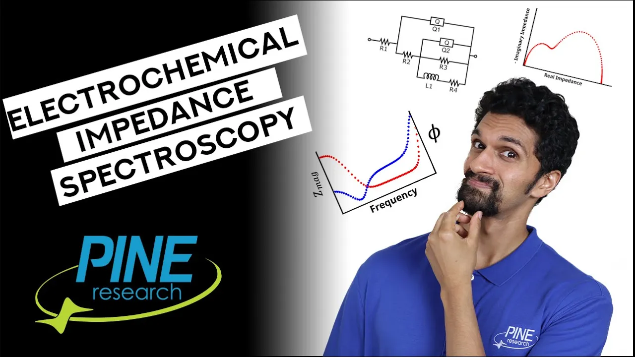
What is Electrochemical Impedance Spectroscopy (EIS) and How Does it Work?
151.7K
Electrochemical Impedance Spectroscopy (EIS) is a technique where a potentiostat applies a sinusoidal potential or current to an electrochemical system and measures the corresponding sinusoidal current or potential response; the technique uses Fourier transform to convert time-domain data to frequency-domain data, calculating impedance (the AC equivalent of resistance) from the ratio of potential and current amplitudes, along with phase shifts, which are then plotted as Bode plots (magnitude and phase vs. frequency) or Nyquist plots (real vs. imaginary impedance), allowing researchers to deconvolute different electrochemical phenomena occurring at characteristic frequencies (such as double-layer charging, solution resistance, charge transfer resistance, and molecular diffusion) that occur simultaneously but on different timescales, enabling both qualitative and quantitative analysis of electrochemical systems through equivalent circuit modeling.
Click to watch →
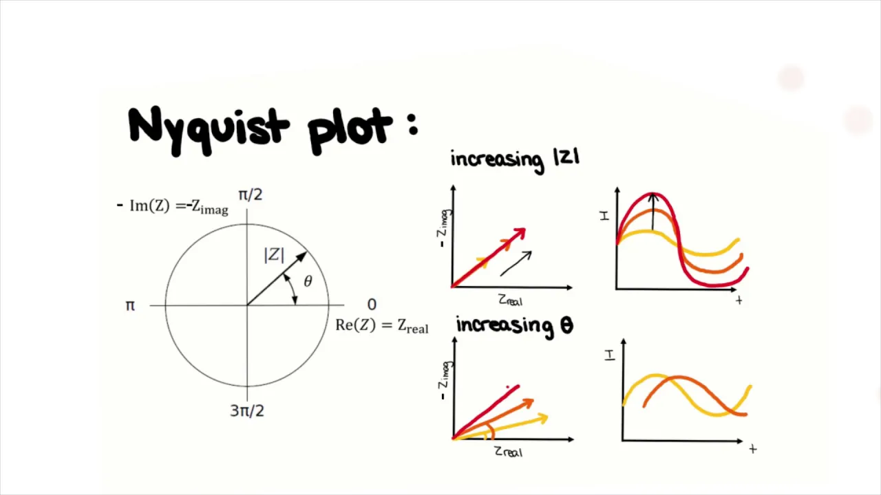
Introduction to Electrochemical Impedance Spectroscopy (EIS)
83.6K
Electrochemical Impedance Spectroscopy (EIS) is a technique that applies alternating current or potential signals across a range of frequencies to an electrochemical system, enabling researchers to separate and analyze different mechanisms (such as double layer formation, charge transfer, and diffusion) based on their characteristic time constants, and then model these processes using equivalent circuit elements consisting of resistors, capacitors, Warburg impedance, and constant phase elements to extract important parameters like diffusion coefficients, kinetic parameters, and electrolyte resistance.
Click to watch →

Intro to Nyquist Plots for Lithium Ion Battery Research
55K
Electrochemical Impedance Spectroscopy (EIS) measures battery impedance by applying a small AC current while maintaining constant voltage (typically 5-10 mV). The technique works by causing electrons and ions to oscillate within the battery at controlled frequencies. High frequencies reveal fast processes like chemical reactions, while low frequencies expose slower processes such as ion diffusion through battery materials. This frequency-dependent measurement acts like shaking ions through an obstacle course, revealing how energy barriers affect ion movement across different battery layers and components.
Click to watch →

Introduction to electrochemical impedance spectroscopy (EIS) for battery research
119.3K
Electrochemical Impedance Spectroscopy (EIS) is a non-destructive technique that measures the impedance of electrochemical systems across a wide range of frequencies (typically 100 kHz to 100 mHz) by applying small alternating current excitations (around 10 mV) to avoid creating concentration gradients and maintain linear response. The resulting data is displayed in Nyquist plots showing real versus imaginary impedance components, where different circuit elements (resistors, capacitors, constant phase elements, and Warburg impedances) produce characteristic shapes that can be fitted to equivalent circuits to extract information about ionic and electronic transport properties, charge transfer resistance, and degradation mechanisms in battery materials. EIS is widely used in battery research to study cathode materials, full cells, and solid electrolyte interfaces without damaging the sample.
Click to watch →
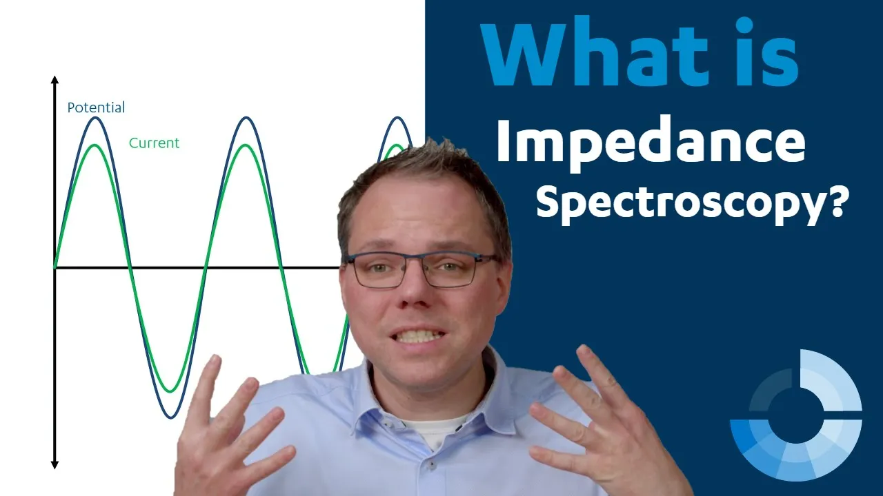
What is Electrochemical Impedance Spectroscopy (EIS)?
3.3K
Electrochemical Impedance Spectroscopy (EIS) is a technique that measures the impedance of electrochemical systems using AC sine waves, where impedance differs from resistance by accounting for phase shifts between voltage and current; scientists simplify complex electrochemical systems by modeling them with equivalent electronic circuits and analyzing how each component's impedance varies with frequency, allowing identification of contributions from capacitive charging, faradaic reactions, diffusion, and solution resistance through frequency-dependent measurements presented in Bode or Nyquist plots, which enables applications such as label-free DNA sensing, coating integrity assessment, and battery health monitoring.
Click to watch →
Deep Dive
Application of TEER-integrated chips in drug permeability assays, pharmacokinetics, and toxicology testing.
TEER Monitoring
554
The Tissue Engineering Laboratory at API Geneva has developed an integrated system that measures trans-epithelial/endothelial electrical resistance (TEER) in real-time and non-invasively to assess the quality of biological barriers, with applications in toxicology testing and pharmacological studies such as drug discovery, addressing the challenge that only one in nine drugs receives regulatory approval due to inability to reach targeted sites.
Click to watch →
Real-Time TEER Monitoring for Organ-on-Chip Barrier Integrity
126
Organ-on-chip technology provides microfluidic-based devices with chambers and channels for living organ substructures, creating controlled microenvironments that mimic in vivo conditions through perfusion systems and biomechanical stimulation. Standard readouts include supernatant sampling, cytotoxicity assessment, tissue recovery, live-cell imaging, and enzyme/protein transporter functionality. Tissue barriers are specialized cellular layers (epithelia/endothelia) acting as selective filters that permit essential nutrients while blocking harmful agents, maintained by tight junctions, adherens junctions, and desmosomes. TEER (Trans-Epithelial/Trans-Endothelial Electrical Resistance) is a non-invasive method measuring electrical resistance across cell monolayers to assess barrier integrity. Compared to permeability assays using labeled tracers (which take 1-24 hours), TEER provides faster results in seconds with less cellular interference, though accuracy depends on electrode type, membrane area, and temperature. Integrated organ-on-chip electrodes offer superior stability compared to movable chopstick electrodes.
Click to watch →

9 Reasons to Trust WPI's New EVOM™ Manual TEER Measurement Meter
1K
Trans-epithelial electrical resistance (TEER) measurement is a fundamental technique for assessing the integrity and barrier function of epithelial cell monolayers in vitro. This method applies a small electrical current across a cell layer and measures the resulting voltage drop to calculate resistance, which correlates with how tightly cells are connected. TEER is essential for studying cellular barrier properties in various applications including drug permeability testing, toxicity assessment, and understanding physiological barriers like the blood-brain barrier. The technique requires specialized electrodes and instruments designed to deliver minimal current while providing accurate, repeatable measurements that reflect true cellular barrier function without disrupting the monolayer.
Click to watch →
Avaliação in vitro da permeabilidade no modelo Caco-2
733
Before permeability testing, monolayer integrity is verified using Transepithelial Electrical Resistance (TEER) measurement with a multimeter and electrodes in each compartment. High TEER values indicate functional tight junctions. The assay procedure involves washing cells with HBSS, measuring TEER, decanting HBSS from the apical side, adding fresh HBSS to collection wells, and introducing test compounds to the apical compartment. Samples are collected separately from both compartments. Analytical methods include liquid chromatography and microplate readers, especially for fluorescent compounds. Results yield the apparent permeability coefficient (Papp), calculated from compound concentrations in both compartments over time. This coefficient correlates with reference compounds to predict human intestinal absorption fractions, enabling informed decisions in drug development programs.
Click to watch →
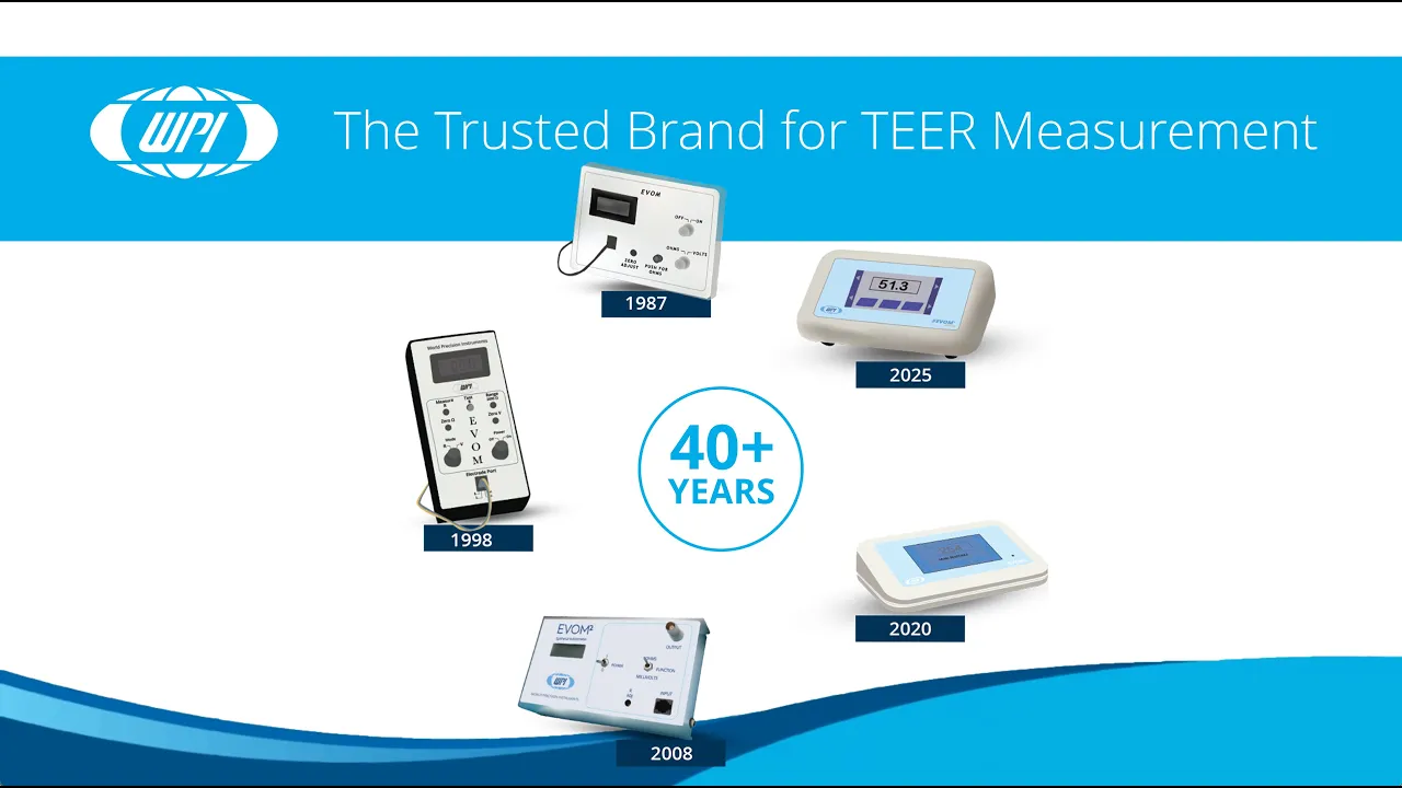
EVOM™ The Trusted Brand for TEER Measurement
229
Transepithelial Electrical Resistance (TEER) is a well-established technique used to assess the barrier function and integrity of epithelial and endothelial cell layers by applying a small AC current to the cell monolayer and measuring the resistance to ion flow, providing valuable insights into tight junction integrity and cell-cell interactions; WPI's EVOM family of meters, including manual and automated systems for 24-well and 96-well plates, serves as the gold standard in TEER technology, having been trusted for nearly 40 years and cited in thousands of peer-reviewed publications for applications in drug absorption studies, tissue engineering, disease modeling, and quality control.
Click to watch →
Deep Dive
Integration of multi-sensor platforms (e.g., incorporating pH, oxygen, and lactate sensors) for real-time, multi-parametric physiological monitoring.

Organ-on-a-Chip, with Dr Ruth Mackay
430
Advanced organ-on-a-chip platforms integrate multiple sensors for comprehensive monitoring: optical sensors for oxygen, CO2, and pH; electrochemical sensors for specific molecule capture; bubble traps; and continuous measurement of temperature, glucose, and lactate. Fully automated systems with MATLAB-based data acquisition can monitor experiments running for days, weeks, or months. Such platforms represent the future direction of the field, though they remain technically challenging to implement.
Click to watch →

2021 IUPAC Analytical Chemistry Medal Lecture: Prof Joseph Wang
907
Modern wearable sensors integrate multiple physiological monitoring modalities within single devices. Advanced systems combine electrochemical sensors for biomarkers like lactate and glucose with acoustic sensors for blood pressure monitoring, or integrate ECG electrodes with chemical sensors for simultaneous cardiac and metabolic assessment. Microfluidic channels integrated with flexible substrates enable continuous sweat sampling and removal, with natural sweat gland activity providing the pumping mechanism. For security applications, printed electrodes on elastic textiles enable flexible biosensors for nerve agent detection, gunshot residue identification, and explosive trace detection. Microneedle arrays penetrate the outer skin layer minimally while delivering sensors into interstitial fluid for detection of multiple biomarkers including glucose, ketones, lactate, and alcohol. Wound healing monitoring uses flexible sensors integrated into dressings to track pH levels and uric acid. For melanoma screening, sensors detect tyrosinase enzyme activity by measuring conversion of substrate catechol to benzoquinone, providing rapid preliminary screening at the point of care.
Click to watch →

Kalmo Smart Monitoring - Demo del servizio di Telemedicina con device multisensore integrato
996
Modern health monitoring devices can simultaneously measure multiple physiological parameters through integrated sensors. The system measures body temperature (36.5-37.5°C normal range), blood oxygen saturation, blood pressure, and heart electrical activity (ECG). Each measurement requires specific user positioning and technique, with the application providing real-time feedback on correct placement. Results are color-coded for quick interpretation and stored for longitudinal health tracking.
Click to watch →
Self-Powered Wearable Sensors for Health and Environmental Monitoring
607
Flexible biochemical sensing platforms combine optical and electrochemical sensing capabilities. The optical patch measures heart rate and oxygenation, while the electrochemical patch detects lactate, cortisol, glucose, pH, and temperature. Multi-analyte electrochemical patches enable simultaneous sensing of multiple biomarkers. For wound monitoring, bi-enzymatic systems for uric acid detection are printed on gauze bandages with spatially distributed sensors to measure changes near and away from the wound, tested in clinical studies at University of Miami Health System.
Click to watch →
Session on Emerging Trends in Wearable Electrochemical Sensors
201
Comprehensive physiological monitoring requires detecting both chemical parameters and electrophysiological signals simultaneously. Combined sensors integrate ECG monitoring electrodes with chemical sensing electrodes (like lactate sensors) on a single PCB. This allows real-time collection of both types of signals simultaneously, providing complete information about underlying physiology rather than isolated measurements. Such integrated systems can be attached to appropriate body regions (like chest for ECG) for comprehensive health monitoring.
Click to watch →
Deep Dive
Scaling up to multi-organ-on-chip systems (Body-on-Chip) to study systemic drug absorption, distribution, metabolism, and excretion (ADME).

3RsC Multi Organ MPS Webinar: Day 1
216
Multi-organ chips can simulate complete drug disposition pathways including absorption, distribution, metabolism, and excretion (ADME). Using Chip 4 with intestine, liver, kidney, brain, and blood-brain barrier models, researchers demonstrated that haloperidol excretion matched patient data (only 1% excreted), while carbamazepine oral application showed expected absorption and metabolism patterns. An antibiotic study revealed that at therapeutic concentrations, the drug was properly metabolized and excreted, but at 1000x higher concentrations, 100% was excreted through the kidney compartment, demonstrating dose-dependent toxicity prediction.
Click to watch →

Don Ingber : "Human Organ Chips: From experimental models to clinical mimicry"
4.1K
An integrated human body-on-chips system connects multiple organ chips (gut, liver, kidney, heart, lung, blood-brain barrier, brain neuronal networks, skin, bone marrow, pancreas) through robotic sampling and shared medium. Drugs pass sequentially through the system to model absorption, metabolism, distribution, and excretion. Computational scaling from chip measurements to in vivo parameters enables prediction of key pharmacokinetic values like Cmax and t1/2. For oral nicotine, predictions matched clinical data from Swedish studies with three formulations. For intravenous cisplatin, predictions matched clinical studies with two infusion protocols, demonstrating potential to shortcut clinical trial design.
Click to watch →

Models of CMT
162
Advanced human-on-a-chip platforms integrate multiple organ systems including blood-brain barrier (BBB), gastrointestinal tract (GIT), kidney, and skin tissues within a single system. These multi-organ models enable study of drug absorption, distribution, metabolism, and excretion (ADME) properties in physiologically relevant contexts. BBB models incorporate tight junction proteins and transporters (glucose transporters and P-glycoprotein), while GIT inserts allow study of oral versus intravenous dosing differences. Recirculating immune system models generate both restorative and inflammatory macrophages within the same system, enabling study of immune responses and immunomodulatory drug effects. Pharmacokinetic-pharmacodynamic (PK-PD) relationships created in these systems correlate well with in vivo data across species, from guinea pigs to non-human primates.
Click to watch →
Journal club | Human organ-on-a-chip technology as a catalyst for drug discovery
109
Organ-on-a-chip technology enables patient-specific drug testing by using cells from individual patients. Since people respond differently to the same drugs due to genetic and physiological differences, testing drugs on patient-derived cells can predict individual responses. This supports precision medicine, which tailors treatment to individual patients—different patients may respond to different doses or require different drugs entirely. Major challenges include scalability (devices are made artesially and cannot be produced at scale), complexity (incorporating immune cells, microbiota, and multiple organ systems), standardization (lack of consistent protocols), cost, and regulatory pathways. Multi-organ-on-a-chip systems connect multiple organ chips (liver, kidney, intestine, heart, bone marrow) to simulate systemic drug effects—for example, testing absorption in intestine, metabolism in liver, excretion in kidney, and systemic toxicity in bone marrow.
Click to watch →

Donald E.Ingber: Human Organ Chips: Revolutionizing Drug Developmentand Personalized medicine
190
Organ chips of virtually every organ have been created, including lymph node, liver, and kidney chips. The vision is connecting multiple organ chips through blood vessel channels to create a human body-on-chips system. A simpler version with gut, liver, and kidney connected by a mixing chamber allows samples to be taken as if from peripheral blood. Computational modeling combined with measurements at every point in linked chips can scale from chip volumes to the human body. When oral nicotine and intravenous cisplatin were tested, predicted levels precisely matched patient observations, demonstrating the system's predictive power.
Click to watch →
Fabrication & TEER
Fabrication & TEER
0:00- 1
Demonstrates microfluidic chip fabrication for TEER measurements.
- 2
Integrated electrodes enable direct barrier function assessment.
- 3
Method applicable to BBB and other organ-chip systems.
Limitations of Integrated TEER and the Case for Macromolecular Tracer Assays
While integrated electrodes for TEER measurement offer real-time, non-destructive monitoring of barrier integrity, this approach faces significant technical and biological criticisms. Critics point out that integrated electrodes often suffer from uneven current density distribution, electrode fouling, and extreme sensitivity to temperature and media fluctuations, which can lead to highly variable and inaccurate resistance readings. Furthermore, the microfabrication of integrated electrodes increases device complexity and cost, while potentially introducing biocompatibility issues or altering fluid flow. Consequently, many researchers advocate for macromolecular tracer assays (using fluorescently labeled dextrans) as a more reliable and biologically relevant alternative. Unlike TEER, which only measures ionic resistance, tracer assays directly quantify the paracellular transport of larger molecules, mimicking the actual physiological barrier function against drugs and pathogens. Additionally, emerging contactless optical profiling and impedance spectroscopy methods provide non-invasive alternatives that avoid the physical and electrochemical artifacts associated with embedded metal electrodes.
Fundamental principles of microfluidics and soft lithography (such as PDMS molding and bonding) used in organ-on-chip fabrication.
The biological significance of cellular barriers (endothelial and epithelial) and the role of tight junctions in tissue permeability.
Basic electrical circuit theory, specifically Ohm's law, electrical resistance, and impedance in the context of biological media.
Standard cell culture techniques, including maintaining sterile environments and seeding cells within microfluidic channels.
Advanced electrochemical impedance spectroscopy (EIS) for detailed, frequency-dependent analysis of barrier tissue dynamics.
Application of TEER-integrated chips in drug permeability assays, pharmacokinetics, and toxicology testing.
Integration of multi-sensor platforms (e.g., incorporating pH, oxygen, and lactate sensors) for real-time, multi-parametric physiological monitoring.
Scaling up to multi-organ-on-chip systems (Body-on-Chip) to study systemic drug absorption, distribution, metabolism, and excretion (ADME).
377 views8likes2:00@JoVEJournalOriginal Release: 2022-05-27
[Music] the overall goal of this video is to show how to fabricate and use microfluidic chips with integrated electrodes for trans-endothelial or trans-epithelial electrical resistance measurements or tear measurements this is demonstrated using a blood brain barrier in a microfluidic chip this method can help answer key questions in the field of organs on chips by enabling direct measurement of the barrier function of for example blood brain barrier tissue using integrated electrodes the main advantages of our technique is that the electrodes are easily integrated into organ and chip systems and that the resulted tier values can be compared among different systems the implications of this technology extend towards understanding blood brain barrier function in health and disease drug discovery and personalized medicine though this method can be used to provide insight into blood-brain barrier function it can also be used in the context of other organ-on-chip systems such as the long-on-chip and the gut-on-chip to begin this procedure mix 27 grams of pdms base agent and 2.7 grams of curing agent thoroughly then degas the mixture in a desiccator for approximately 45 minutes to remove air bubbles meanwhile prepare the mold for the liquid pdms mixture by sticking clear tape around the mold or place the mold in a suitable wafer holder pour the degast pdms mixture onto the mold next cure the pdms mixture in an oven at 60 degrees celsius for 4 hours and allow it to cool down afterward in a cross flow hood pull the cured pdms from the mold cut the pdms replica into separate top and bottom chip parts using cutting lines in the pdf
Up Next
12:09
How to Build a Cloud Chamber Particle Detector
@ElectroBOOM
2.7M views•2019-11-14

12:54
Triumph of Orthodoxy Icon: Byzantine Art & History Explained
@BenCallan
2.1K views•2024-08-06

2:00
E. coli Cell-Free Protein Synthesis Protocol Overview
@JoVEJournal
996 views•2022-05-30

1:42
Game of Thrones Opening Credits: A Cinematic Analysis
@gameofthrones
46.3M views•2011-04-18
Related Study Plans & Knowledge Roadmaps
Structured learning paths in General & Interdisciplinary Studies
general-studies
Elevator Engineering: History, Physics & Cities
01From Hoists to Safety Brakes: The Dawn of Vertical Lift
02Hydraulic vs. Traction: The Engineering Physics of Elevators
03Modern Speed, Safety, and Algorithms
+ 2 more learning modules
12h5 modules
Explore Roadmap
general-studies
Rubber: Material Science, History & Geopolitics
01Origins of Natural Rubber & Mesoamerican History
02The Chemistry of Natural Rubber & Vulcanization
03The Geopolitics of the Rubber Boom & Colonization
+ 2 more learning modules
12h5 modules
Explore Roadmap
general-studies
Containerization: Evolution, Logistics & Trade
01Ancient Trade & Bulk Cargo: Amphorae to Barrels
02The Industrial Revolution & Cardboard Packaging
03The Containerization Revolution: Malcom McLean
+ 2 more learning modules
12h5 modules
Explore Roadmap
general-studies
Textile Tech: Evolution, Labor & Global Impact
01Pre-Industrial Textiles: Handloom & Cottage Industry
02Mechanization: The Spinning Jenny to the Power Loom
03The Factory System & Global Labor Transformation
+ 2 more learning modules
10h5 modules
Explore Roadmap