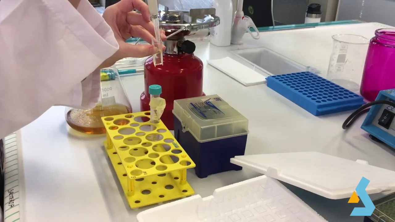
This video demonstrates a simplified protocol for E. coli-based cell-free protein synthesis, which provides a robust, flexible, and accessible platform technology for rapid protein production. The key steps include growing E. coli cultures to OD600 = 3.0, balancing centrifuge bottles with equal volumes of culture and water, centrifuging at 5000g for 10 minutes at 10°C, resuspending the cell pellet in cold S30 buffer supplemented with 2mM DTT, and vortexing in short bursts. This method enables applications in functional genomics, high-throughput testing, biosensors, educational kits, metabolic engineering, and genetic code expansion within 4 days or less.
E. coli Cell-Free Protein Synthesis Protocol Overview
Added:Deep Dive
The Central Dogma of Molecular Biology: A solid understanding of DNA transcription and RNA translation mechanisms, including the functions of ribosomes, tRNAs, and RNA polymerases.
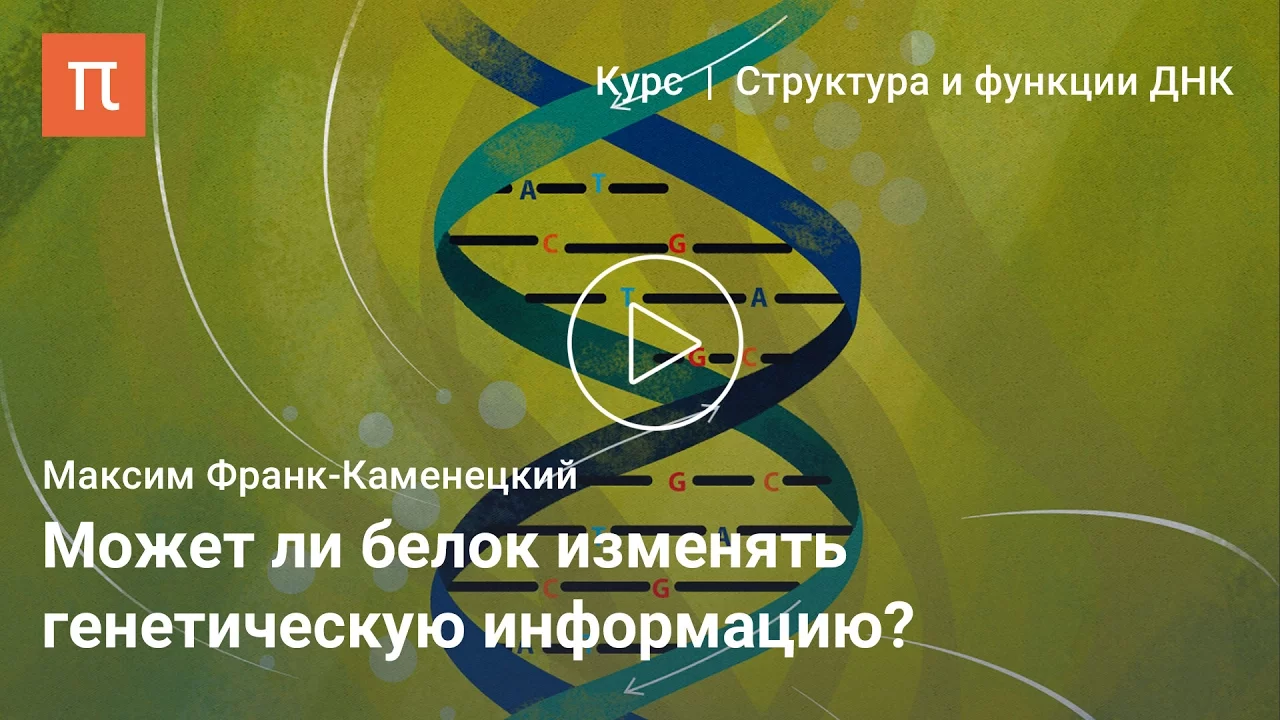
Центральная догма молекулярной биологии — Максим Франк-Каменецкий
23.3K
The Central Dogma, formulated by Francis Crick in 1958, describes the directional flow of genetic information in cells: DNA → RNA → protein. DNA stores genetic information using four nucleotide bases (A, T, G, C). During transcription, this information is copied into RNA (with U replacing T). Translation then converts RNA sequences into proteins using the genetic code, where triplets of nucleotides (codons) specify 20 amino acids. The dogma is fundamentally a negative statement: it prohibits information flow from protein back to nucleic acids. This principle established the foundation for understanding how genetic instructions are used to build and maintain living organisms.
Click to watch →

DOGMA CENTRAL DA BIOLOGIA MOLECULAR | Thaís Magalhães
2.7K
The Central Dogma of Molecular Biology, proposed by Francis Crick in 1958, describes the fundamental flow of genetic information in living organisms. A dogma is defined as an unquestionable doctrine or firmly established opinion. The dogma states that DNA can replicate itself and produce RNA, and RNA can produce proteins, but the reverse is not possible - proteins cannot produce DNA. This represents the one-directional flow of genetic information from DNA to RNA to protein. The processes involved are DNA replication (DNA making copies of itself), transcription (DNA generating RNA), and translation (RNA producing proteins).
Click to watch →

central dogma
233
The Central Dogma describes the flow of genetic information from DNA to RNA to protein. Proposed by Francis Crick in 1958, it explains how genetic instructions are used to build functional proteins. The process involves transcription (DNA to mRNA) and translation (mRNA to protein). DNA contains all information needed for protein synthesis, while mRNA acts as the messenger carrying this information to ribosomes. Some viruses like HIV can reverse this process through reverse transcription, converting RNA back to DNA.
Click to watch →

RSSB LDC Science Classes 2026 | सेंट्रल डोग्मा सिद्धांत (Central Dogma) | LDC Online Classes | #07
14K
The Central Dogma describes the flow of genetic information: DNA → RNA → Protein. Francis Crick proposed this principle. DNA replication (DNA-dependent DNA polymerase) copies DNA. Transcription (DNA-dependent RNA polymerase) creates RNA from DNA. Translation (ribosomes) synthesizes proteins from RNA. Howard Temin and David Baltimore later discovered reverse transcription (RNA → DNA) in retroviruses like HIV, expanding the Central Dogma.
Click to watch →

7. Clase 10 marzo 2021
1.2K
The Central Dogma of Molecular Biology, proposed by Francis Crick, describes the flow of genetic information in cells: DNA replicates itself, DNA is transcribed into RNA, and RNA is translated into protein. Crick originally called this a 'dogma' (a principle that cannot be questioned), but later acknowledged that information can flow in reverse (from RNA to DNA) through reverse transcription in retroviruses.
Click to watch →
Deep Dive
E. coli Biology: Familiarity with E. coli as a model organism, specifically its growth phases (such as the exponential phase optimal for harvesting) and cellular machinery.

E coli | Escherichia coli | Infection | Intoxication | Pathology | Hindi | Food Microbiology
11.2K
Escherichia coli is a facultative anaerobe that can grow in both aerobic and anaerobic conditions. As a gram-negative bacterium, it stains pink during Gram staining due to its thin peptidoglycan layer surrounded by an outer membrane. Five major pathogenic types exist: Enteropathogenic E. coli (EPEC) causes persistent childhood diarrhea through adherence factors; Enterotoxigenic E. coli (ETEC) produces LT and ST toxins causing traveler's diarrhea; Enteroinvasive E. coli (EIEC) invades intestinal cells causing dysentery; Enterohemorrhagic E. coli (EHEC), particularly O157:H7, causes bloody diarrhea and hemolytic uremic syndrome; Enteroaggregative E. coli (EAEC) causes persistent diarrhea through aggregative adherence. Each type employs distinct virulence mechanisms targeting different stages of intestinal infection.
Click to watch →
New Study: Chicken Causes 1 in 5 UTIs — Dr. Neal Barnard Explains the Hidden Risk
9.4K
E. coli, discovered in 1885 by Theodor Escherich, derives its name from 'Escherich' and 'coli' (meaning colon). While some strains are harmless, others cause disease. Contrary to belief, stomach acid doesn't always kill bacteria—Helicobacter pylori survives in the stomach, causing ulcers. E. coli cannot grow on plant products like spinach or beans; contamination occurs externally through irrigation water or handling. The bacterium thrives only in animal intestinal tracts. During meat processing, chickens are slaughtered in conditions where fecal contamination is inevitable, and sanitizing solutions primarily cool carcasses rather than kill bacteria. This creates a continuous pathway from farm to fork where E. coli can reach consumers.
Click to watch →

E coli bacteria microbiology | pathogenesis, Symptoms and disease
32.9K
This comprehensive section covers E. coli's fundamental biology and disease mechanisms. E. coli is a Gram-negative, facultative anaerobic gut bacterium characterized by short rod morphology, flagellar motility, and capsule formation. Its defining feature is lactose fermentation ability, distinguishing it from other enterobacter species. Classification relies on O, K, and H antigens, with O157:H7 representing a dangerous serotype. Four major pathogenic types cause distinct diseases: Enterotoxigenic E. coli (ETEC) causes traveler's diarrhea through heat-labile toxin; Enteropathogenic E. coli (EPEC) causes persistent infantile diarrhea; Enterohemorrhagic E. coli (EHEC) causes hemorrhagic colitis and potentially fatal hemolytic uremic syndrome; Enteroinvasive E. coli (EIEC) causes invasive disease requiring bloodstream entry. All types share oral-fecal transmission routes and predominantly affect developing regions with inadequate sanitation.
Click to watch →
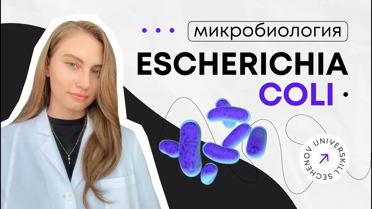
Кишечная палочка | Escherichia coli
26.6K
E. coli includes both commensal and pathogenic strains. Pathogenic strains are classified into five categories: Enterotoxigenic E. coli (ETEC) causes watery diarrhea via heat-labile and heat-stable enterotoxins; Enteroinvasive E. coli (EIEC) invades colon epithelium; Enteropathogenic E. coli (EPEC) causes infant diarrhea through intimin protein; Enterohemorrhagic E. coli (EHEC) produces Shiga toxin causing bloody diarrhea; Enteraggregative E. coli (EAEC) forms biofilms causing persistent diarrhea.
Click to watch →

CONVERTERAM A BACTÉRIA PARA CONSUMIR CO2! | Canal do Slow 79
20.9K
E. coli (Escherichia coli) represents approximately 0.1% of human gut flora and is the largest part of the harmless bacterial lineage. It has become one of the most studied bacteria due to its simple metabolism, ease of laboratory maintenance, and rapid reproduction cycle of about 20 minutes, allowing scientists to generate approximately 72 generations in 72 hours. E. coli possesses plasmids—small DNA molecules containing exotic genes that can be transferred between bacteria, enabling the spread of traits like antibiotic resistance. This genetic flexibility makes E. coli ideal for biotechnology applications, including the production of therapeutic proteins like insulin through genetic engineering.
Click to watch →
Deep Dive
Basic Biochemistry Lab Techniques: Prior knowledge of laboratory processes such as cell lysis (e.g., sonication, homogenization), high-speed centrifugation, dialysis, and precise buffer preparation.
All Practical sections | general biochemistry
181
This comprehensive section covers essential biochemical laboratory techniques for detecting biomolecules and quantitative analysis. Three key tests are demonstrated: Biuret test detects proteins by adding copper-containing reagent, causing blue-to-purple color change; Iodine test identifies starch by turning orange/yellow solutions blue-black; Benedict's test detects reducing sugars by heating sample with reagent, producing color changes from blue to brick red. Proper technique requires accurate volume measurement using graduated pipettes, reading menisci at eye level, and heating at 45°C for approximately 2 minutes. Spectrophotometry uses instruments with light source, monochromator, sample holder, detector, amplifier, and display meter. The Beer-Lambert Law (A = εCL) enables quantitative analysis, with unknown concentrations calculated using C₁/A₁ = C₂/A₂. Calibration with blank solution is essential before measurements.
Click to watch →

BIOCHEMISTRY SPOTTER PART 2 FOR MBBS AND MEDICAL STUDENTS|BIOCHEMISTRY PRACTICAL 1 YEAR /MBBS NEETPG
4.3K
This segment covers practical biochemistry laboratory techniques. Albinism is identified as a condition caused by tyrosinase enzyme deficiency, resulting in white skin, hair, and eyes. Glucosazone crystals appear needle-shaped and are formed by glucose, fructose, or mannose reacting with phenylhydrazine. Vitamin A (found in carrots) has an RDA of 900 micrograms RAE for males and 700 micrograms RAE for females, increasing during pregnancy and lactation. Gel electrophoresis separates DNA fragments by size, with negatively charged DNA moving toward the positive electrode, useful for diagnosing genetic disorders.
Click to watch →
BIOCHEMIST- 232/2024 // New Syllabus // Medical Education Department// BIOCHEM-MENTOR //
547
This comprehensive section covers essential biochemistry laboratory techniques including microscopy (bright field, phase contrast, fluorescence, confocal, electron microscopy, atomic force microscopy), histology, electrophoresis (agarose gel, SDS-PAGE, capillary, pulse field), PCR and immunological techniques, spectroscopy (UV, visible, IR, flame photometry, atomic absorption), NMR and mass spectrometry, and chromatography (partition, absorption, TLC, ion exchange, gel filtration, gas chromatography, HPLC). It also covers enzyme fundamentals including classification, activation energy, specificity, co-enzymes, mechanisms of enzyme action, and catalysis.
Click to watch →
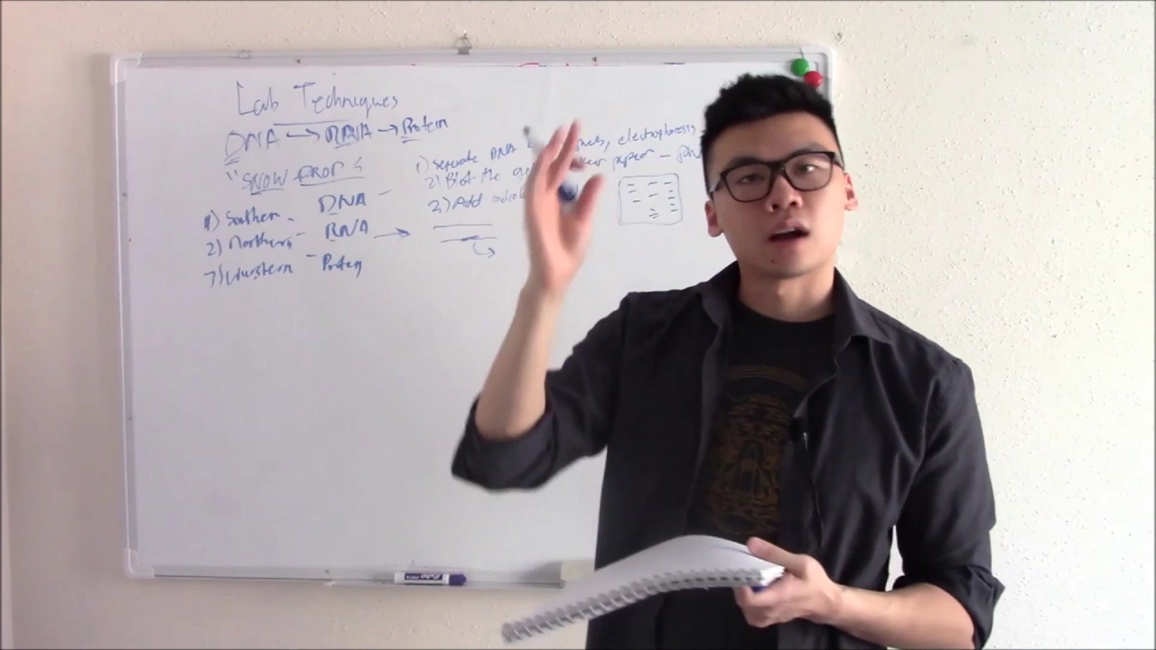
USMLE Biochemistry 23 Lab Techniques
13.5K
This video covers essential laboratory techniques for USMLE Step 1 biochemistry, including PCR (DNA amplification through denaturation, annealing, and elongation cycles), gel electrophoresis (separating biomolecules by size and charge), blotting techniques (Southern blot for DNA, Northern blot for RNA, Western blot for proteins, and Southwestern blot for DNA-binding proteins), flow cytometry (analyzing cell characteristics using lasers and fluorescent markers), microarray (simultaneously analyzing thousands of genetic sequences), ELISA (detecting proteins/antibodies using enzyme-linked antibodies), karyotyping (chromosome analysis for detecting abnormalities like translocations, duplications, and deletions), and gene therapy approaches including CRISPR-Cas9 and RNA interference.
Click to watch →
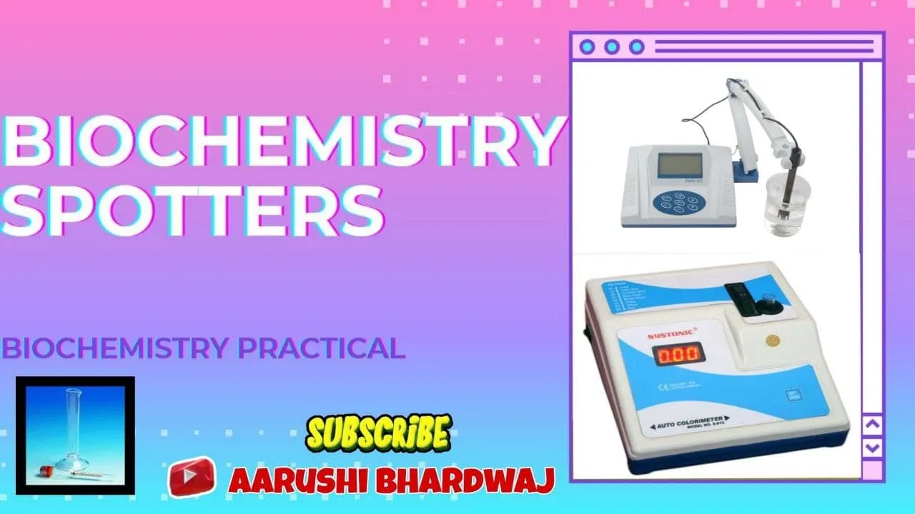
Biochemistry Spotters (First year MBBS biochemistry practical)
102.8K
This comprehensive video covers essential laboratory instruments and techniques used in biochemistry education and practice. The content is organized into three main categories: (1) Fundamental glassware including pipettes for precise liquid transfer, test tube holders for safe handling, borosilicate glassware for heat resistance, and measuring cylinders for volume measurement; (2) Analytical instruments such as urinometers for specific gravity measurement, colorimeters for optical density analysis, micropipettes for small volumes, pH meters for acidity measurement, and spectrophotometers based on Beer-Lambert law; (3) Clinical biochemistry techniques including serum protein electrophoresis for diagnosing conditions like multiple myeloma. The video emphasizes the importance of understanding instrument principles, proper handling techniques, and their applications in laboratory experiments and clinical diagnostics.
Click to watch →
Deep Dive
Recombinant DNA and Plasmid Design: Understanding how target genes are cloned into expression vectors containing specific promoters (e.g., T7 promoter) and ribosome binding sites.

Biotechnology : Principles and Processes 01| Introduction | Class 12th/CUET
153.8K
Recombinant DNA was first constructed in 1972 by Stanley Cohen and Herbert Boyer using salmonella bacteria. Plasmids serve as vectors in this process - they are double-stranded, circular DNA molecules that exist independently of the bacterial chromosome and can self-replicate. The process involves: (1) Attaching desired genes to plasmids, (2) Introducing recombinant DNA into host cells, (3) Allowing host cells to divide, (4) The plasmid replicates along with host DNA, creating multiple cells each containing the desired gene. This enables large-scale production of target genetic material.
Click to watch →

35 Years PYQs Biotechnology : Principles & Processes | NEET Biology 2025 | Dr Gargi
18.2K
Restriction enzymes recognize specific DNA sequences (typically 4-6 base pairs) and cut at precise locations. EcoRI and EcoRV both have 6-base-pair recognition sequences. PBR322 plasmid contains origin of replication (controls copy number), selectable marker genes (antibiotic resistance), and multiple cloning sites. The correct sequence for recombinant DNA formation is: isolation of desired DNA fragment, cutting at specific locations by restriction enzymes, separation of fragments, and amplification. Cloning vectors (plasmids, bacteriophages, cosmids) carry foreign DNA into host cells, while probes are detection tools tagged with radioactive markers.
Click to watch →

Biotechnology Principles & Process | Class 12 Board & NEET | UP Board English Medium RWA
2.1K
Plasmids are ideal vectors because they are self-replicating, can be easily isolated and transferred, and carry antibiotic-resistant genes for selection. Recombinant DNA is constructed by cutting both the gene of interest and plasmid with the same restriction enzyme, then joining fragments using DNA ligase. The resulting molecule combines DNA from different sources to create new genetic combinations.
Click to watch →

Complete Biotechnology in One Shot | with Handwritten Notes and PYQs | NEET 2021 | Ritu Rattewal
132.9K
Restriction enzymes were discovered in 1963 and are part of the bacterial immune system, protecting bacteria from viral infections by cutting foreign DNA at specific sites. These enzymes recognize specific DNA sequences called recognition sites, which are typically palindromic. Restriction enzymes are named according to the bacterium from which they were isolated, including the genus, species, and strain number. Plasmid vectors used in genetic engineering must have an origin of replication, multiple cloning sites, and selectable markers. Recombinant DNA molecules are formed when foreign genetic material is inserted into a host vector, combining host vector DNA with inserted foreign DNA.
Click to watch →
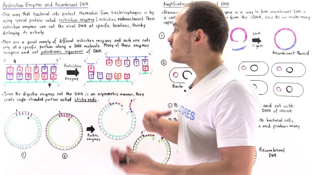
Restriction Enzymes and Recombinant DNA
330.7K
Restriction enzymes are bacterial proteins that protect cells by cutting viral DNA at specific palindromic sequences, creating complementary sticky ends; scientists exploit this property to form recombinant DNA molecules by cutting two different DNA sources with the same enzyme and joining them with DNA ligase, then amplifying these recombinant molecules using plasmids in bacterial cells.
Click to watch →
Prerequisite Knowledge
- Concept 01The Central Dogma of Molecular Biology: A solid understanding of DNA transcription and RNA translation mechanisms, including the functions of ribosomes, tRNAs, and RNA polymerases.
- Concept 02E. coli Biology: Familiarity with E. coli as a model organism, specifically its growth phases (such as the exponential phase optimal for harvesting) and cellular machinery.
- Concept 03Basic Biochemistry Lab Techniques: Prior knowledge of laboratory processes such as cell lysis (e.g., sonication, homogenization), high-speed centrifugation, dialysis, and precise buffer preparation.
- Concept 04Recombinant DNA and Plasmid Design: Understanding how target genes are cloned into expression vectors containing specific promoters (e.g., T7 promoter) and ribosome binding sites.
Subsequent Learning
- Step 01Optimization of CFPS Reaction Parameters: Learning how to fine-tune concentrations of magnesium, potassium, amino acids, and energy regeneration sources (e.g., PEP, creatine phosphate) to maximize protein yield.
- Step 02Incorporation of Non-Canonical Amino Acids (ncAAs): Exploring how cell-free systems can be modified to incorporate synthetic amino acids for site-specific protein labeling and engineering.
- Step 03Downstream Purification and Characterization: Mastering methods to isolate the synthesized proteins (e.g., affinity chromatography) and analyze their yield, purity, and functional activity.
- Step 04High-Throughput Screening and Synthetic Biology: Applying cell-free protocols to rapid prototyping of genetic circuits, metabolic pathway engineering, and automated protein synthesis arrays.
Cell-Free Synthesis
Cell-Free Synthesis
0:01Playing Section
- 1
Protocol enables non-experts to implement cell-free protein synthesis.
- 2
Method offers speed, cost-effectiveness, and ease of setup.
- 3
Cell culture processing includes centrifugation and resuspension steps.
Limitations of E. coli Cell-Free Synthesis compared to Eukaryotic and In Vivo Systems
While E. coli cell-free protein synthesis (CFPS) offers high yields and rapid prototyping, it has significant limitations compared to traditional in vivo expression and eukaryotic cell-free systems. First, E. coli lysates lack the complex machinery required for essential post-translational modifications, such as glycosylation and disulfide bond formation, which are critical for the functionality of many eukaryotic proteins. Second, for industrial-scale production, traditional in vivo fermentation remains far more cost-effective and scalable than cell-free methods. Finally, when folding complex, multi-domain, or membrane-associated proteins, researchers often favor eukaryotic cell-free platforms (such as wheat germ or mammalian lysates) because they contain specialized chaperones and lipid environments that E. coli systems lack.
Deep Dive
Optimization of CFPS Reaction Parameters: Learning how to fine-tune concentrations of magnesium, potassium, amino acids, and energy regeneration sources (e.g., PEP, creatine phosphate) to maximize protein yield.

Build-a-Cell seminar Karen Polizzi: Developing eukaryotic cell-free protein synthesis systems
359
For Pichia pastoris CFPS, lysis method selection is critical—high-pressure homogenization provides highest protein concentration, while sonication yields lower concentrations. Harvest timing affects ribosome content; biosensor strains tracking ribosomal gene transcription show transcription peaks and declines as OD increases, with optimal harvest at higher OD values (18-20). Translation initiation challenges due to loss of 5' cap structure are addressed using Internal Ribosome Entry Sites (IRES), with Cricket Paralysis Virus IRES showing broad host specificity. Luciferase is preferred over GFP as reporter due to better performance in static conditions. Overexpressing global regulators like Fhl1 increases ribosome content and luciferase synthesis by five-fold. Reaction mix optimization uses definitive screening design, achieving 3-4 fold increases in protein yield.
Click to watch →

Cell Free Protein Synthesis | Cell Free Protein Expression | CFPS | Cell Free Technology |
5.6K
Cell-free protein synthesis (CFPS) is a technique that produces recombinant proteins in solution using extracted cellular translation machinery, enabling direct control of the translation environment for applications such as membrane protein solubilization, protein production optimization, and stable isotope labeling for mass spectrometry analysis.
Click to watch →

2026년 대한민국이 수천억 원을 쏟아붓는 '이 기술', 모르면 무조건 손해입니다
2.4K
Cell-free Protein Synthesis (CFPS) is a technology that enables protein production outside living cells. Scientists extract cellular components like ribosomes and enzymes from cells, creating a lysate. When DNA, energy sources (ATP), and amino acids are added, transcription and translation occur, producing desired proteins. This method can complete experiments in hours that previously required weeks or months.
Click to watch →
Computer Assisted Synthesis Planning 3 Applications
255
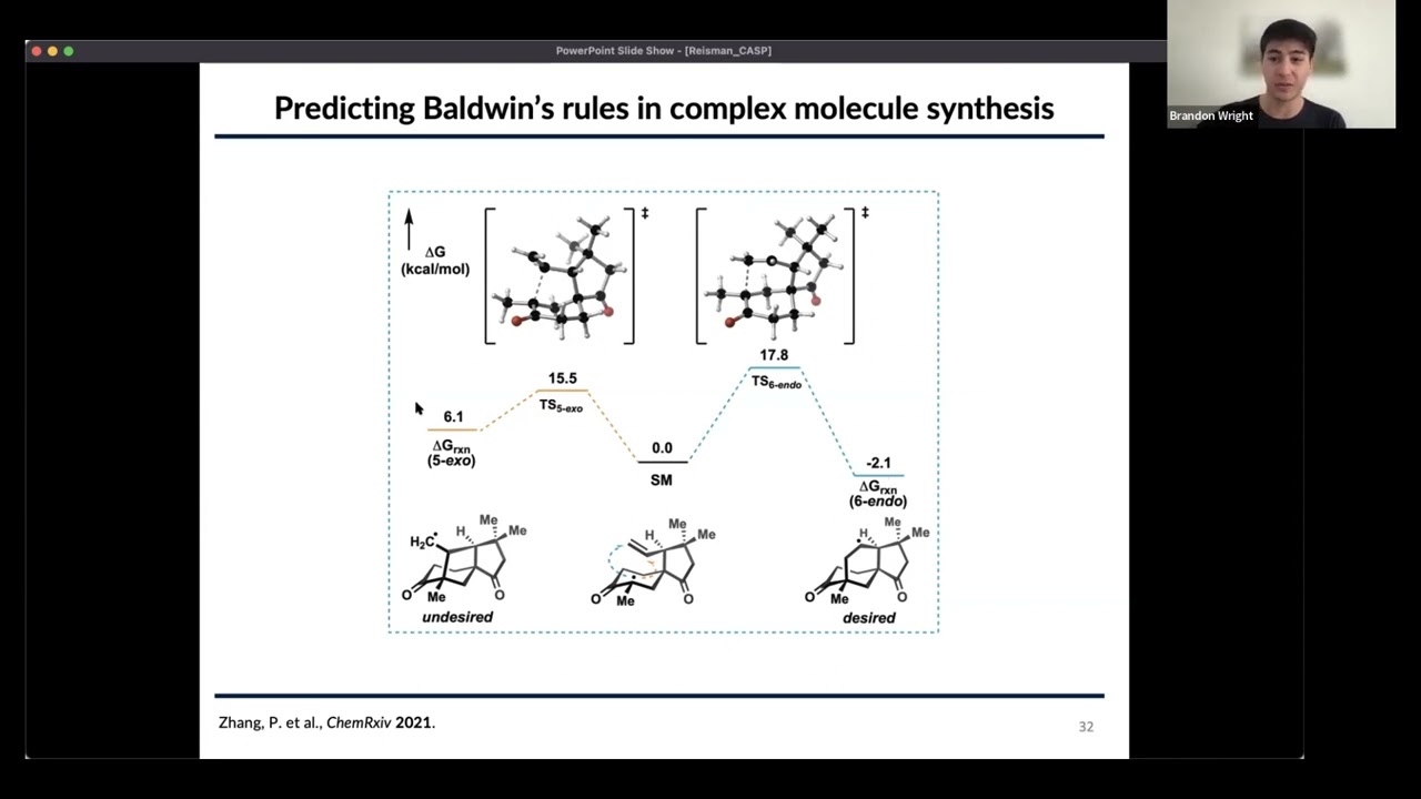
Bayesian optimization is a machine learning algorithm designed to balance exploration and exploitation when optimizing reaction parameters. The method uses an acquisition function to guide the search through parameter space: exploitation-focused functions prioritize finding maximum yields in already-explored regions, while exploration-focused functions prioritize testing conditions with high uncertainty to discover new regions of the parameter space. The algorithm iteratively updates its model based on experimental results, deciding which conditions to try next. This approach can find optimal reaction conditions in far fewer experiments than random or purely systematic approaches.
Click to watch →

Route Scouting for Kilogram-Scale Manufacturing of API’s - Dr Martin Strack - MercachemSyncom
1.5K
Successful reaction optimization requires systematic screening of multiple parameters simultaneously. Key variables include coupling agents/activators, bases, solvents, temperature ranges, and concentrations. Early-stage chemistry typically uses excess solvent compared to scale-up conditions, so optimization must account for higher concentrations used in production. Reaction times become critical at larger scales, and addition sequences and work-up procedures must be developed to maximize efficiency and yield while minimizing side reactions.
Click to watch →
Deep Dive
Incorporation of Non-Canonical Amino Acids (ncAAs): Exploring how cell-free systems can be modified to incorporate synthetic amino acids for site-specific protein labeling and engineering.

Antimicrobial Peptides Produced: Non-Canonical Amino Acids l Protocol Preview
411
This video demonstrates an Escherichia coli-based methodology for incorporating non-canonical amino acids (ncAAs) into ribosomally synthesized antimicrobial peptides like nisin, enabling parallel testing of multiple ncAA modifications without genetic alterations; the process involves growing transformed bacterial cultures in defined media, inducing expression, and producing modified peptides that can be screened for antimicrobial activity using agar diffusion assays to study peptide function and develop novel antimicrobial agents against drug-resistant pathogens.
Click to watch →

L6 || Amino acids I (2025)
148
Non-standard amino acids include selenocysteine and pyrrolysine, which are incorporated into proteins during translation through unique mechanisms. Selenocysteine is encoded by the UGA stop codon and contains selenium instead of sulfur. Pyrrolysine is encoded by the UAG stop codon and is found in certain methanogenic archaea. These amino acids require specific tRNA and release factors for incorporation. Additionally, beta and gamma amino acids have the amino group attached to the beta or gamma carbon respectively, rather than the alpha carbon. These non-standard amino acids expand the chemical diversity of proteins beyond the 20 standard amino acids.
Click to watch →

EP 122: Building genome-scale engineered cells with Leslie Mitchell | The Genetics Podcast
105
To incorporate non-canonical amino acids into proteins, researchers must globally eliminate one or more codons from the genome and repurpose them for non-standard amino acid incorporation. This requires orthogonal translation machinery including synthetic tRNAs and aminoacyl-tRNA synthetases that work exclusively with non-canonical amino acids while excluding all canonical amino acids.
Click to watch →

Protein expression & purification techniques - overview of some common techniques & typical workflow
4.4K
Cell-free systems enable incorporation of non-canonical amino acids that do not exist in natural proteins. These modified amino acids can carry chemical handles for bioorthogonal reactions, click chemistry, and other chemical biology applications. This capability allows researchers to introduce specific chemical functionalities into proteins for structural studies or functional assays.
Click to watch →

Scripps Research - Organometallics 2021 (Renata) - Day 20
848
Amber suppression (developed by Schultz) allows incorporation of non-canonical amino acids like acetophenylalanine into proteins. This enables site-specific anchoring of metal complexes to protein scaffolds. The Lewis group used this technique to incorporate acetophenylalanine into carboxypeptidase A, then used click chemistry to attach rhodium complexes.
Click to watch →
Deep Dive
Downstream Purification and Characterization: Mastering methods to isolate the synthesized proteins (e.g., affinity chromatography) and analyze their yield, purity, and functional activity.

Downstream Lec 1
5.9K
Downstream processing typically involves four main stages: (1) Removal of insolubles - separating cells, cell debris, and particulate matter from the liquid, (2) Product isolation - concentrating the product from a large volume of liquid (typically 5-25% concentration), (3) Product purification - achieving high purity (90-99% for pharmaceuticals), and (4) Product polishing - adding stabilizers to improve shelf life and storage properties. Product isolation involves extraction using precipitation, solvent extraction, or supercritical extraction. Product purification uses chromatography techniques including affinity chromatography, ion exchange chromatography, and reverse phase chromatography. Polishing involves crystallization, lyophilization, or spray drying. For proteins, techniques involve extraction and precipitation; for metabolites, distillation is suitable.
Click to watch →
Bio-processing overview (Upstream and downstream process)
239.7K
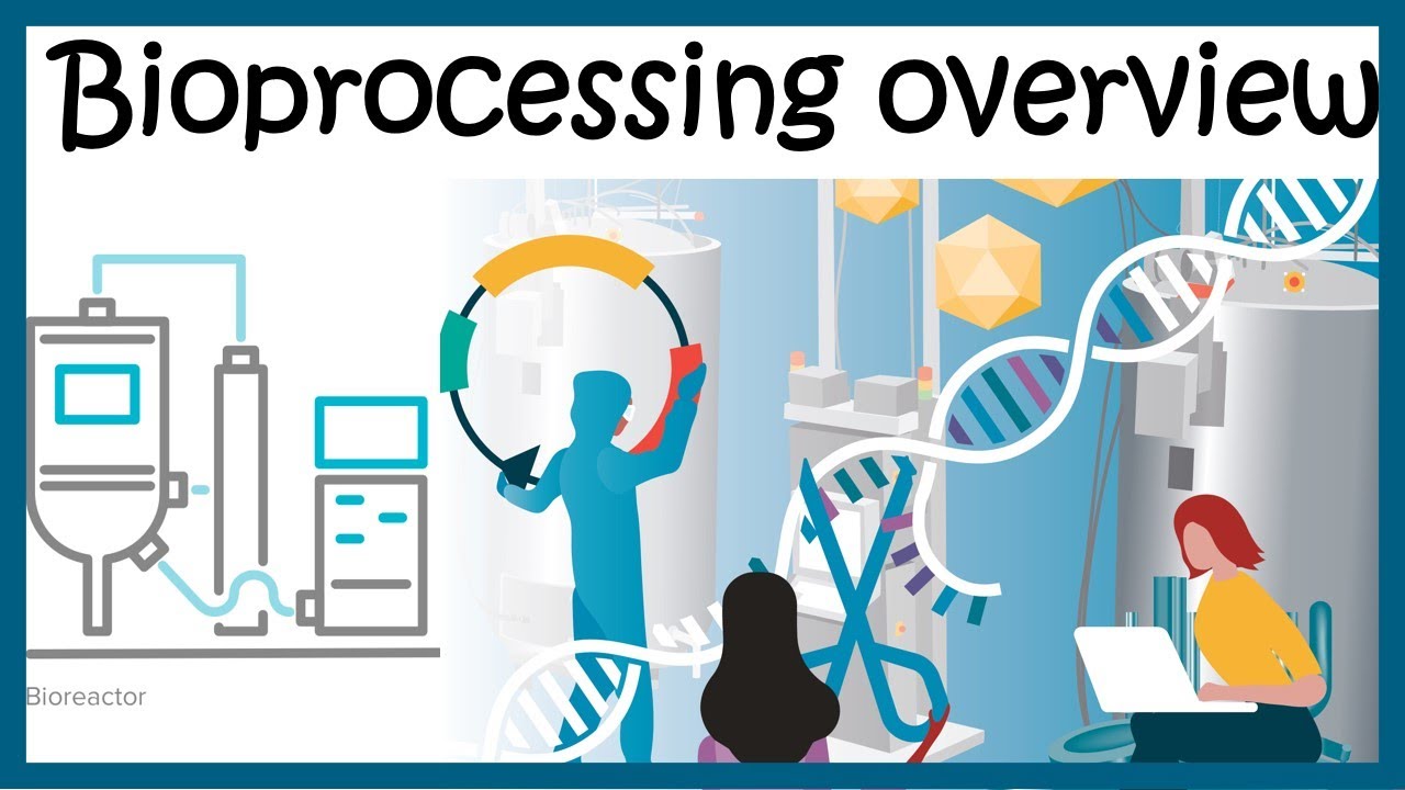
Downstream processing encompasses recovery, purification, polishing, quality control, and packaging. For extracellular products, centrifugation separates cells, followed by filtration and column chromatography. For intracellular products, mechanical disruption releases contents before purification. Due to large broth volumes, concentration via vacuum drying reduces processing burden. High-throughput chromatographic methods including HPLC, hydrophobic interaction, and ion exchange columns achieve initial purification. Final polishing ensures 98-100% purity, and inert additives enhance stability. Rigorous quality control verifies product functionality and safety before market release and distribution.
Click to watch →

Microbial Cell Culture and its Applications - 5 #CH22SP #swayamprabha
121
Downstream processing (bioseparation) converts fermentation broth into marketable products through four sequential steps: removal of insolubles, product isolation, purification/up-concentration, and polishing/packing. The fundamental principle exploits differences in physical and chemical properties between products and impurities, including solubility, hydrophobicity, electrostatic interactions, and stability under different conditions. This process accounts for approximately 60% of total bioprocess costs. Required purity levels vary by application: 99.99% for therapeutics, 98-99% for diagnostics, and 90-92% for industrial products. Analytical tools essential for characterization include HPLC, ICPMS, FTIR, NMR, SEC, and LC-MS/MS, providing both qualitative and quantitative information about molecular properties.
Click to watch →
Downstream Processing - S5 | Purification Methods - Dr. Deepika Malik - Learn Microbiology With Me
1.5K
Purification in downstream processing employs multiple techniques including chromatography (adsorption, ion-exchange, gel permeation, affinity, HPLC, and partition chromatography), membrane processes (reverse osmosis and ultrafiltration), liquid membranes, and crystallization to isolate and concentrate target products from complex biological mixtures by exploiting differences in molecular properties such as charge, size, solubility, and binding affinity.
Click to watch →

Plus Two Biology | Biotechnology: Principles and Processes - Chapter 11 | Eduport plus two
54.1K
Downstream processing recovers and purifies products from bioreactors. It involves cell separation, product extraction, and purification to isolate the desired molecule. Quality control ensures products meet specifications for safety and efficacy. This final stage transforms biological production into usable products for research, medicine, or industry.
Click to watch →
Deep Dive
High-Throughput Screening and Synthetic Biology: Applying cell-free protocols to rapid prototyping of genetic circuits, metabolic pathway engineering, and automated protein synthesis arrays.

Developing biosensors for high-throughput screening in synthetic biology
653
Biosensors enable rapid, cost-effective screening of microbial cell factories by genetically reprogramming microorganisms to produce fluorescent signals proportional to target molecule concentration, thereby overcoming the limitations of traditional analytical techniques like HPLC and mass spectrometry in the design-build-test-learn cycle of synthetic biology.
Click to watch →
“Full Steam Ahead!” – taking synthetic biology to the high throughput era
752
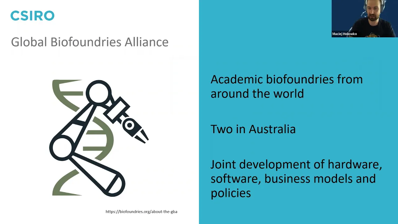
Synthetic biology has evolved from artisanal, low-throughput laboratory work to high-throughput automated platforms called biofoundries, which use standardized protocols and modular workflows to accelerate the design-build-test-learn cycle for DNA and strain engineering, enabling faster identification of candidate strains and reducing time-to-market for biotechnological products.
Click to watch →

The Future of Synthetic Biology with Prof. George Church (Harvard Medical School) || HAE
577
Machine learning combined with protein barcoding allows simultaneous testing of thousands to millions of different experimental conditions in single organisms. For example, 1.2 million different viral capsid designs for gene therapy delivery can be tested in one animal, enabling rapid optimization of delivery systems.
Click to watch →

Synthetic Biology: High throughput synthetic biology and biosensors - Group 4
3.1K
Synthetic biology enables the creation of inexpensive, portable biosensors by using engineered microbes that detect environmental signals (like arsenic in drinking water) and produce visible outputs (such as light), with high-throughput robotics allowing researchers to efficiently test thousands of genetic combinations to identify optimal biosensor designs.
Click to watch →

Panel: The Past, Present & Future of Gene Delivery
448
Modern synthetic biology platforms enable rapid exploration of design space through automation and high-throughput screening. Companies like Ginkgo Bioworks can design between 5,000-20,000 genes monthly, representing a dramatic increase from earlier eras when researchers designed only a handful of genes during their PhD work. This brute-force approach combined with automation allows customers to identify optimal designs quickly. However, an equally important consideration is connecting high-throughput screening to manufacturability—ensuring new designs are tested in manufacturing contexts early to avoid scalability issues that often arise when promising hits from screens prove difficult to scale up.
Click to watch →
Cell-Free Synthesis
Cell-Free Synthesis
0:01- 1
Protocol enables non-experts to implement cell-free protein synthesis.
- 2
Method offers speed, cost-effectiveness, and ease of setup.
- 3
Cell culture processing includes centrifugation and resuspension steps.
Limitations of E. coli Cell-Free Synthesis compared to Eukaryotic and In Vivo Systems
While E. coli cell-free protein synthesis (CFPS) offers high yields and rapid prototyping, it has significant limitations compared to traditional in vivo expression and eukaryotic cell-free systems. First, E. coli lysates lack the complex machinery required for essential post-translational modifications, such as glycosylation and disulfide bond formation, which are critical for the functionality of many eukaryotic proteins. Second, for industrial-scale production, traditional in vivo fermentation remains far more cost-effective and scalable than cell-free methods. Finally, when folding complex, multi-domain, or membrane-associated proteins, researchers often favor eukaryotic cell-free platforms (such as wheat germ or mammalian lysates) because they contain specialized chaperones and lipid environments that E. coli systems lack.
The Central Dogma of Molecular Biology: A solid understanding of DNA transcription and RNA translation mechanisms, including the functions of ribosomes, tRNAs, and RNA polymerases.
E. coli Biology: Familiarity with E. coli as a model organism, specifically its growth phases (such as the exponential phase optimal for harvesting) and cellular machinery.
Basic Biochemistry Lab Techniques: Prior knowledge of laboratory processes such as cell lysis (e.g., sonication, homogenization), high-speed centrifugation, dialysis, and precise buffer preparation.
Recombinant DNA and Plasmid Design: Understanding how target genes are cloned into expression vectors containing specific promoters (e.g., T7 promoter) and ribosome binding sites.
Optimization of CFPS Reaction Parameters: Learning how to fine-tune concentrations of magnesium, potassium, amino acids, and energy regeneration sources (e.g., PEP, creatine phosphate) to maximize protein yield.
Incorporation of Non-Canonical Amino Acids (ncAAs): Exploring how cell-free systems can be modified to incorporate synthetic amino acids for site-specific protein labeling and engineering.
Downstream Purification and Characterization: Mastering methods to isolate the synthesized proteins (e.g., affinity chromatography) and analyze their yield, purity, and functional activity.
High-Throughput Screening and Synthetic Biology: Applying cell-free protocols to rapid prototyping of genetic circuits, metabolic pathway engineering, and automated protein synthesis arrays.
996 views5likes2:00@JoVEJournalOriginal Release: 2022-05-30
[Music] this protocol simplifies and clarifies the methods for implementing cell-free protein synthesis by non-experts improved access to these methods will help democratize the platform and the broad set of applications that it enables the main advantages of this technique are the speed cost effectiveness and the ease of reaction setup compared to other cell-free protein synthesis platforms our platform can enable a number of applications including functional genomics high throughput testing biosensors educational kits and with minor modifications metabolic engineering and genetic code expansion while this technique only requires basic laboratory training new users should plan to familiarize themselves with techniques like sonication for successful execution of the protocol to begin this procedure prepare all media and culture e coli cells as outlined in the text protocol place a one liter centrifuge bottle into an ice water bath once the culture's od 600 reaches 3.0 pour the cell culture into the chilled bottle using a double beam balance add water to a second one liter centrifuge bottle until it weighs the same as the first to create a balance for the centrifuge after pre-cooling the centrifuge to 4 degrees celsius centrifuge the bottles at 5000 g and at 10 degrees celsius for 10 minutes to pellet the cells after this slowly pour off and dispose of the supernatant place the cell pellet on ice using a sterile spatula scrape the cell pellet out of the centrifuge bottle and transfer it to a cold 50 milliliter conical tube add 30 milliliters of cold s30 buffer supplemented with 2molar dtt and resuspend the pellet by vortexing in short bursts
Up Next
24:50
Bacterial Protein Expression & His-Tag Purification
@eroglulab7013
41.8K views•2020-12-02

12:54
Triumph of Orthodoxy Icon: Byzantine Art & History Explained
@BenCallan
2.1K views•2024-08-06

2:00
Organ-on-Chip Fabrication with Integrated Electrodes for TEER | Protocol Preview
@JoVEJournal
377 views•2022-05-27

1:42
Game of Thrones Opening Credits: A Cinematic Analysis
@gameofthrones
46.3M views•2011-04-18
Related Study Plans & Knowledge Roadmaps
Structured learning paths in General & Interdisciplinary Studies
general-studies
Elevator Engineering: History, Physics & Cities
01From Hoists to Safety Brakes: The Dawn of Vertical Lift
02Hydraulic vs. Traction: The Engineering Physics of Elevators
03Modern Speed, Safety, and Algorithms
+ 2 more learning modules
12h5 modules
Explore Roadmap
general-studies
Rubber: Material Science, History & Geopolitics
01Origins of Natural Rubber & Mesoamerican History
02The Chemistry of Natural Rubber & Vulcanization
03The Geopolitics of the Rubber Boom & Colonization
+ 2 more learning modules
12h5 modules
Explore Roadmap
general-studies
Containerization: Evolution, Logistics & Trade
01Ancient Trade & Bulk Cargo: Amphorae to Barrels
02The Industrial Revolution & Cardboard Packaging
03The Containerization Revolution: Malcom McLean
+ 2 more learning modules
12h5 modules
Explore Roadmap
general-studies
Textile Tech: Evolution, Labor & Global Impact
01Pre-Industrial Textiles: Handloom & Cottage Industry
02Mechanization: The Spinning Jenny to the Power Loom
03The Factory System & Global Labor Transformation
+ 2 more learning modules
10h5 modules
Explore Roadmap