Haematococcus PBR: Design, Stress & Astaxanthin
Learning Goal: Designing and optimizing a bench-scale photobioreactor system for the cultivation of Haematococcus pluvialis to induce astaxanthin accumulation, covering culture media formulation, lighting and aeration parameter control, stress-induced carotenogenesis, and spectrophotometric quantification of the extracted pigment.
Prerequisites
- Fundamental Biology: Undergraduate-level cell biology or botany (specifically understanding photosynthesis, cellular organelles, and basic algal life cycles).
- General Chemistry: Basic preparation of solutions, molar calculations, and operating laboratory instruments such as spectrophotometers.
- Engineering Basics: High school physics or introductory bioprocess engineering concepts (fluid dynamics, mass balance, and light properties) will facilitate comprehension of the photobioreactor transport mechanics.
Estimated Study Time
- Total Course Duration: ~15 Hours (including video material, analytical reading, mathematical problem sets, and practical design exercises).
Module 1: Biology and Life Cycle of Haematococcus pluvialis
Overview
Haematococcus pluvialis is a freshwater unicellular green microalgae globally renowned for its capacity to synthesize and accumulate massive amounts of the red ketocarotenoid astaxanthin (up to 4–5% of its dry biomass). Understanding the morphology and biology of H. pluvialis requires an in-depth exploration of its highly dynamic biphasic life cycle and the complex metabolic pathways driving carotenogenesis.
[Macrozoid / Microzoid] (Green flagellated motile vegetative cell) │ ▼ (Environmental stress / Nutrient limitation) [Palmella Phase] (Non-motile vegetative cell; loses flagella) │ ▼ (Severe stress: high light, nitrogen starvation) [Aplanospore / Cyst Phase] (Tough secondary wall; high astaxanthin accumulation)
Biochemical Pathway of Carotenogenesis
During favorable growth conditions, H. pluvialis partitions carbon into primary metabolites (proteins and structural lipids) inside its green vegetative cells. When stressed, it activates the mevalonate-independent methylerythritol phosphate (MEP) pathway within the plastids to synthesize carotenoids:
- Isopentenyl Pyrophosphate (IPP) and Dimethylallyl Pyrophosphate (DMAPP) serve as the 5-carbon isoprenoid building blocks.
- Sequential condensation steps yield Geranylgeranyl Pyrophosphate (GGPP) ().
- Two GGPP molecules are coupled by Phytoene Synthase (PSY) to form Phytoene (), the first committed carotenoid precursor.
- Sequential desaturations by Phytoene Desaturase (PDS) and -Carotene Desaturase (ZDS) yield Lycopene.
- Lycopene -cyclase (LCYb) cyclizes the end-groups of lycopene to form -Carotene.
- Finally, -Carotene Ketolase (BKT) introduces keto groups at the 4,4' positions of the ionone rings (forming intermediate canthaxanthin), while -Carotene Hydroxylase (CHYb) introduces hydroxyl groups at the 3,3' positions, yielding the highly stable, symmetric end-product: Astaxanthin (3,3'-dihydroxy--carotene-4,4'-dione).
The resulting astaxanthin is subsequently esterified with fatty acids and sequestered into cytosolic lipid droplets, protecting the cell's photosynthetic apparatus from photo-oxidative damage.
Recommended Videos

- Why this video is valuable: This video provides an direct microscopic visualization of the microalgal morphology. It allows learners to visually differentiate between the active, green vegetative cells (macrozoids) and the transition stages leading to immotile cysts.
- Knowledge Checkpoint:
- Identify the anatomical differences between flagellated macrozoids and immotile palmella cells.
- Locate the red-pigmented astaxanthin accumulation zones inside the maturing cyst under light microscopy.
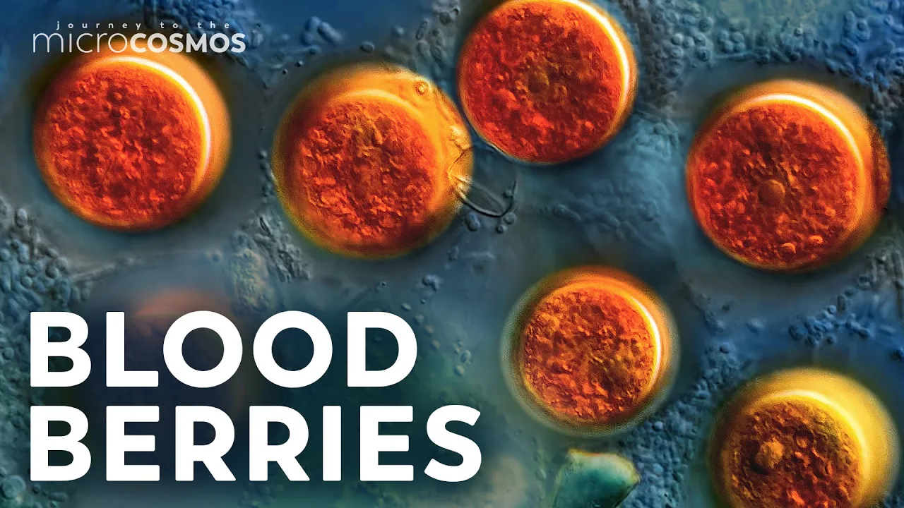
- Why this video is valuable: An exceptionally detailed micro-cinematography video illustrating how H. pluvialis reacts to environmental changes. It shows the morphological transformation into defensive cysts, illustrating the evolutionary purpose of carotenogenesis as a photoprotection mechanism.
- Knowledge Checkpoint:
- Explain the ecological conditions that trigger H. pluvialis to transition from vegetative growth to cyst formation.
- Describe the cellular defense mechanism of accumulating astaxanthin against ultraviolet (UV) radiation and reactive oxygen species (ROS).

- Why this video is valuable: A rigorous academic lecture outlining the commercial cultivation steps, life-cycle transitions, biochemistry, and physiological significance of astaxanthin synthesis. This bridges laboratory biology with industrial-scale processing applications.
- Knowledge Checkpoint:
- List the four key morphotypes in the H. pluvialis life cycle: macrozoids, microzoids, palmella, and hematocysts (aplanospores).
- Identify the key precursor molecules and enzymatic steps leading to the synthesis of astaxanthin from -carotene.
Module 2: Culture Media Formulation and Inoculation
Overview
Cultivating H. pluvialis requires an exact nutritional profile. BG-11 or Bold's Basal Medium (BBM) are the most popular media variants used to satisfy macronutrient and micronutrient demands.
BG-11 Media Composition & Preparation
BG-11 contains critical salts that supply nitrogen, phosphorus, magnesium, calcium, and trace elements.
| Compound | Purpose | Concentration in Final Medium (mg/L) |
|---|---|---|
| Nitrogen source (for green protein synthesis) | 1500 | |
| Phosphorus (nucleic acids, ATP) | 40 | |
| Magnesium (chlorophyll core molecule) | 75 | |
| Calcium (cell signaling and wall structural integrity) | 36 | |
| Citric Acid / Ammonium Ferric Citrate | Iron chelation (photosynthetic electron transport chain) | 6 / 6 |
| Metal chelator (preventing trace metal precipitation) | 1 | |
| Trace Metal Mix () | Micronutrient cofactors for cellular enzymes | Variable ( level) |
During preparation, all components must be dissolved in deionized water, adjusted to a starting pH of 6.8 to 7.2, and sterilized via autoclaving at 121°C for 20 minutes to eliminate bacterial and fungal contaminants.
Inoculation and Cell Density Quantification
Aseptic inoculation protocols must be performed under a Laminar Airflow Hood to maintain culture purity. Algal growth kinetics are tracked daily using cell concentration calculations via a Hemocytometer (Levy-Hausser slide).
To ensure statistical accuracy, count the four corner large squares ( area, depth, containing 16 smaller squares) and calculate the average before applying the formula.
Recommended Videos

- Why this video is valuable: A concise step-by-step preparation tutorial showing the physical addition, dissolution, and stabilization steps of BG-11 components.
- Knowledge Checkpoint:
- Describe the sequential mixing steps required to prevent precipitation of mineral components when preparing stock solutions.
- State the targeted final pH of the autoclaved BG-11 solution prior to inoculation.
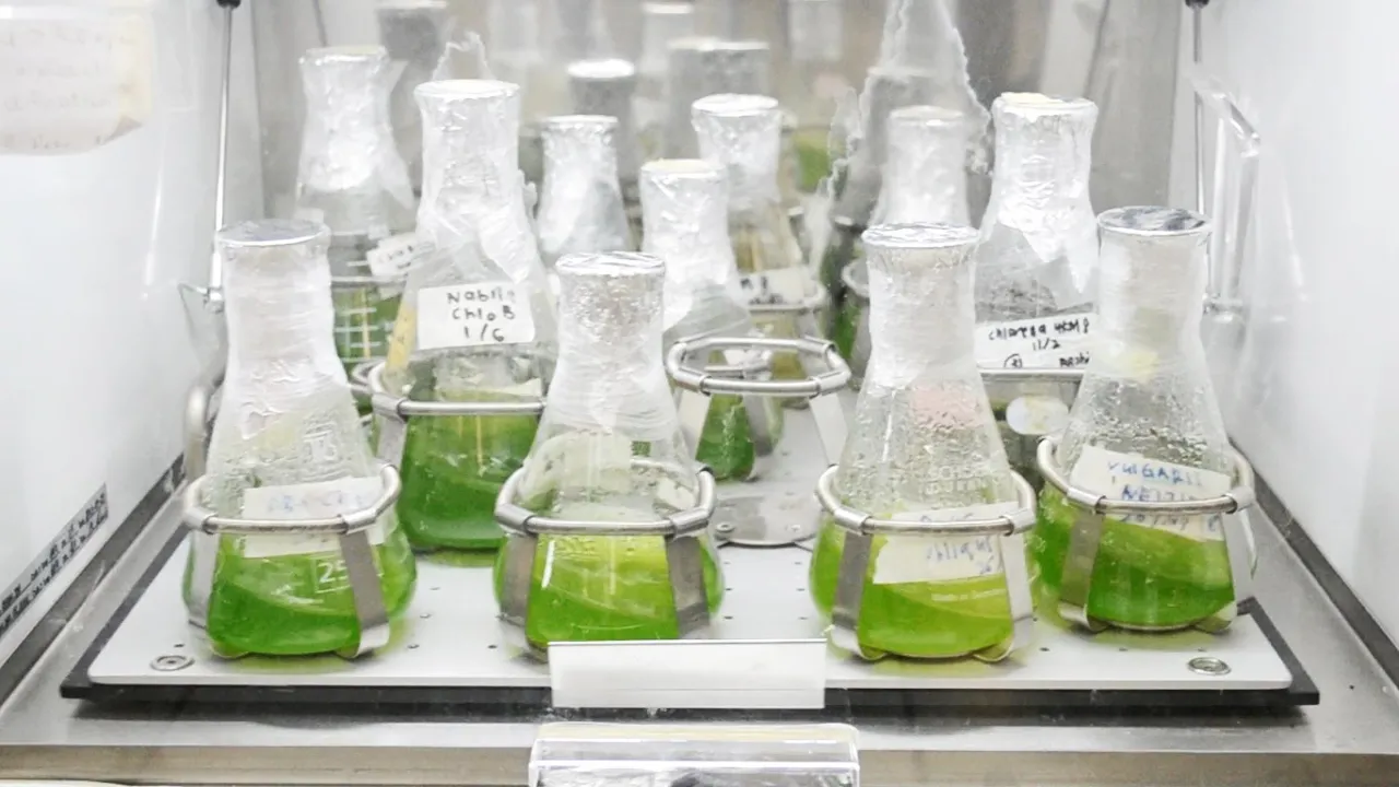
- Why this video is valuable: Demonstrates professional aseptic techniques. It covers how to light and use a Bunsen burner, sterile hood operations, and serial dilutions to transition a starter microalgae culture into fresh media without introducing contaminants.
- Knowledge Checkpoint:
- Name three common sources of biological contamination in flask-level microalgae cultures.
- Describe the "laminar airflow" protocol to maintain sterile conditions during culture transfer.
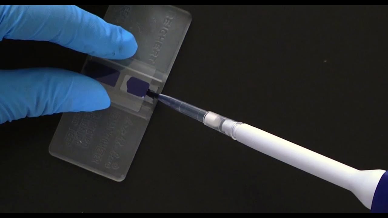
- Why this video is valuable: Provides a thorough guide to the structure of a hemocytometer slide, proper sample loading via capillary action, and cell counting rules (e.g., handling cells on the boundary lines).
- Knowledge Checkpoint:
- Explain which boundary lines (e.g., top/left vs. bottom/right) to include when counting cells to prevent double-counting.
- Calculate the cell concentration of a sample diluted 1:1 with trypan blue if 120 cells are counted across 4 large corner squares.
Module 3: Bench-Scale Photobioreactor (PBR) Design and Engineering
Overview
Optimizing H. pluvialis biomass generation requires controlled parameters. A bench-scale Photobioreactor (PBR) manages light delivery, temperature stability, and mass transfer dynamics.
   [Vent/Filter]
        ▲
┌───────┴───────┐
│  PBR Vessel   │ ◄─── [External LED Light Panel]
│ (Glass/Acrylic│      (Controllable Photosynthetic Photon Flux Density)
│  with Algae)  │
└───────┬───────┘
        ▲
        │ (Continuous Aeration: 1.5-2.0% CO2 in air)
 [Sparger/Nozzle]
Light Path Attenuation
Light intensity decreases exponentially as it penetrates the culture depth due to absorption by the algal cells and scattering by suspended particles. This phenomenon is modeled by a modified version of the Beer-Lambert Law:
Where:
- = Light intensity at depth ().
- = Incident light intensity at the reactor surface.
- = Specific light attenuation/absorption coefficient ().
- = Biomass concentration ().
- = Distance from the illuminated surface ().
To minimize self-shading, bench-scale reactors are engineered with a short light path length (typically for tubular or flat-panel designs).
Mass Transfer Coefficient ()
Mixing provides uniform light exposure (averaging the light/dark cycles) and maintains dissolved gas equilibrium. Liquid-to-gas mass transfer is governed by:
Where:
- = Rate of gas concentration change in the liquid ().
- = Instantaneous concentration of dissolved gas ( or ) in the culture bulk.
- = Saturation concentration of the gas in equilibrium with the gas phase.
- = Volumetric mass transfer coefficient ().
Efficient PBRs require bubble spargers that generate bubbles with a high surface-area-to-volume ratio. This increases to provide adequate dissolved carbon dioxide () while stripping out toxic photosynthetic dissolved oxygen (), which can inhibit Rubisco and promote cellular photo-oxidation.
Recommended Videos

- Why this video is valuable: A detailed, hands-on construction guide showing how to build an automated turbidostat photobioreactor. This build features physical vessel geometries, LED lighting arrays, sensor integrations, and fluid pumping systems.
- Knowledge Checkpoint:
- Explain how a turbidostat uses optical density (OD) measurements to automate media dilution and maintain constant cell density.
- Identify the physical trade-offs when selecting a narrow tube diameter versus a wide diameter regarding light attenuation.

- Why this video is valuable: Outlines the core classifications of closed systems (bubble columns, flat panels, tubular loops, airlift reactors). It highlights their respective hydrodynamic properties, gas exchange efficiency, and sterilization challenges.
- Knowledge Checkpoint:
- Contrast the hydrodynamic mixing mechanisms of a bubble column reactor and an airlift loop reactor.
- State how bubble size distribution affects both the gas-liquid interfacial area () and the mass transfer coefficient ().

- Why this video is valuable: A clear bioprocess engineering presentation focused on optimizing fluid dynamics, shear stress, and mixing parameters. This content is crucial for protecting shear-sensitive vegetative H. pluvialis flagellates.
- Knowledge Checkpoint:
- Define "shear stress" and explain its physiological effects on motile macrozoids during aggressive agitation.
- Describe how the gas flow rate controls the liquid circulation speed and mixing efficiency in a vertical column.
Module 4: Two-Stage Cultivation and Stress Induction
Overview
Industrial production of astaxanthin from H. pluvialis relies on a Two-Stage Cultivation Strategy. This approach physically separates the biological growth phase from the pigment-accumulation stress phase.
STAGE 1: GREEN PHASE
┌────────────────────────────────────────────────────────┐ │ - Nutrient-rich BG-11 │ │ - Low to moderate light (40-80 μmol photons/m²/s) │ │ - Rapid cell division & biomass accumulation │ └──────────────────────────┬─────────────────────────────┘ │ ▼ (Wash / Nitrogen Starvation) STAGE 2: RED PHASE ┌────────────────────────────────────────────────────────┐ │ - Nitrogen-free media (BG-11_0) │ │ - High light intensity (>200-500 μmol photons/m²/s) │ │ - Carotenogenesis & cyst formation (Aplanospores) │ └────────────────────────────────────────────────────────┘
Step-by-Step Stress Induction Protocol
- Stage 1: Harvest and Centrifugation: Cultivate H. pluvialis under optimal green-phase conditions until the culture reaches the late-exponential phase (). Harvest the biomass via centrifugation at () for 5 minutes.
- Stage 2: Washing: Decant the nutrient-rich supernatant. Resuspend the cell pellet in sterile distilled water or nitrogen-free BG-11 () to wash out residual nitrate. Centrifuge a second time.
- Stage 3: Resuspension under Nutrient Starvation: Resuspend the washed cells in media containing zero nitrogen ().
- Stage 4: Photo-stress and Salinity Synergism: Transfer the culture back into the photobioreactor. Increase the light intensity from the standard vegetative range () up to high-stress levels (). Optionally, add or apply a temperature shift () to accelerate the transition to red cysts.
- Monitoring: Trace the morphological change from green flagellated cells to large, non-motile red aplanospores over a 5 to 7-day period.
Recommended Videos

- Why this video is valuable: Demonstrates a modern, state-of-the-art facility operating a two-stage cultivation scheme. It highlights the transition from pristine green indoor cultures to outdoor photobioreactor stress arrays.
- Knowledge Checkpoint:
- Outline the steps taken to prevent biological contamination when transitioning from the indoor green phase to the high-exposure red stress phase.
- Contrast the environmental requirements of the green phase (growth, nutrients) with those of the red phase (intense light, nutrient deprivation).

- Why this video is valuable: A research lecture segment that explains the evolutionary and cellular biology of microalgae under stress conditions, specifically exploring how nitrogen starvation redirects carbon pathways toward storage lipids and secondary metabolites.
- Knowledge Checkpoint:
- Explain why nitrogen starvation stops cell division and promotes carbohydrate/lipid accumulation.
- Name the organelle where newly synthesized astaxanthin esters are sequestered inside the cell.

- Why this video is valuable: Explains the physical process of microalgae stress transformation. It provides simple visual transitions showing green vegetative cells turning into deep-red thick-walled cysts.
- Knowledge Checkpoint:
- Identify the color progression timeline as vegetative cells lose chlorophyll and accumulate astaxanthin.
- List the physical stress factors used in industrial production facilities to induce this pigment transition.
Module 5: Cell Disruption, Extraction, and Spectrophotometric Assay
Overview
Harvested red aplanospores possess a thick, multi-layered cell wall containing algaenan, an insoluble lipid-based biopolymer. This wall is resistant to chemical attack and physical lysis, making effective cell disruption a critical requirement for extraction.
Physical and Chemical Disruption Protocols
- Physical (Bead Beating / Ball Milling): High-speed collision with glass or zirconium beads ( diameter) shears the tough aplanospore cell wall without thermally degrading the heat-sensitive astaxanthin.
- Chemical (DMSO Treatment): Pre-treating the biomass with dimethyl sulfoxide (DMSO) at permeabilizes the cell wall, allowing organic solvents (e.g., acetone, ethanol) to reach the internal lipid vacuoles.
Extraction & Spectrophotometric Quantification
Following cell disruption, astaxanthin is dissolved in organic solvents. Because residual green chlorophyll pigments absorb light in overlapping wavelengths, using simple direct absorption readings will lead to concentration errors. To accurately calculate concentrations, we must apply multi-wavelength spectrophotometric equations (such as the Lichtenthaler or specialized carotenoid-correcting formulas in organic solvents like 90% acetone):
Where:
- = Measured absorbances at (carotenoid maximum), (chlorophyll ), and (chlorophyll ).
- = Corrected absorbance representing isolated carotenoids (primarily astaxanthin).
The quantitative yield of extracted astaxanthin can then be calculated using the beer-lambert relationship with the specific extinction coefficient:
Where:
- = Specific extinction coefficient of astaxanthin in acetone ().
- = Cuvette optical path length ().
Recommended Videos

- Why this video is valuable: Demonstrates the physical mechanisms of bead milling and ball milling. This provides the engineering principles behind utilizing bead diameter, vibration frequency, and filling ratios to disrupt highly resilient cell walls.
- Knowledge Checkpoint:
- Explain how bead size and sample density affect the mechanical shear stress generated during milling.
- Identify potential issues with excessive milling times regarding temperature management and pigment degradation.
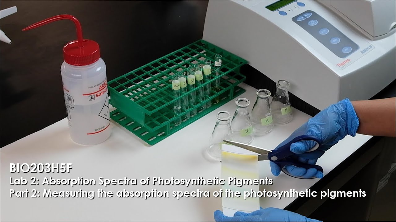
- Why this video is valuable: A practical demonstration of using a UV-Vis spectrophotometer to generate absorption spectra. It covers how to blank the device, calibrate wavelengths, and measure absorbance peaks for chlorophylls and carotenoids.
- Knowledge Checkpoint:
- Identify the primary absorption peak wavelengths for carotenoids versus chlorophylls.
- Describe the step-by-step process of preparing a reference blank using pure solvent before reading a sample.
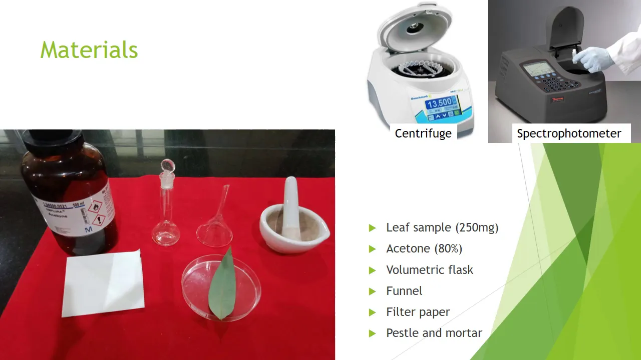
- Why this video is valuable: Shows a classic laboratory extraction protocol. It details how to use organic solvents (acetone), perform centrifugation steps to separate cellular debris, and run subsequent spectrophotometric assays.
- Knowledge Checkpoint:
- Explain why centrifugation is necessary after extraction before reading optical density on a spectrophotometer.
- Describe how the solvent concentration (e.g., 80% vs. 100% acetone) changes the extraction efficiency of non-polar plant pigments.
Course Map
Key People Index
- Dr. Gerald Cysewski
- Context: A pioneer in microalgae biotechnology and the co-founder of Cyanotech Corporation in Hawaii. He was instrumental in developing industrial-scale, closed-system photobioreactors and open raceway cultivation designs optimized for commercializing H. pluvialis astaxanthin.
- Dr. Jonathan Trent
- Context: Director of the OMEGA (Offshore Membrane Enclosures for Growing Algae) project at NASA Ames Research Center. His research contributions focus on large-scale photobioreactor geometries, gas-liquid mass transfer, and utilizing microalgae for carbon capture.
- Dr. Hartmut K. Lichtenthaler
- Context: A botanist and plant physiologist whose published works established the standard spectrophotometric multi-wavelength equations. His formulas are used globally to quantify carotenoids, chlorophyll , and chlorophyll from mixed solvent extracts.
Final Self-Assessment
Complete this practical self-assessment checklist before completing the course:
- Morphology Identification: Can you visually differentiate between green vegetative macrozoids, intermediate palmella cells, and red thick-walled hematocysts under a light microscope?
- Carotenogenesis Pathways: Can you map the enzymatic progression from Phytoene down to Astaxanthin, noting the roles of both the -carotene ketolase (BKT) and hydroxylase (CHYb) enzymes?
- Media Calculation: Can you calculate and prepare a 5-liter batch of BG-11 growth media from concentrated macronutrient and micronutrient stock solutions without generating chemical precipitation?
- Cell Count Calculations: Are you able to perform a complete hemocytometer calculation, accounting for dilutions and boundary cell exclusions?
- Beer-Lambert Estimations: Can you apply the modified Beer-Lambert equation to estimate the light intensity profile inside a flat-panel photobioreactor at a biomass density of ?
- Management: Can you explain how changing the bubble diameter (sparger pore size) and gas flow rate alters the volumetric mass transfer coefficient ()?
- Two-Stage Execution: Can you design a step-by-step laboratory SOP detailing how to transition a high-density green culture into a nitrogen-depleted, high-light stress environment?
- Cell Lysis Operations: Can you explain why physical bead beating is more effective than simple solvent washing for extracting pigments from H. pluvialis cysts?
- Spectrophotometric Extraction Assay: Can you solve the multi-wavelength Lichtenthaler-based correction equations to accurately calculate astaxanthin concentrations from a mixed chlorophyll-carotenoid sample?