























A hemocytometer is a specialized microscope slide with two chambers containing a grid of 9 squares, each holding 10^-4 mL of cell suspension; cells are counted by loading a diluted suspension (typically 1:1 ratio with Trypan blue dye to distinguish live from dead cells), counting cells in specific squares following boundary rules (counting cells touching top and left boundaries but ignoring those on right and bottom), then calculating viable cell concentration using the formula: (average cells per square × dilution factor × 10^4) cells/mL.
How to Count Cells Using a Hemocytometer | Protocol & Calculations
Added:Basic principles of cell biology, including the difference between suspension and adherent cell lines in a laboratory setting.
Fundamental microscopy skills, specifically how to operate and focus a standard compound light microscope.
Mathematical proficiency in calculating dilution factors, concentrations, and converting small volume units (e.g., microliters to milliliters).
The concept of cell viability and the biochemical principle behind dye-exclusion assays, such as how Trypan blue interacts with damaged cell membranes.
Calculating and plotting cell growth curves over time to determine the doubling time of a specific cell line.
Applying cell count data to downstream culture workflows, such as seeding multi-well plates at precise densities for experimental assays.
Understanding cryopreservation protocols, specifically how to calculate the correct freezing media volume based on post-harvest cell yield.
Comparing manual hemocytometer counting with automated cell counting technologies, including flow cytometry and image-based counters.
1.5M views18.1Klikes6:30Original Release: 2012-07-31
A hemocytometer is a device that is used for counting cells.
It's a modified microscope slide, containing two identical wells, or chambers, into which a small volume of a cell suspension is pipetted.
We have already removed 100 µL of our cell suspension and placed it in a micro-centrifuge tube.
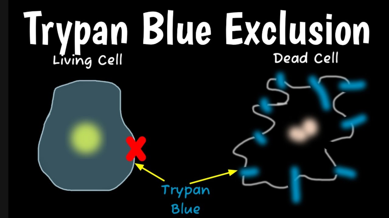
Dilute the suspension by adding 100 µL of Trypan blue. Trypan blue is a dye that helps us distinguish between living and dead cells. The dye passes through the membranes of dead cells so they will appear blue under a microscope.
Living cells exclude and will appear mostly clear.
Load both chambers by pipetting the suspension under the cover slip.
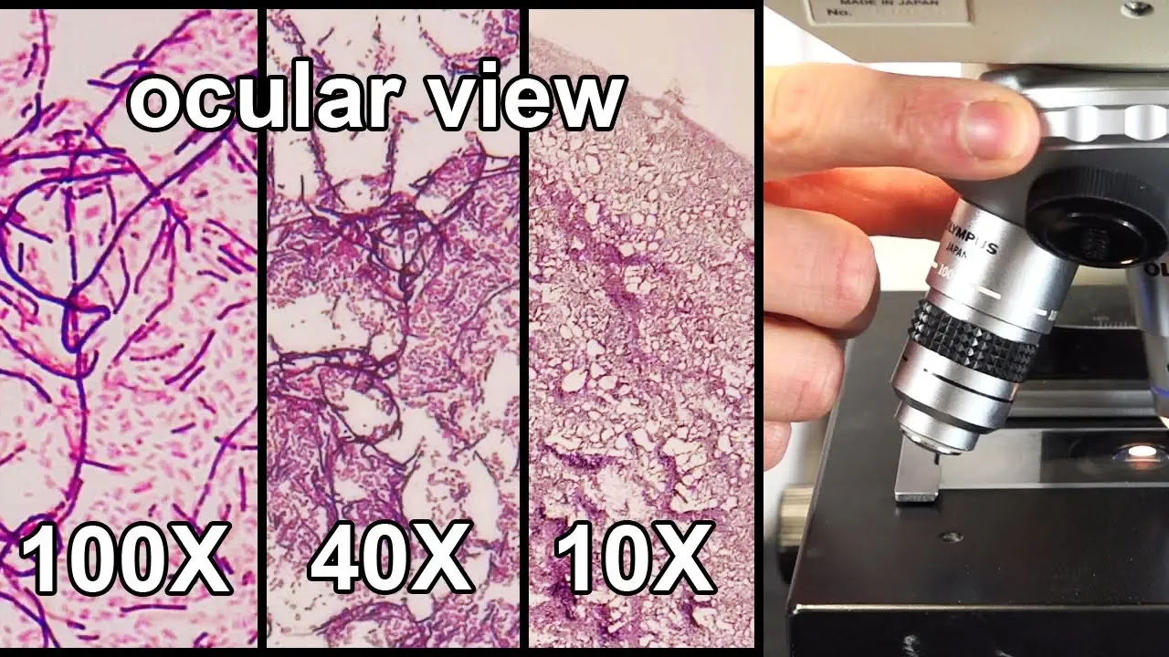
Now place the hemocytometer under the microscope.
Each chamber is divided into a grid pattern, consisting of 9 large squares.
Each square has the same dimensions and contains 10 to the negative-fourth power mL of suspension.
The rules for counting cells sometimes differ from lab-to-lab.
In our lab, we count cells in the 4 large corner squares and the center square.
Let's count the cells in the first square.
One, Two, Three, Four, Five, Six, Seven, Eight... So what about the cells that are touching the outside boundaries of the square?
In our lab we count the cells that touch the top and left boundaries, and we ignore the cells that touch the right and bottom boundaries.
Nine. Ten.
We need to count the number of both living and dead cells. Remember, the dead cells will appear blue.
Occasionally you will see artifacts - objects or debris that appear blurry and don't have a well-defined shape.
This is an example of an artifact. We won't include it in our count.
Proper storage, cleaning, and handling of the hemocytometer will minimize the number of artifacts.
There are 10 viable cells and 1 non-viable cell in the first square.
Now, the top-right square.
There are 9 viable cells and no non-viable cells.
Next let's count the bottom-right square.
There are 11 viable cells and no non-viable cells.
And now the bottom-left square.
There are 10 viable cells and 2 non-viable cells.
And finally, let's count the cells in the center square.
Sometimes cells will appear as clumps or small groups.
It may be difficult to determine exactly how many cells are in a group.
The method of counting clumps of cells differs from lab to lab, so be sure to follow the procedure in your lab.
We will count this clump as 2 cells.
There are 14 viable cells and no non-viable cells in the center square.
The total number of viable, or living cells from all 5 squares is 54.
The total number of non-viable cells is 3.
Now that we have counted our cells, there are several calculations we need to perform.
First, let's calculate the percentage of viable cells.
Here's the formula. 54 viable cells, divided by 57Éthe total number of cellsâ gives us 0.947.
Multiply by 100 and the percentage of viable cells is 94.7%.
Next, let's determine the average number of cells-per-square.
We counted 54 viable cells. We divide 54 by 5, because we counted in 5 squares.
The average number of cells-per-square is 10.8 cells.
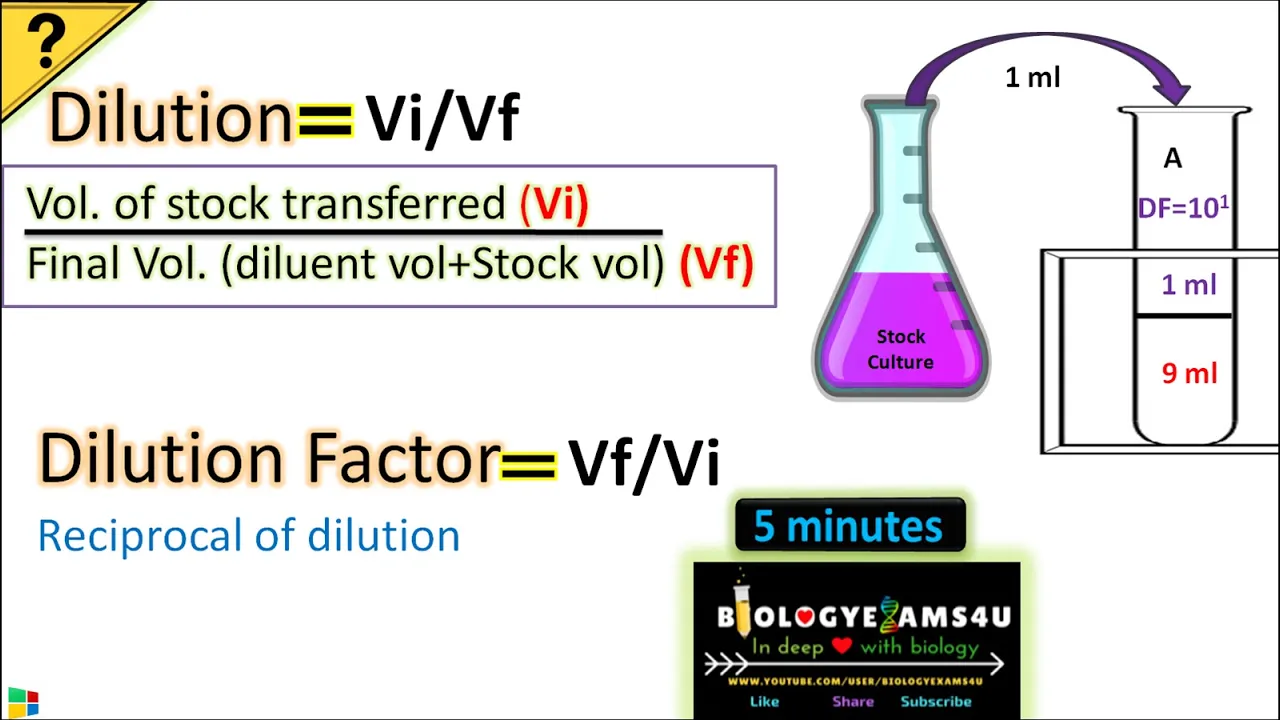
Now let's calculate the dilution factor.
The dilution equals the final volume divided by the volume of cells.
Our final volume is 200 µL, because we started with 100 µL of cells and added another 100 µL of trypan blue.
200 divided by 100 is 2. Therefore the dilution factor is 2.
Next we need to calculate the concentration of viable cells - the number of living cells/mL Our average count-per-square is 10.8.
The dilution factor is 2.
10.8 times 2 times 10-to-the-fourth-power equals 216,000 cells/mL.
We can write the concentration using scientific notation as 2.16 times 10-to-the-fifth power cells/mL.
From our calculations, we now know the concentration of cells in our culture is 216,000 cells-per-milliliter and approximately 94.7 percent of those cells are viable, living cells.
Up Next
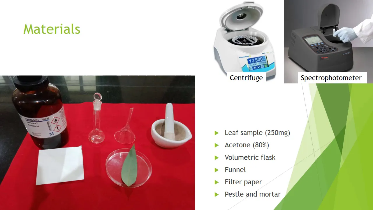
5:08
Chlorophyll Extraction and Estimation Using 80% Acetone Method
@delphyrocha3594
67.7K views•2020-07-08

50:12
Bacterial Communication: Quorum Sensing and Biofilm Formation Explained
@JHUAAP
12.6K views•2011-06-28

5:35
Enteric Nervous System Explained: The Gut's Brain | Neurobiology Lecture
@alumniu6029
438 views•2018-09-12

7:08
Bacteriophages: Earth's Deadliest Killers and Future Antibiotics
@kurzgesagt
34.6M views•2018-05-13
Related Study Plans & Knowledge Roadmaps
Structured learning paths in Biology
biology
RNAi: Biogenesis, RISC & Gene Silencing
01Molecular Biology Foundations: DNA to Protein
02Introduction to RNA Interference (RNAi)
03Molecular Biogenesis of miRNA and siRNA
+ 2 more learning modules
10h5 modules
Explore Roadmap
biology
Mycorrhizae: Signaling, Biophysics & Ecology
01Foundations of Mycorrhizal Symbiosis
02Molecular Signaling and Root Colonization
03Biophysics and Bioenergetics of Nutrient Exchange
+ 1 more learning modules
11h4 modules
Explore Roadmap
biology
Circadian Clocks: TTFL, SCN & Periphery
01Foundations of Molecular Biology & Gene Expression
02Introduction to Chronobiology and Actograms
03The Core TTFL: Molecular Architecture of the Clock
+ 3 more learning modules
14h6 modules
Explore Roadmap
biology
Population Genetics: Math, Drift & Selection
01Genetic & Probability Foundations
02The Hardy-Weinberg Principle and Baseline Models
03Mathematical Modeling of Natural Selection
+ 3 more learning modules
18h6 modules
Explore Roadmap