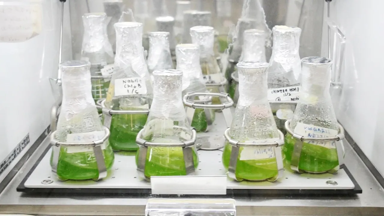
This video demonstrates how to prepare BG11 medium, a nutrient solution commonly used in plant tissue culture experiments for growing plant cells and tissues.
BG11 Medium Preparation: Step-by-Step Protocol Guide
Added:Deep Dive
Aseptic laboratory techniques, including sterilization methods like autoclaving, to prevent contamination during media preparation.

BIOL2421 Practical I Review (Microbiology for Science Majors)
2.1K
Media types include TSA plates, TSA slants, TSA deeps, and TSB broth. Autoclaving sterilizes media. Plates must be incubated upside down to prevent condensation and contamination. Aseptic transfers prevent contamination; contaminants appear outside streak lines rather than along them. Inoculation techniques include streak plates (isolate colonies), spread plates (even surface distribution), and pour plates (colonies grow within agar).
Click to watch →

Culture Media
258.2K
All microbiological media should be prepared in a sterile environment employing proper aseptic techniques. Essential precautions include: dedicating a separate room for media preparation, pouring, and sterilization; disinfecting the work area; washing hands thoroughly; wearing gloves, lab coat, and tying back hair; reading manufacturer's instructions carefully; sterilizing all glassware; keeping distilled water and powdered medium ready before beginning; and following all safety protocols to prevent contamination.
Click to watch →

Teknik Sterilisasi, Aseptik, dan Pembuatan Media Kultur Mikrobiologi
1.8K
This video explains essential laboratory techniques for microbiology: aseptic technique involves continuous procedures to prevent microorganism contamination (cleaning with 70% alcohol, using separate tips for different solutions, closing containers tightly, and working quickly), while sterilization eliminates all microorganisms through physical methods (filtration with 0.2-micron filters, autoclaving at 121°C, dry heat in ovens/incinerators, flame sterilization with Bunsen burners, and UV radiation in laminar airflow cabinets) or chemical methods (gas and liquid sterilization). Microbiology media are nutrient mixtures containing water, carbon sources, minerals, and growth factors, classified by composition (synthetic, complex, inorganic, organic), consistency (solid, liquid, semi-solid), and function (enrichment, selective, differential, test media), with common examples including nutrient agar, nutrient broth, blood agar, potato dextrose agar, lactose broth, and malt extract agar.
Click to watch →

Lab 1 - Lab and Home Experiment (Media Preparation, sterilization and aseptic technique)
225
This video demonstrates the fundamental laboratory techniques for preparing culture media, sterilizing it using pressure cookers, and performing aseptic techniques to prevent contamination during microbiological experiments. The protocol covers preparing nutrient agar by weighing 3 grams, dissolving in distilled water, and pouring into petri dishes and universal bottles while working close to a Bunsen burner flame. The experiment includes a hand washing demonstration comparing contamination on agar plates before and after proper hand sanitization, with plates incubated upside down to observe bacterial growth. The video also shows how to prepare agar at home using common kitchen supplies like water, agar powder, sugar, and skim milk, then sterilizing the mixture in a pressure cooker for 15 minutes at high pressure to ensure complete sterilization.
Click to watch →

Unit 3: Microbiology Lab Media
227
Culture media are growth substrates used in microbiology labs to cultivate microorganisms, with agar plates being the most common solid medium prepared by mixing powder with water, boiling, autoclaving, and pouring into plates; aseptic technique is essential for preventing contamination, involving sterilization of inoculating loops with a Bunsen burner until they glow orange-red, proper tube handling by heating barrel openings, and avoiding contact between sterile equipment and non-sterile surfaces; streaking for isolation separates mixed cultures into pure cultures by progressively diluting microbes across an agar plate to obtain individual colonies for study.
Click to watch →
Deep Dive
Basic chemistry skills, specifically preparing stock solutions, performing serial dilutions, calculating molarity, and adjusting pH.
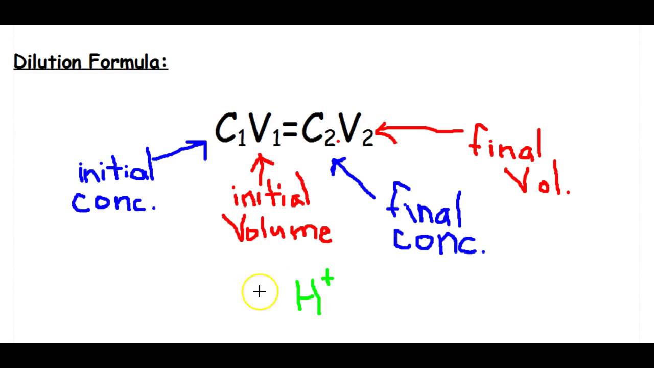
Stock Solutions & Dilutions
121.5K
A stock solution is a highly concentrated solution that can be diluted to create solutions of lower concentrations; when diluting, the number of moles of solute remains constant while the volume changes, and this relationship is calculated using the formula C₁V₁ = C₂V₂, where C represents concentration and V represents volume. When preparing a dilute solution, always add acid to water (not water to acid) for safety, and use a volumetric flask to measure the final volume precisely.
Click to watch →

série de dilutions 01
1.3K
Serial dilution is a laboratory technique where a series of solutions with progressively lower concentrations are prepared from a single stock solution by transferring specific volumes of the previous solution into new containers and diluting to a known final volume; the concentration of each solution is calculated using the formula C1V1 = C2V2, where C1 and V1 are the concentration and volume of the source solution, and C2 and V2 are the concentration and volume of the diluted solution.
Click to watch →

Stock Solution| Dilution Formula| How to prepare Stock Solutions|Numericals|URDU\HINDISAAD ANWAR
28.5K
Serial dilutions involve preparing a series of solutions with decreasing concentrations from a single stock solution. For example, preparing solutions of 10^-3 M, 2×10^-3 M, 5×10^-3 M, and 10×10^-3 M from a 0.10 M stock solution requires calculating V1 for each: 1 mL, 2 mL, 5 mL, and 10 mL respectively, each diluted to 100 mL total volume. Practical laboratory applications include preparing 2 M sulfuric acid from 5 M stock solution (requiring 4 mL of stock for 10 mL total volume). The general procedure involves measuring the calculated volume of stock solution using appropriate glassware (graduated cylinder or pipette), transferring to a volumetric flask, and adding solvent until the total volume reaches the target. This systematic approach ensures accurate solution preparation for analytical chemistry applications.
Click to watch →
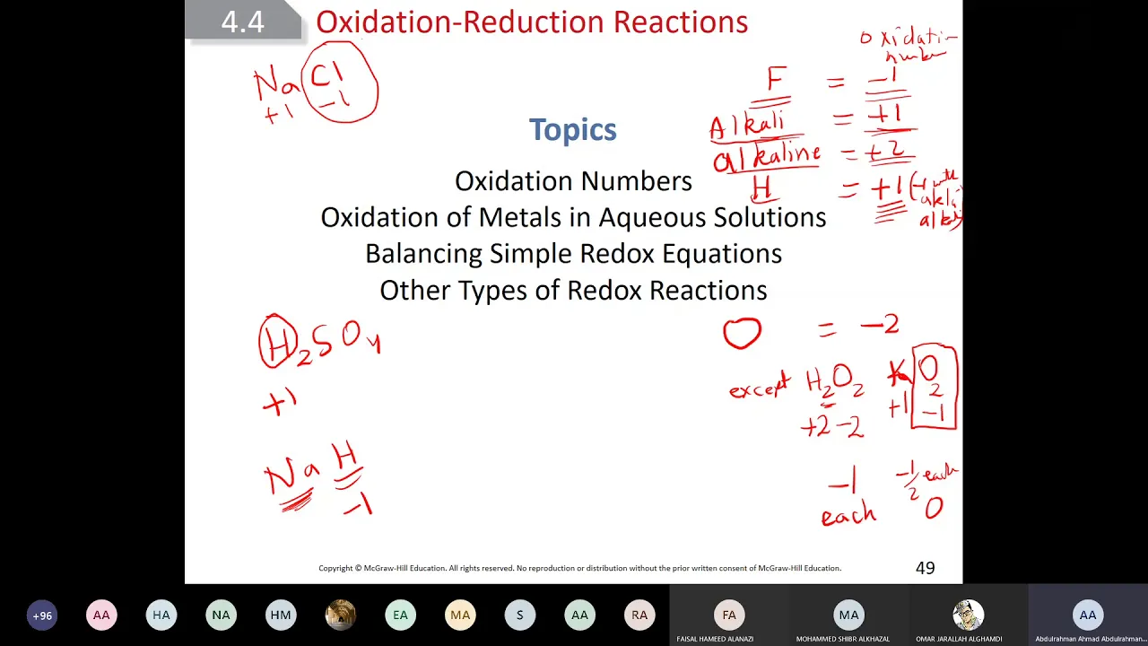
chem101 9 lecture
7K
Molarity (M) is moles of solute per liter of solution. To calculate: find molar mass, calculate moles (mass/molar mass), divide by volume in liters. For 50g glucose (180.2 g/mol) in 2L: moles = 0.278, M = 0.139. To find moles: multiply M by V (0.14M × 0.500L = 0.07 mol). The dilution formula M1V1 = M2V2 relates concentrated and diluted solutions. For 12M HCl to 0.125M in 250mL: V1 = (0.125 × 0.250)/12 = 2.6mL. Serial dilution involves repeatedly diluting portions of previous solutions to prepare solutions of decreasing concentration. This technique is essential for preparing solutions of precise concentrations in laboratory settings.
Click to watch →

Advanced Higher Chemistry 2025 (Section 1 And Section 2)
268
To prepare a diluted solution: (1) Calculate required volume of stock solution using C₁V₁ = C₂V₂, (2) Pipette calculated volume into volumetric flask, (3) Dilute to mark with distilled water. For 0.1 M from 0.2 M: V₁ = (0.1 × 500)/0.2 = 250 cm³ of stock diluted to 500 cm³.
Click to watch →
Deep Dive
The nutritional requirements of photoautotrophs, understanding the roles of macronutrients and trace micronutrients in microbial growth.

O Que São Macronutrientes e Micronutrientes, Para Que Servem | Funções | Diferenças | Resumo
342K
Macronutrients (carbohydrates, lipids/grease, and proteins) are nutrients needed in larger quantities for energy and structural functions, while micronutrients (vitamins and minerals) are required in smaller amounts but essential for regulating bodily processes; carbohydrates provide primary energy for daily activities and brain function, lipids serve as secondary energy sources and provide thermal insulation, proteins support muscle structure and immune defense, and vitamins/minerals regulate health aspects including bones, skin, hair, nails, teeth, and immune function.
Click to watch →

Complete Plant Physiology | Revise with Keynotes 2.0 | NEET 2022 | Dr. Anand Mani
125.4K
Macronutrients are essential elements required in larger amounts: Carbon, Hydrogen, Oxygen, Nitrogen, Phosphorus, Potassium, Calcium, Magnesium, and Sulfur. Micronutrients are essential elements required in smaller amounts: Iron, Manganese, Boron, Zinc, Copper, Molybdenum, Chlorine, and Nickel. Calcium and Magnesium are sometimes classified separately because they have structural roles (calcium in cell walls, magnesium in chlorophyll) despite being required in smaller amounts than some macronutrients. The distinction between macronutrients and micronutrients is based on the concentration required in plant tissues, not on their relative importance.
Click to watch →

My Cheat Code to Faster Fat Loss (Lazy Tracking)
147.8K
Nutrients encompass macronutrients (proteins, fats, carbohydrates) and micronutrients (vitamins and minerals). Macronutrients provide energy and build the body: proteins help build and repair the body, while fats and carbohydrates fuel it. Micronutrients are like different parts of a car—vitamins and minerals help cells and organs function properly. When tracking food, entering accurate age, gender, height, and weight is essential because men and women have different nutritional requirements (e.g., women need more iron due to menstruation). The RDAs serve as minimum recommendations to avoid deficiencies, not optimal health targets.
Click to watch →

INTRODUCTION | CHEMISTRY OF LIFE
296.9K
Minerals are dietary inorganic chemicals essential for life, categorized into macronutrients (required in large amounts) and micronutrients or trace minerals (required in smaller amounts). Plants obtain minerals from soil while animals must obtain them through diet. Humans cannot synthesize these minerals and must absorb them from food sources. Key minerals include calcium (found in cheese, milk, green vegetables; important for bone development, blood clotting, nerve impulses, and plant cell walls), magnesium (found in fish, beans, leafy vegetables, whole wheat; important for bone formation, muscles, nerves, and chlorophyll in plants), potassium (found in fruits, vegetables, grains; relaxes muscles and supports nervous system and photosynthesis), iron (found in liver, beans, peas, potatoes, leafy vegetables; required for hemoglobin synthesis in red blood cells and chlorophyll formation), and zinc (found in dairy products, liver, wheat; important for enzymes and fetal brain development).
Click to watch →

ဒေါက်တာအောင်မြင့် (ပါမောက္ခ) " ကျန်းမာရေးနှင့်ညီညွတ်သော အစားအစာများ "
74.9K
Nutrients are categorized into macronutrients (needed in larger amounts) and micronutrients (vitamins and minerals needed in smaller amounts).
Click to watch →
Deep Dive
General biology of cyanobacteria and microalgae, highlighting their photosynthetic nature and lack of requirement for organic carbon sources.

Introduction of Microbiology |lect-1|Microbiology| Mycology | Plant Pathology|Botany|iSTUDY Online
25K
Cyanobacteria (blue-green algae) contain chlorophyll and phycobilin pigments, enabling photosynthesis. They store food as cyanophycin granules and fix atmospheric nitrogen. Algae are autotrophic plants containing chlorophyll, storing excess food as starch. Cell wall composition differs: cyanobacteria have peptidoglycan, algae have cellulose.
Click to watch →

Class 11 BIOLOGY I Biological Classification I Kingdom Monera
139
Some bacteria are photosynthetic and can make their own food using light energy. These bacteria convert light energy into chemical energy to produce organic compounds. Cyanobacteria are photosynthetic bacteria that can make their own food using light energy. They are also called blue-green algae. Cyanobacteria cells contain chlorophyll and can perform photosynthesis. They have a cell wall, cell membrane, and cytoplasm. They do not have a true nucleus. Cyanobacteria grow in aquatic environments where they can access light and nutrients. They require light for photosynthesis and can grow on water surfaces.
Click to watch →

Cianobacterias como Bioinsumos y agroecologia
401
Microalgae and cyanobacteria are microscopic organisms that utilize light, water, minerals, and CO2 for carbon fixation, making them valuable for biofertilizer production. They grow faster than vegetable crops and do not compete with food grain production. Cyanobacteria possess specialized cells called heterocysts that enable nitrogen fixation, making atmospheric nitrogen available for plants. They form resistant structures called akinetes when environmental conditions are unfavorable. As pioneer organisms, they enrich agricultural soil through exudates that add organic matter and create favorable conditions for other photosynthetic microorganisms.
Click to watch →

Cyanobacteria।Blue Green Algae। Photosynthetic bacteria। Agricultural Microbiology।ICAR/ASRB Exam।
409
Cyanobacteria, also known as blue-green algae, are among the oldest known fossils dating back over 3.5 billion years. They play important roles in antiviral, antibacterial, antifungal, and anti-cancerous activities. Cyanobacteria use photosynthesis to convert sunlight into energy, using blue-green pigments unlike plants. They are found naturally in fresh water and can form blooms under certain conditions. Chloroplasts, the organelles where photosynthesis occurs, contain chlorophyll that captures sunlight energy. These organelles contain their own DNA inherited from ancestral cyanobacteria and must be inherited during cell division. Cyanobacteria have a prokaryotic cell structure with naked DNA, 70s ribosomes, and a 4-layered peptidoglycan cell wall. They range from 1-10 microns in size and contain thylakoid membranes for photosynthesis, carboxysomes, glycogen granules, and lipid bodies.
Click to watch →
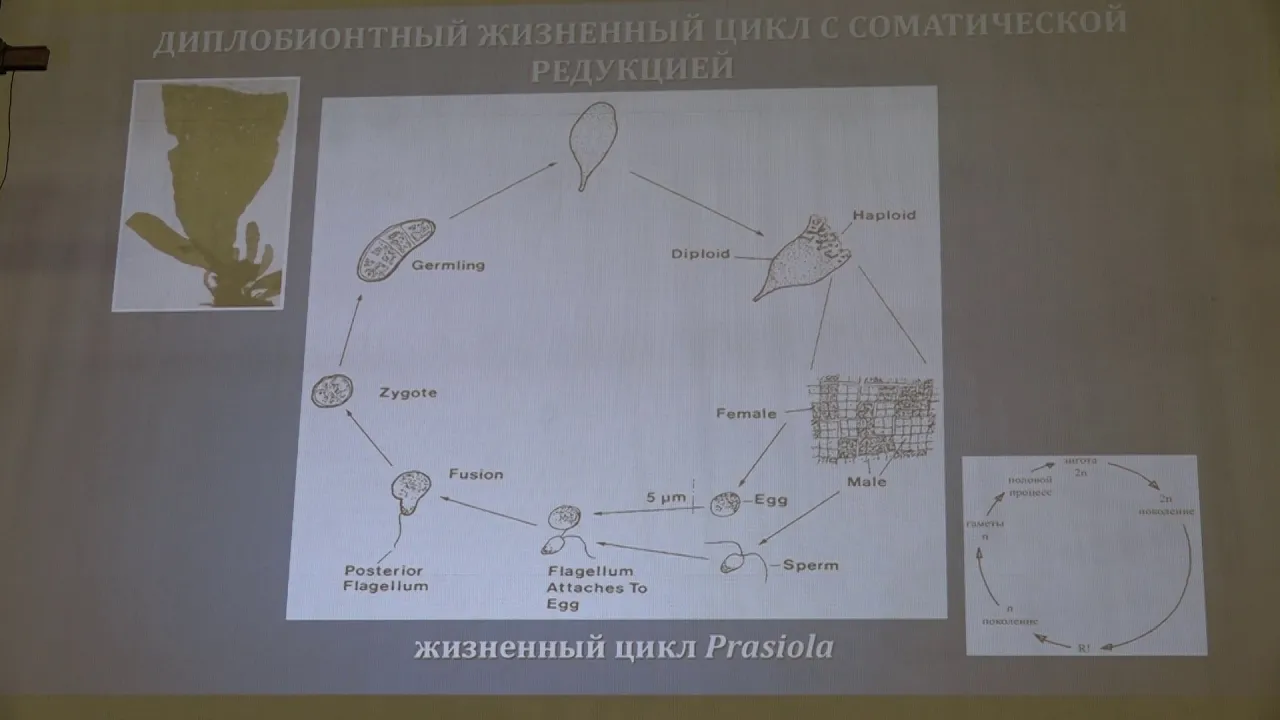
Гололобова М. А. - Низшие растения - 3. Жизненные циклы водорослей. Цианобактерии
6.4K
Cyanobacteria (blue-green algae) are prokaryotic organisms that perform oxygenic photosynthesis. They lack a true nucleus and membrane-bound organelles, with genetic material located in the nucleoid region. Their cell walls contain peptidoglycan, similar to gram-negative bacteria. The term 'cyanobacteria' is preferred over 'blue-green algae' because it accurately reflects their prokaryotic nature. They are classified as bacteria within the domain Bacteria, despite their ecological importance as photosynthetic organisms.
Click to watch →
Prerequisite Knowledge
- Concept 01Aseptic laboratory techniques, including sterilization methods like autoclaving, to prevent contamination during media preparation.
- Concept 02Basic chemistry skills, specifically preparing stock solutions, performing serial dilutions, calculating molarity, and adjusting pH.
- Concept 03The nutritional requirements of photoautotrophs, understanding the roles of macronutrients and trace micronutrients in microbial growth.
- Concept 04General biology of cyanobacteria and microalgae, highlighting their photosynthetic nature and lack of requirement for organic carbon sources.
Subsequent Learning
- Step 01Inoculation and maintenance protocols for specific cyanobacterial model organisms (such as Synechocystis or Anabaena) under controlled light and temperature.
- Step 02Formulating modified BG11 media, such as nitrogen-free BG11 (BG11_0) to study diazotrophic growth and heterocyst differentiation.
- Step 03Methods for monitoring and quantifying algal growth, including optical density measurements, cell counting, and chlorophyll-a quantification.
- Step 04Biotechnological applications of cyanobacterial cultures, such as metabolic engineering, biofuel production, and wastewater bioremediation.
Opening
Music Intro
Continued Music
Acknowledgment
Music Returns
Foreign End
Opening
0:02Playing Section
- 1
Video begins with a foreign language segment.
- 2
Initial audio sets the overall tone.
- 3
No substantive content introduced yet.
Limitations of BG11 and the Advocacy for Ecologically Relevant Media
While BG11 is the standard medium for culturing cyanobacteria and microalgae, critics argue its highly concentrated, hyper-eutrophic formulation does not reflect natural aquatic environments. This can induce physiological artifacts, select for atypical lab strains, and inhibit the growth of sensitive or oligotrophic species. Additionally, BG11 is prone to nutrient precipitation during autoclaving. Consequently, many researchers advocate for alternative media, such as diluted formulations (e.g., 1/10 BG11), natural water-based media, or specialized alternatives like WC and ASN-III to achieve more ecologically relevant results.
Deep Dive
Inoculation and maintenance protocols for specific cyanobacterial model organisms (such as Synechocystis or Anabaena) under controlled light and temperature.
LC-MS/MS Diagnostic Fragmentation Filtering: Application-Microcystin Analysis l Protocol Preview
125
Cultivate cyanobacteria under photoautotrophic conditions with specific environmental parameters: illuminate cultures with cool white fluorescent light using a 12-hour light-dark regime, maintain temperature at 27 degrees Celsius, swirl flasks once daily, and allow cultivation for 26 days to achieve sufficient biomass for microcystin extraction and analysis.
Click to watch →

Isolation and characterization of Cyanobacteria with examples and diagram | MSc practical series
2.2K
Cyanobacteria are photosynthetic, oxygenic bacteria that require sunlight, warmth, and nutrient-rich environments (high phosphorus and nitrogen) to grow and form blooms; they can be isolated using BG-11 media with or without nitrogen, employing agar digging for filamentous types and streak plate for unicellular types, with incubation at 1500-2000 lux light intensity, 25-30°C temperature, and 10-12 hour photoperiod; key morphological features for identification include heterocysts (for nitrogen fixation), akinetes (spore-like cells), and aerotopes (gas vesicles), with different genera exhibiting distinct characteristics such as Anabaena's cylindrical cells with intercalary heterocysts, Synechococcus's unicellular spherical form, Calothrix's parallel filaments, Microcoleus's sheath-bound trichomes, Oscillatoria's motile trichomes without sheaths, and Merismopedia's planar cell arrangements.
Click to watch →

光合成細菌で農業を始めよう!やぶ田秀行氏講演会イン八ヶ岳2026年4月4日
603
Cyanobacteria require specific conditions for optimal growth: (1) Sunlight exposure, particularly UV radiation, which they evolved to withstand, (2) Water/moisture, (3) Nutrients (such as rice bran or Ebiyos vitamins), and (4) Temperature around 35°C. Unlike other microorganisms, cyanobacteria thrive in these conditions and can be cultivated in transparent containers that allow sunlight penetration. The bacteria prefer natural environments similar to their original habitat. The cultivation method involves taking 500cc of cyanobacteria starter culture, adding 4.5 liters of water to make a 10% dilution, and adding 5 grams of rice bran. The ratio of cyanobacteria to water and nutrients is critical - too much cyanobacteria can cause the culture to fail.
Click to watch →

Isolation of Photosynthetic bacteria | Cyanobacteria | Purple and Green bacteria
592
For isolating cyanobacteria: (1) Collect sample from gardens or ponds; (2) Transfer a small amount onto sterile Petri plates containing BG11 agar; (3) Incubate at optimal temperature (25-35°C) with continuous light exposure; (4) After several days, observe plates for characteristic colonies exhibiting green, red, or purple colors based on pigment content; (5) Perform careful microscopic examination of isolated colonies to check cell size, shape, arrangement, and confirm they are photosynthetic bacteria; (6) Conduct colony morphology studies, gram staining, and biochemical tests (catalase, oxidase) for identification; (7) Confirm photosynthetic nature through appropriate testing.
Click to watch →

Glenn PARTSCH: To what extent is there a relationship between phosphorus and nitrogen...
166
Cyanobacterial growth was measured by monitoring changes in light transmission using a calorimeter set at 490 nanometers. The experiment tested five nitrogen and phosphorus concentration ranges: no nutrients added (control), and concentrations of 1x, 2x, 3x, and 4x times the standard culture solution. Temperature and light intensity were controlled by exposing cultures to Luxembourgish weather outdoors from June to July, while CO2 concentration was maintained by regularly opening bottle caps.
Click to watch →
Deep Dive
Formulating modified BG11 media, such as nitrogen-free BG11 (BG11_0) to study diazotrophic growth and heterocyst differentiation.

Lecture 03 Nitrogen Cycle: The Molecular Biology of N Fixation 3 of 3
418
Diazotrophs are microorganisms (bacteria and archaea) capable of fixing atmospheric nitrogen gas into more usable forms such as ammonia. They have the ability to grow without an external nitrogen source, utilizing nitrogen from the air present in the headspace of their growth media. Examples include Azotobacter, Rhizobia, Frankia, and Azospirillum. These organisms can be heterotrophs or phototrophs, and may be aerobic, facultative anaerobic, or anaerobic depending on the species.
Click to watch →
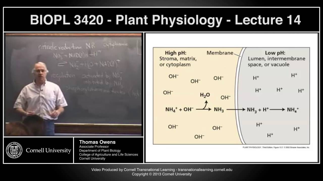
BIOPL3420 - Plant Physiology - Lecture 14
9.7K
Cyanobacteria (blue-green algae) are photosynthetic prokaryotes that can perform both oxygenic photosynthesis and nitrogen fixation. Under nitrogen-limiting conditions, certain cells in the filament differentiate into specialized heterocysts. These heterocysts are structurally modified to create an anaerobic environment suitable for nitrogenase activity while the surrounding vegetative cells continue normal photosynthesis. This differentiation allows the organism to maintain both photosynthetic and nitrogen-fixing capabilities simultaneously.
Click to watch →

REVISION DE TÉCNICAS DE AILSAMIENTO MEDIOS DE CULTIVO Y CARACTERIZACIÓN DE CEPAS
218
N-free broth (nitrogen-free medium) is used to isolate nitrogen-fixing bacteria like Azospirillum. The medium contains specific nutrients including malic acid, dipotassium phosphate, ferrous sulfate, magnesium sulfate, manganese sulfate, sodium chloride, and bromothymol blue indicator. The indicator changes color from blue to yellow as bacteria metabolize malic acid, producing organic acids that lower pH. Only bacteria capable of fixing nitrogen can grow in this medium. This selective approach allows isolation of specific nitrogen-fixing bacteria from environmental samples.
Click to watch →
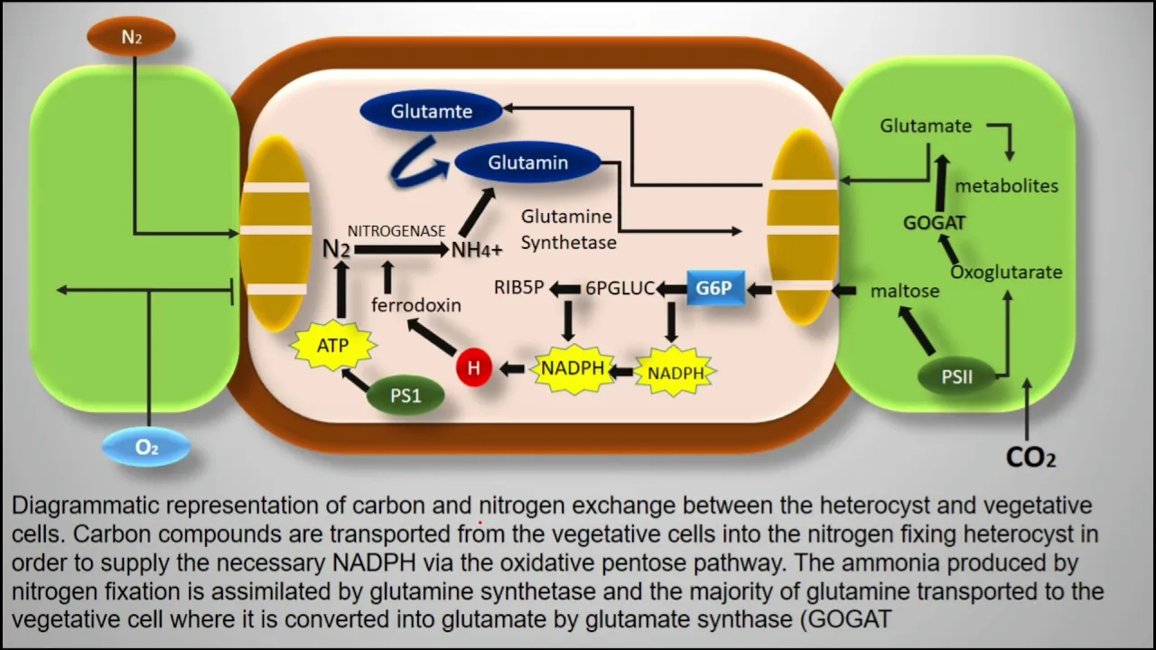
Heterocyst | Cyanobacteria | Nostoc
2.8K
Heterocysts are specialized cells in cyanobacteria (blue-green algae) that are larger and lighter in color than normal vegetative cells; they lack Photosystem II and the Calvin cycle, which prevents oxygen release that would inactivate nitrogenase, enabling efficient nitrogen fixation into ammonia that is then converted to amino acids like glutamine and transported to vegetative cells for growth, while receiving carbohydrates in return.
Click to watch →

LECTURE 2 || NITROGEN FIXATION || BY ANJU MAM
9.8K
When cyanobacteria face nitrogen deficiency, certain cells differentiate into specialized structures called heterocysts. These heterocysts have a thickened cell wall that prevents oxygen entry, which is essential because nitrogenase enzyme is oxygen-sensitive and cannot function in the presence of oxygen.
Click to watch →
Deep Dive
Methods for monitoring and quantifying algal growth, including optical density measurements, cell counting, and chlorophyll-a quantification.

KNOW YOUR ALGAE- Algal Biorefineries: A Wastewater Perspective | #EWRG_Environment_Education
388
Four primary methods exist for measuring algal growth in laboratory and field settings. First, solids measurement determines biomass concentration through gravimetric analysis of filtered samples. Second, optical density (OD) measurements provide rapid, non-destructive estimates of cell concentration based on light absorption characteristics. Third, chlorophyll content measurement offers a direct assessment of photosynthetic pigment concentration, correlating with biomass and physiological status. Fourth, direct cell counting using microscopy provides absolute quantification of cell numbers. Seven well-established mathematical models capture the sigmoidal growth curves characteristic of algal populations, enabling prediction of growth kinetics and optimization of cultivation parameters for maximum productivity.
Click to watch →

Basic Algae Culture Class for Nannochloropsis
2.3K
Three main methods quantify algae biomass: (1) Hach stick optical density measurements, (2) Spectrophotometer readings at 540nm or 750nm wavelengths (outside photosynthetic range for accuracy), and (3) Hemocytometer counting. Dry weight measurements provide additional verification.
Click to watch →

Batch culture of marine microalgae and growth measurement
2.1K
This section presents comprehensive techniques for quantifying algal growth: (1) Dry weight measurement involves filtering 50ml samples onto microfiber filters, drying at 65°C for 48 hours, ashing at 540°C for 4 hours, and calculating ash-free dry weight; (2) Cell density calculation uses the formula Gamma = (Total cells × Total area) ÷ (Counting unit area × Counting units × Sample volume); (3) Optical density measurement at 604nm correlates with cell concentration using standard curves. These complementary methods enable precise biomass assessment and growth rate determination.
Click to watch →

The Dirty Lab Coat - Brainy Briny Beaker Bag (BBBB-00015)
2.9K
Optimal microalgae growth requires about 100 micro einsteins per meter squared second of light (sunlight provides 3,000, which is too bright). Temperatures above 100°F kill algae. After about 8 days, brine shrimp cysts hatch and swim in the culture, visible against the grid. The seshi stick uses optical density to measure biomass: fold it on the dotted line, submerge until the black and white target disappears, then use the provided graph to determine cells per milliliter. This allows tracking cell density over time and creating growth curves to monitor culture development.
Click to watch →

Algal Innovation Centre University of Cambridge algal biotech scale up
334
Monitoring microalgae cultures involves measuring optical density (OD) using spectrophotometers and pH using pH meters, as different cultures grow only at specific pH levels. Light microscopes monitor cell quality including physiology, vacuoles, cell wall degradation, and bacterial load. The AIC uses shock bottles for 2.5L cultures with filtered air bubbling, five-liter bags (industry standard for oyster hatcheries) scalable from 2L to 20L, and tubular upright reactors for 10L controlled environments. Three species are studied: Nanochloropsis (marine, excellent fatty acids), Chlorella (pigments), and Phaeodactylum (fucoxanthin). Samples are taken through sterile syringes for metabolite analysis.
Click to watch →
Deep Dive
Biotechnological applications of cyanobacterial cultures, such as metabolic engineering, biofuel production, and wastewater bioremediation.

Lecture 10 Cyanobacteria I 230530 complete
288
Cyanobacteria in wastewater treatment face challenges from eutrophication and harmful algal blooms (HABs) producing cyanotoxins including neurotoxins, hepatotoxins, and cytotoxins. HABs cost millions annually and can force water treatment shutdowns, as occurred in Toledo, Ohio in 2011. However, cyanobacteria offer significant biotechnological potential: high membrane lipid content, rapid growth rates, and amenability to genetic manipulation enable production of biofuels (biodiesel, bioethanol, biogas, hydrogen) and industrial chemicals. Spirulina is FDA-recognized as safe for food supplements. Different species can be optimized for protein or carbohydrate production.
Click to watch →

SSB / OPSC PGT Botany 2026–27 | Cyanobacteria Class-2 | Important Concepts for Exam
275
Cyanobacteria have significant biotechnological potential including production of bioactive compounds with pharmaceutical properties (antibiotics, antivirals, anti-cancer agents), and research into biofuel production from hydrogen, lipids, and carbon dioxide fixation capabilities.
Click to watch →
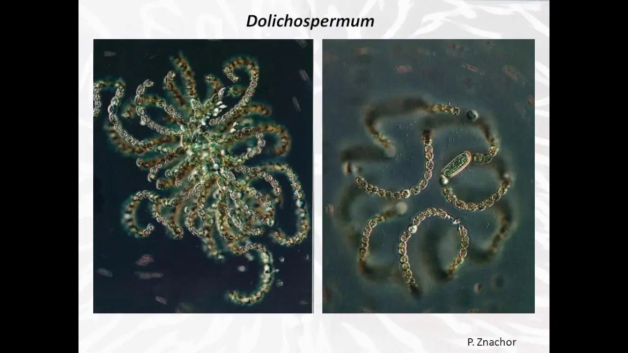
Les cyanobactéries, microscopiques mais fondamentales !
3.3K
Cyanobacteria have significant applications in biotechnology and sustainable systems. Spirulina (Arthrospira) is consumed as a nutritional supplement worldwide, containing high-quality proteins (up to 70% of dry weight) with essential amino acids, vitamins, and minerals. Production has grown to approximately 5,000 tons annually across 23 countries, with over 50% produced in China. Phycocyanin, a blue pigment from spirulina, is the only blue food colorant approved for use in the food industry. Cyanobacteria can accumulate polyhydroxybutyrate (PHB), a biodegradable polymer with applications in packaging and biomedical fields, though current production yields (5-20% of dry weight) are much lower than specialized bacteria. They also possess hydrogenase enzymes enabling hydrogen production, a potential future biofuel. Non-ribosomal peptide synthesis (NRPS) produces diverse secondary metabolites with pharmaceutical value, including penicillin and cyclosporine. Cyanobacteria contribute over 10% of known NRPS-derived molecules. Culturing requires light, CO2, nitrogen, phosphorus, sulfur, and trace elements, with careful light management to prevent photoinhibition. Open systems (raceways) and photobioreactors (bags, cylinders, tubular systems) offer different approaches, with productivity ranging from 1 to 1000 kg biomass per hectare per year.
Click to watch →
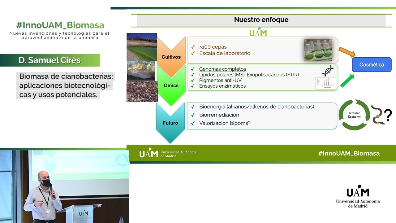
Biomasa de cianobacterias: aplicaciones biotecnológicas y usos potenciales - InnoUAM_Biomasa
616
Cyanobacteria, ancient prokaryotic organisms responsible for oxygenic photosynthesis with over 1,500 described species, are being researched at the Autonomous University of Madrid for their diverse applications in bioenergy, biomedicine, agriculture, and cosmetics; researchers are working with over 100 strains isolated from diverse environments including polar regions and deserts to produce valuable metabolites like polar lipids, exopolysaccharides, and UV-protective pigments, while also exploring innovative approaches to valorize cyanobacterial blooms (harmful algal blooms) through anaerobic digestion, hydrothermal liquefaction, and hydrothermal carbonization for biogas, biodiesel, and hydrochar production, contributing to circular economy initiatives.
Click to watch →

Julie Zedler | Cyanobacteria for Biotechnology - Can SynBio Make It Happen?
1.9K
Modern cyanobacterial metabolic engineering employs sophisticated strategies beyond simple pathway introduction. Iterative optimization cycles systematically identify and eliminate bottlenecks, as demonstrated in 29 rounds of astaxanthin pathway improvement. In-cell derivatization makes products more hydrophilic or hydrophobic, reducing toxicity and enabling higher accumulation before chemical recovery. Light-driven catalysis channels photosynthetic electrons directly to P450 enzymes in thylakoid membranes, bypassing cellular biomass production. Metabolic channeling through enzyme co-localization reduces intermediate diffusion losses and provides natural detoxification. These approaches represent paradigm shifts requiring deeper understanding of cyanobacterial cellular mechanisms while demonstrating pathways to industrial-scale implementation.
Click to watch →
Opening
Music Intro
Continued Music
Acknowledgment
Music Returns
Foreign End
Opening
0:02- 1
Video begins with a foreign language segment.
- 2
Initial audio sets the overall tone.
- 3
No substantive content introduced yet.
Limitations of BG11 and the Advocacy for Ecologically Relevant Media
While BG11 is the standard medium for culturing cyanobacteria and microalgae, critics argue its highly concentrated, hyper-eutrophic formulation does not reflect natural aquatic environments. This can induce physiological artifacts, select for atypical lab strains, and inhibit the growth of sensitive or oligotrophic species. Additionally, BG11 is prone to nutrient precipitation during autoclaving. Consequently, many researchers advocate for alternative media, such as diluted formulations (e.g., 1/10 BG11), natural water-based media, or specialized alternatives like WC and ASN-III to achieve more ecologically relevant results.
Aseptic laboratory techniques, including sterilization methods like autoclaving, to prevent contamination during media preparation.
Basic chemistry skills, specifically preparing stock solutions, performing serial dilutions, calculating molarity, and adjusting pH.
The nutritional requirements of photoautotrophs, understanding the roles of macronutrients and trace micronutrients in microbial growth.
General biology of cyanobacteria and microalgae, highlighting their photosynthetic nature and lack of requirement for organic carbon sources.
Inoculation and maintenance protocols for specific cyanobacterial model organisms (such as Synechocystis or Anabaena) under controlled light and temperature.
Formulating modified BG11 media, such as nitrogen-free BG11 (BG11_0) to study diazotrophic growth and heterocyst differentiation.
Methods for monitoring and quantifying algal growth, including optical density measurements, cell counting, and chlorophyll-a quantification.
Biotechnological applications of cyanobacterial cultures, such as metabolic engineering, biofuel production, and wastewater bioremediation.
1.3K views5likes1:18@Hey_DelphiOriginal Release: 2023-07-07
foreign [Music] [Music] thank you [Music] foreign
Up Next
7:42
Microalgae Streaking and Inoculation: Aseptic Lab Protocol
@bioproject2705
67.8K views•2018-10-29

12:54
Triumph of Orthodoxy Icon: Byzantine Art & History Explained
@BenCallan
2.1K views•2024-08-06

9:49
FastAPI vs Flask vs Django: Choosing the Right Python Web Framework
@TechWithTim
302.5K views•2024-05-26

1:42
Game of Thrones Opening Credits: A Cinematic Analysis
@gameofthrones
46.3M views•2011-04-18
Related Study Plans & Knowledge Roadmaps
Structured learning paths in General & Interdisciplinary Studies
general-studies
Elevator Engineering: History, Physics & Cities
01From Hoists to Safety Brakes: The Dawn of Vertical Lift
02Hydraulic vs. Traction: The Engineering Physics of Elevators
03Modern Speed, Safety, and Algorithms
+ 2 more learning modules
12h5 modules
Explore Roadmap
general-studies
Rubber: Material Science, History & Geopolitics
01Origins of Natural Rubber & Mesoamerican History
02The Chemistry of Natural Rubber & Vulcanization
03The Geopolitics of the Rubber Boom & Colonization
+ 2 more learning modules
12h5 modules
Explore Roadmap
general-studies
Containerization: Evolution, Logistics & Trade
01Ancient Trade & Bulk Cargo: Amphorae to Barrels
02The Industrial Revolution & Cardboard Packaging
03The Containerization Revolution: Malcom McLean
+ 2 more learning modules
12h5 modules
Explore Roadmap
general-studies
Textile Tech: Evolution, Labor & Global Impact
01Pre-Industrial Textiles: Handloom & Cottage Industry
02Mechanization: The Spinning Jenny to the Power Loom
03The Factory System & Global Labor Transformation
+ 2 more learning modules
10h5 modules
Explore Roadmap