This video demonstrates the complete process of inoculating microalgae, including aseptic techniques using a Bunsen burner and laminar airflow cabinet, quadrant streaking method for isolating pure colonies on agar plates, and inoculation into suspension media for culture growth, with specific parameters of 139 revolutions per minute and 23.9°C temperature for optimal microalgae cultivation.
Microalgae Streaking and Inoculation: Aseptic Lab Protocol
Added:Deep Dive
Fundamental principles of aseptic technique, including the correct usage of laminar flow hoods, Bunsen burners, and sterile handling tools.

Principles of aseptic technique
604
Aseptic technique prevents contamination of both the worker and experimental work. The four main contamination sources are: (1) the environment, requiring disinfection of the work station with lab-grade disinfectant and wiping beyond the work area; (2) the worker, requiring thorough hand washing or disinfecting gloves; (3) tools, sterilized by heating inoculation loops in a Bunsen burner flame until glowing red, then counting 1001 seconds before cooling near the flame; (4) airborne particles, mitigated by working near the flame where hot air rises and creates a sterile zone. Key practices include positioning the flame directly in front of the worker, bringing materials to the flame area, opening containers only when necessary, and keeping all manipulations within the flame zone. These principles apply across all laboratory protocols.
Click to watch →

Laminar Flow Hood
47.2K
A laminar flow hood (safety cabinet) must be prepared by warming media to 37°C, turning on the blower and germicidal lamp for 15 minutes, then switching to fluorescent light; the hood and all materials must be sterilized with 70% ethanol before use, and aseptic technique requires flaming bottle necks and pipettes before opening them to maintain a sterile environment.
Click to watch →
@JJChemistry panscht mit Bakterien. Sorry - das ist so unglaublicher Blödsinn. Joe reagiert (nie)
265
Sterile technique requires working in a laminar flow hood or creating a sterile atmosphere using a Bunsen burner. The burner creates an upward air current preventing airborne contamination. All materials must be sterilized before use through autoclaving, flaming, or UV treatment. Working without proper sterile technique introduces environmental contaminants that compromise results. This distinction between sterile and non-sterile work is fundamental to microbiological analysis.
Click to watch →

TECNICA ASEPTICA
28K
Aseptic technique prevents contamination during microbial culture handling by creating a sterile working area approximately 12 cm in radius using a Bunsen burner. Essential sterile instruments include serological pipettes, Pasteur pipettes, automatic pipettes, bacteriological loops, and inoculation needles. The bacteriological loop is sterilized by heating until it glows red, then cooled within the aseptic zone. When inoculating from test tubes, flame the tube mouth, insert the loop, withdraw the inoculum without touching walls, and flame the mouth again before capping. For culture plates, hold the base on your palm, lift the lid, introduce the loop, and close the plate with the lid facing down. Serological pipettes require propipettes or bulbs and must never be used with the mouth. Pasteur pipettes need appropriate bulbs. Automatic pipettes use sterile interchangeable tips. After use, sterilize loops from base to tip to prevent aerosol formation. Final steps include disinfecting the work surface and washing hands. For biosafety level 2 laboratories, laminar flow hoods replace basic aseptic technique for potentially pathogenic microorganisms.
Click to watch →
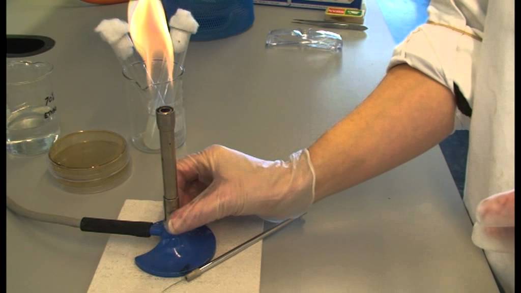
Aseptic techniques in the biology lab
136.9K
Aseptic techniques prevent contamination when working with microorganisms by maintaining sterility in both apparatus and the work environment. Key principles include: closing windows and doors to prevent drafts, turning off fans, and avoiding door openings during work. Gather all equipment beforehand and work efficiently near the Bunsen burner. Personal protection requires rolled-up sleeves secured with rubber bands, previously sterilized glassware, removed jewelry, tied-back hair, and thorough handwashing with soap and hot water. The work area should be swabbed with 70% alcohol (keeping flammable ethanol away from open flames). Flaming equipment involves holding items in a Bunsen flame until red-hot, drawing them through slowly, allowing cooling, and repeating to ensure sterility while preventing airborne contaminants from settling.
Click to watch →
Deep Dive
Basic biology and physiology of microalgae, including their photosynthetic mechanisms, growth phases, and standard culture media requirements.
WEBINAR BIOTEKNOLOGI KELAUTAN DI ERA NEW NORMAL
3.2K
Microalgae are microscopic algae that cannot be seen with the naked eye and require magnification of 100-400 times for identification. They are classified into 9 divisions based on their pigments and are phototrophic (using light for energy), though some can also be heterotrophic. Microalgae growth follows distinct phases: (1) Lag phase - initial adaptation period, (2) Exponential phase - rapid growth when nutrients are abundant, (3) Stationary phase - growth slows as nutrients deplete, (4) Death phase - cell death when nutrients are exhausted. Harvesting for valuable compounds typically occurs during the stationary phase.
Click to watch →

Microalgae Culture | Microalgae Cultivation |
7.6K
Microalgae are primary producers that convert water and carbon dioxide into biomass and oxygen through photosynthesis, serving as valuable sources of bioactive compounds (lipids, proteins, carbohydrates, carotenoids, vitamins) for nutrition and aquaculture applications; successful microalgae culture requires careful management of key environmental parameters including light intensity (with blue and red wavelengths being most effective), pH (maintained between 7-9), temperature (optimal range 20-24°C), salinity, and aeration, along with proper nutrient enrichment using macronutrients (nitrate, phosphate, silicate) and micronutrients (vitamins and trace metals), while growth dynamics follow five distinct phases: lag phase, exponential growth phase, declining growth rate phase, stationary phase, and death/crash phase.
Click to watch →
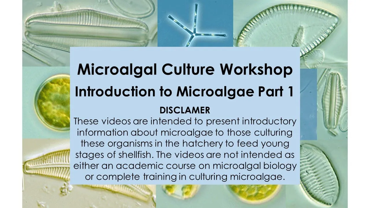
2 Microalgal Culture Workshop Introduction to Microalgae
11.9K
Photosynthesis converts water and carbon dioxide into sugar and oxygen using photopigments like chlorophyll a, b, and c. Microalgae exhibit two cellular organizations: prokaryotic cyanobacteria (blue-green algae) with simple designs, and eukaryotic algae with true nuclei and compartmentalized organelles. Major groups include green algae (evolutionary origin of land plants), dinoflagellates (flagellated with diverse ecological roles), diatoms (glass-shelled with planktonic and benthic forms), cryptophytes (complex internal structures), and haptophytes (small green with potential nutritional value). Each group varies dramatically in morphology, ecology, and utility for aquaculture applications.
Click to watch →

Curso 4 - Módulo III: Cultivo de microalgas marinas como alimento para larvas de moluscos bivalvos
793
This section covers the different cultivation systems, growth phases, and complete production flow in microalgae cultivation. Three main cultivation systems exist: discontinuous (batch) where nutrients are added once and entire culture is harvested after 7-8 days; semi-continuous where partial harvesting with nutrient replenishment reduces cycle time to 3-4 days; and continuous where constant nutrient input and harvest maintain steady-state conditions. The choice depends on scale, contamination risk, and production requirements. Microalgae cultivation follows a characteristic growth curve with distinct phases: lag phase (day 0) where microalgae adapt to new conditions; acceleration phase where growth begins; exponential phase where optimal growth and nutritional value occur, ideal for harvesting; deceleration phase where growth slows; stationary phase where density stabilizes; and death phase where cells begin dying. For production, harvesting occurs during the exponential phase (around day 7) when microalgae have optimal nutritional value. The complete production flow involves: maintaining stock cultures on agar plates; selecting healthy colonies for inoculation; initial cultivation in test tubes (7 days); transfer to 150mL flasks (7 days); transfer to 12L bottles (7 days); transfer to 18L volumes; and final scale-up to 500L tanks. Each transfer serves as inoculum for the next phase. Cultures should be renewed after 4-5 repiquings to maintain vigor.
Click to watch →

The Dirty Lab Coat - Brainy Briny Beaker Bag (BBBB-00015)
2.9K
Optimal microalgae growth requires about 100 micro einsteins per meter squared second of light (sunlight provides 3,000, which is too bright). Temperatures above 100°F kill algae. After about 8 days, brine shrimp cysts hatch and swim in the culture, visible against the grid. The seshi stick uses optical density to measure biomass: fold it on the dotted line, submerge until the black and white target disappears, then use the provided graph to determine cells per milliliter. This allows tracking cell density over time and creating growth curves to monitor culture development.
Click to watch →
Deep Dive
The theoretical concept of dilution streaking and how it isolates individual cells on solid agar to produce clonal colonies.

Aseptic Technique & Streak Plate
110.5K
The streak plate technique dilutes cells to obtain isolated colonies. Steps include: sterilizing the loop in blue flame until orange-hot, cooling by touching agar (no sizzling), collecting a single colony, performing primary streaks gently, flaming between sets, turning the plate to intersect lines, and creating final vertical/zigzag patterns. The loop must be cooled between transfers to prevent killing organisms. Only the primary streak receives inoculum—the technique relies on progressive dilution.
Click to watch →

Microbiology: the streak plate technique
16K
The streak plate technique is a fundamental microbiology method used to isolate individual bacterial colonies on an agar plate by spatially diluting bacteria across multiple sectors; a single bacterial cell undergoes binary fission to form a visible colony containing approximately one million bacteria, and this technique allows researchers to determine if a culture is pure or contaminated by observing whether multiple distinct colony types appear, with each sector showing progressively lighter growth until isolated colonies form in the final sector.
Click to watch →
Streaking for Isolation
48.1K
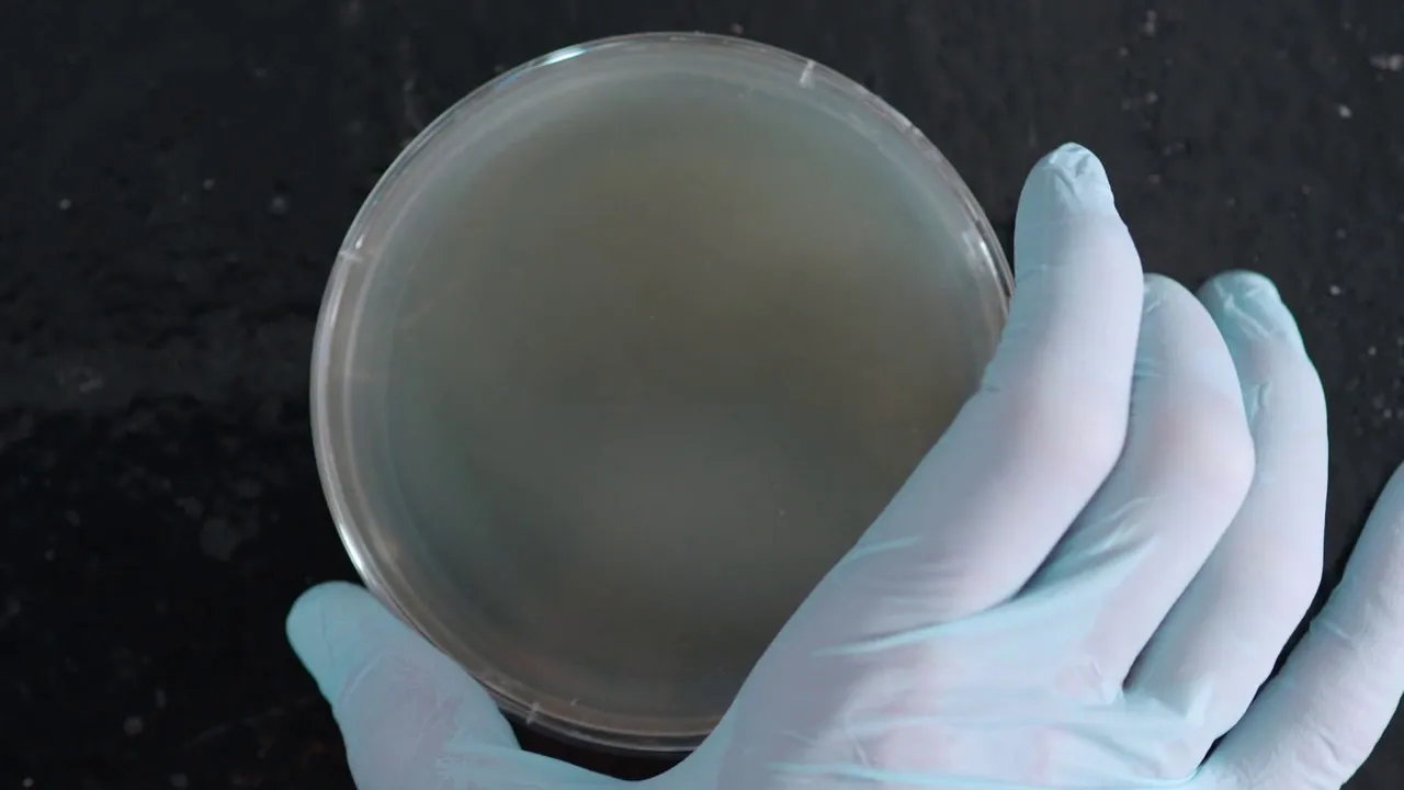
Isolation streaking is a microbiology technique that uses progressive dilution to create individual bacterial colonies on an agar plate; the process involves dividing the plate into quadrants and making zigzag streaks where each subsequent streak crosses the previous one, ensuring that later streaks contain fewer bacteria and allowing for the isolation of pure colonies after incubation.
Click to watch →
How to Streak for Single Colonies
54.7K
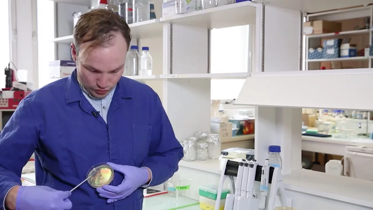
To successfully streak for single colonies on a petri dish, use a disposable inoculating loop to spread cells across the plate in progressively diluting regions, replacing the loop between each streak to ensure cells are spread far enough to isolate individual colonies; always store petri dishes upside down to prevent condensation from dripping onto the agar and smearing the cells.
Click to watch →

Streaking
1.2K
Streaking is a dilution method used in microbiology to isolate pure bacterial colonies by progressively diluting bacteria across a petri dish through repeated sterilization of the inoculation loop and spreading bacteria in multiple zones, allowing individual bacterial cells to grow into isolated colonies that can be identified and studied.
Click to watch →
Deep Dive
An introductory understanding of high-value bioproducts, specifically carotenoids like lutein, and why they are synthesized by microalgae.

Vídeo aula 1_Unidade 4_ Cultivo de Microalgas
2.7K
Microalgae products range from low-value biomass to high-value compounds. Low-value products include carbohydrates, lipids, and biomass with large market sizes. High-value products include pigments (carotenoids, astaxanthin), omega-3 and omega-6 fatty acids, and amino acids, used in cosmetics, nutrition, and biomedicine. Advantages include 10-100 times higher productivity than traditional crops, efficient carbon capture, high lipid content for biodiesel, and ability to grow on non-arable land using wastewater. Challenges include high production and extraction costs requiring innovative solutions.
Click to watch →

BrisScience (June 2019): Algae - From seafood to solar
1.1K
Beyond omega-3 fatty acids, algae produce valuable carotenoids (pigments with antioxidant properties) and phytosterols (compounds that lower cholesterol levels). One particular microalgae species, Pavlova lutherii, produces omega-3 fatty acids, carotenoids, and phytosterols together at concentrations up to 5% of its dry weight—making it exceptionally valuable for health applications. These compounds represent premium market opportunities that can subsidize lower-margin fuel production, creating economically attractive business models for algae cultivation.
Click to watch →

Metabolites of marine micro algae
557
Carotenoids are lipophilic components soluble in lipids, found in higher plants and microalgae, with proven anti-inflammatory and anti-cancer activities representing a billion-dollar global market. Beta-carotene, produced by Dunaliella salina, serves as vitamin A precursor and is preferred over synthetic forms for its better bioavailability. Astaxanthin, produced by Hematococcus pluvialis through stress-induced thickening of cell walls, costs $2500/kg and offers UV protection and therapeutic benefits for cancer and inflammatory diseases, crossing blood-brain barriers without eye crystal formation. Canthaxanthin enhances poultry liver vitamin E content. Lutein, essential for retinal and lens health, reduces disease risk when present at significant levels in human serum.
Click to watch →
1 łyżeczka uodparnia na pasożyty, powstrzymuje choroby, rozbija trójglicerydy
1.2M
Spirulina and chlorella are excellent dietary sources of carotenoids lutein and zeaxanthin, which are essential for eye health. One teaspoon of these microalgae provides clinically significant amounts of these nutrients. Lutein and zeaxanthin protect the retina from UV radiation damage and oxidative stress from free radicals. Scientific evidence shows these carotenoids help prevent age-related macular degeneration and cataracts, and may also improve visual acuity in conditions such as diabetic retinopathy.
Click to watch →
Prof Dr Laurent Picot , Rochelle University, France
116
This section covers the discovery of carotenoids during microalgae research for anti-cancer compounds. Carotenoids are liquid pigments belonging to the terpene family, typically containing 40 or 45 carbon atoms, appearing in red, orange, and yellow colors. Over 1,000 carotenoids have been identified. Examples include lycopene (tomatoes), beta-carotene (carrots), astaxanthin (microalgae), and fucoxanthin (brown seaweed). Some carotenoids contain complex structures like epoxy functions and glycosylation, making them difficult to synthesize chemically and requiring purification from biological sources. Carotenoids serve multiple biological functions: photoreception for vision and photosynthesis, phototropism, membrane stabilization, antioxidant activity against reactive oxygen species, photoprotection against high light, allelopathy, chemoattractant functions, and regulation of cell cycle and metabolism. Retinoic acid, a carotenoid derivative, is important in embryonic development.
Click to watch →
Prerequisite Knowledge
- Concept 01Fundamental principles of aseptic technique, including the correct usage of laminar flow hoods, Bunsen burners, and sterile handling tools.
- Concept 02Basic biology and physiology of microalgae, including their photosynthetic mechanisms, growth phases, and standard culture media requirements.
- Concept 03The theoretical concept of dilution streaking and how it isolates individual cells on solid agar to produce clonal colonies.
- Concept 04An introductory understanding of high-value bioproducts, specifically carotenoids like lutein, and why they are synthesized by microalgae.
Subsequent Learning
- Step 01Scale-up protocols transitioning microalgae from agar plates to liquid flasks, and eventually to large-scale photobioreactors (PBRs).
- Step 02Environmental stress optimization techniques (such as nitrogen starvation or light intensity variation) to trigger and maximize lutein accumulation.
- Step 03Downstream processing methods for microalgal biomass harvesting, cell disruption, and solvent extraction of lipophilic pigments.
- Step 04Analytical methods, such as High-Performance Liquid Chromatography (HPLC) and spectrophotometry, to quantify lutein yield and purity.
Streaking
Plate Sealing
Inoculation
Incubation Setup
Streaking
0:15Playing Section
- 1
Demonstrates aseptic quadrant streaking on agar to isolate colonies.
- 2
Flaming loop, picking colony, and diluting via successive touches.
Co-Cultivation and Non-Axenic Production Systems
While aseptic protocol is fundamental for maintaining pure (axenic) master cultures in a laboratory, strict sterility is often impractical, costly, and energy-intensive at an industrial scale. A major counterpoint to the pure-culture paradigm is the use of non-axenic cultivation and engineered co-cultures. In industrial biotechnology, microalgae are increasingly grown alongside beneficial bacteria or fungi (microbiome consortia). These symbiotic relationships can enhance microalgal growth, improve harvesting efficiency, and boost the yield of valuable metabolites like lutein through natural growth promotion and nutrient recycling. Furthermore, large-scale commercial production often utilizes open ponds or non-sterile photobioreactors, relying on selective environmental conditions (such as specific pH, salinity, or rapid growth rates) rather than sterile techniques to prevent contamination. Teaching students only aseptic monoculture overlooks these ecologically-driven, economically viable industrial alternatives that challenge the necessity of absolute sterility in microalgae biotechnology.
Deep Dive
Scale-up protocols transitioning microalgae from agar plates to liquid flasks, and eventually to large-scale photobioreactors (PBRs).

Biofuels from Algae Project - Brunswick Community College Center for Aquaculture & Biotechnology
224.7K
Scaling up algal cultures involves progressively increasing the volume from small maintenance tubes to larger volumes in sterile culturing flasks. Before inoculation, cultures should be homogenized to ensure even distribution, which is particularly important for non-motile species that tend to congregate at the bottom. The scaled-up cultures are then placed in controlled conditions within the cell laboratory for growth periods of 2-4 weeks before being transferred to photobioreactors.
Click to watch →

The Role of Microalgae in Raising Queen Conch
328
Scaling up microalgae culture involves transferring cultures from smaller to larger vessels following aseptic techniques. The process begins by lighting an alcohol burner and swirling source cultures to ensure uniform cell suspension. The receiving vessel's mouth is flamed, followed by the source vessel's mouth. A sterile pipette is inserted near the flame to draw a sample, then transferred to the new vessel. The receiving vessel is swirled to mix new cells into the media. Vessels must be labeled with inoculation dates to track growth stages. This systematic scaling ensures sufficient microalgae biomass accumulates for daily larval feeding requirements.
Click to watch →

Inoculation Of Microalgae | Culturing Microalgae | Growing Microalgae |
2.4K
This video demonstrates the complete process of inoculating microalgae, beginning with aseptic agar plate streaking where a sterilized wire loop picks a well-isolated colony and creates parallel streaks on solidified agar, followed by transferring the isolated colony to a conical flask containing prepared suspension media, which is then incubated in an orbital shaker at 140 rpm and 24°C for 5 days; success is confirmed by observing the green coloration of the suspension media indicating microalgae growth.
Click to watch →
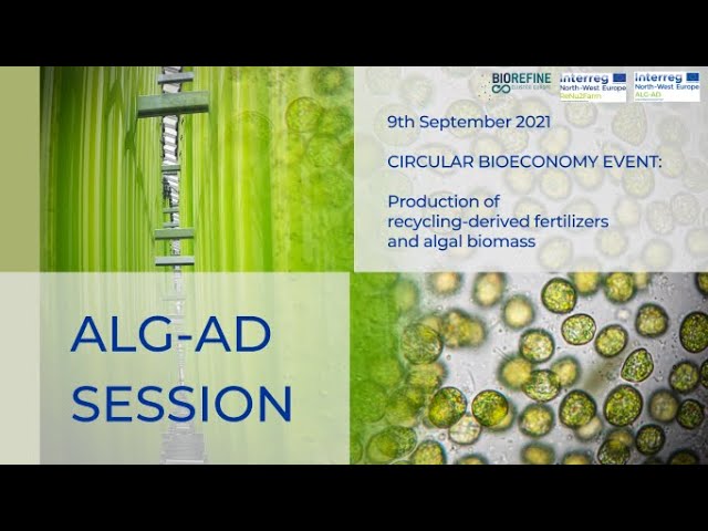
9/9/2021 - ALG-AD & ReNu2Farm event - ALG-AD session
260
This comprehensive process covers the complete microalgae cultivation workflow: (1) Photobioreactor design with 7500L capacity, modular system, temperature control, and artificial lighting; (2) Scaling up from 100ml to 1L in laboratory with digestate concentration adaptation from 0.5% to 2.5%; (3) Greenhouse inoculation of 25L culture to inoculate 80L bags; (4) Contamination prevention through membrane filtration, bleach treatment, and neutralization; (5) PBR inoculation with 400L culture from university; (6) Harvesting with membrane concentration to 6-7g/L for Cenedesmus and 2-3g/L for Chlorella; (7) Mixotrophic culture model with 72-hour aeration and carbon supplementation; (8) Daily monitoring of pH, dissolved oxygen, temperature, and microscopic contamination checks.
Click to watch →
Curso 4 - Módulo III: Cultivo de microalgas marinas como alimento para larvas de moluscos bivalvos
793
This section covers microalgae culture maintenance, medium preparation, and the systematic scaling-up process. Microalgae strains are maintained on solid media (agar) in Petri dishes for long-term preservation, allowing researchers to periodically check strain health and detect contamination. The F/2 medium is a standard formulated medium for marine microalgae cultivation, serving as the control medium in research. Guillard's medium (f/2) requires preparation of multiple components: nitrate and phosphate solution (75g/L for nitrate), silicate solution (for diatom cultivation), trace metal solution (containing seven metals: copper, iron, manganese, zinc, cobalt, boron, nickel), and vitamin solution (containing vitamins B1, B2, B6, B12, and others). Each component is prepared separately and stored in amber bottles to protect light-sensitive compounds. Scaling up requires a systematic approach: starting with small volumes (10mL) in test tubes, progressing through intermediate volumes (150mL flasks, 12L bottles), and reaching large-scale production (500L tanks). Each phase requires specific handling and monitoring. Repiquing (transferring cultures) is performed when volumes are small (under 250mL) to maintain cell density. Cultures should not be repiqued more than 4-5 times before renewal. Three main cultivation systems exist: discontinuous (batch) where nutrients are added once and entire culture is harvested after 7-8 days; semi-continuous where partial harvesting with nutrient replenishment reduces cycle time to 3-4 days; and continuous where constant nutrient input and harvest maintain steady-state conditions. Microalgae cultivation follows a characteristic growth curve with distinct phases: lag phase (day 0) where microalgae adapt to new conditions; acceleration phase where growth begins; exponential phase where optimal growth and nutritional value occur, ideal for harvesting; deceleration phase where growth slows; stationary phase where density stabilizes; and death phase where cells begin dying. For production, harvesting occurs during the exponential phase (around day 7) when microalgae have optimal nutritional value. The complete production flow involves: maintaining stock cultures on agar plates; selecting healthy colonies for inoculation; initial cultivation in test tubes (7 days); transfer to 150mL flasks (7 days); transfer to 12L bottles (7 days); transfer to 18L volumes; and final scale-up to 500L tanks. Each transfer serves as inoculum for the next phase.
Click to watch →
Deep Dive
Environmental stress optimization techniques (such as nitrogen starvation or light intensity variation) to trigger and maximize lutein accumulation.

Logee's Longhouse Greenhouse Tour — Ep 171
293.1K
Flowering in many plants can be triggered by environmental stress. When plants receive too much nitrogen fertilizer, they may not flower. To encourage flowering, plants sometimes need to be stressed in specific ways, such as reducing water or fertilizer. This stress signals the plant to shift from vegetative growth to reproductive growth. The greenhouse uses grow lights for 12 hours daily, and plants are also placed in south-west facing windows where they receive maximum light. However, some plants still require additional stress to initiate flowering.
Click to watch →

Bioprocess Engineering International Guest Lecture: Microalgal Biotechnology and Production
238
Haematococcus pluvialis demonstrates a remarkable stress response mechanism where environmental stressors trigger metabolic changes. Under normal conditions, the algae produces lutein (important for eye health). When subjected to high light intensity and nitrogen depletion stress, the algae converts lutein to astaxanthin, resulting in a dramatic color change from green to bright red. This stress-induced conversion is exploited commercially for astaxanthin production, demonstrating how controlled environmental conditions can maximize the production of specific high-value compounds.
Click to watch →

99%の眼科医がおすすめするこの栄養素をとるべき!~ルテイン~
83.3K
Lutein is a nutrient that eliminates oxidative damage and free radicals. Unlike other antioxidants that circulate throughout the body, lutein accumulates specifically in the eyes, particularly in the lens and macula. The macular pigment containing lutein naturally decreases with age, especially after 40. Lutein acts as a natural blue light filter, protecting eyes from blue light damage from sunlight, computers, and smartphones. Lutein is found in green-yellow vegetables like kale and bitter melon, eggs (yolk), and chrysanthemum flowers.
Click to watch →

лютеин рабочая добавка для зрения и здоровья глаз
142.9K
Lutein is a carotenoid pigment that accumulates in the macula of the retina at concentrations 10,000 times higher than in blood plasma, where it serves as a powerful antioxidant by absorbing harmful blue and violet light wavelengths that would otherwise generate free radicals and damage retinal cells; this protective function helps prevent age-related macular degeneration, reduces eye strain from digital screens by minimizing chromatic aberration, and may slow cataract progression, with a recommended daily dosage of 10mg providing approximately 80% bioavailability for effective eye health maintenance.
Click to watch →
![[2022 KAIST 연구성과] 미생물 이용한 루테인 생산 기술 개발 - 생명화학공학과 이상엽 연구책임자 (KOR)](https://i.ytimg.com/vi/DD-eye6UrG8/maxresdefault.jpg)
[2022 KAIST 연구성과] 미생물 이용한 루테인 생산 기술 개발 - 생명화학공학과 이상엽 연구책임자 (KOR)
182
KAIST researchers developed a novel method to produce lutein, an eye-protective carotenoid, by engineering E. coli bacteria to express the lutein biosynthetic pathway using glycerol as a carbon source; they achieved this breakthrough by implementing two key strategies: substrate channeling (clustering enzymes like Lcb lut2 using CPA scaffold proteins to improve alpha-carotene precursor production) and electron channeling (fusing P450 enzymes LUT5, LTT1 with P450 reductase ATL2 to the CPB scaffold to facilitate electron transfer), resulting in a record-high yield of 218 mg/liter of lutein in just 48 hours—dramatically outperforming the 200-day cultivation period required for traditional marigold flower extraction.
Click to watch →
Deep Dive
Downstream processing methods for microalgal biomass harvesting, cell disruption, and solvent extraction of lipophilic pigments.
Bioprocess Engineering International Guest Lecture: Microalgal Biotechnology and Production
238
Downstream processing accounts for 50-60% of total microalgae production costs, making it the most critical area for optimization. Harvesting methods include bio-based flocculation using natural polymers (such as tannic acid-derived products that work in just 10 minutes), centrifugation providing high-quality concentrates but requiring frequent manual intervention, and membrane filtration operating continuously for 12-14 hours with lower operating costs. Drying technologies present trade-offs: freeze-drying preserves the highest pigment quality but is expensive; spray drying and drum drying offer the most economical options but may degrade heat-sensitive compounds; solar drying is the least expensive but causes significant degradation through prolonged heating. Cell disruption methods include bead beating (effective but generates heat that degrades sensitive compounds) and wet processing techniques. For food-grade applications, supercritical carbon dioxide or pressurized liquid extraction provides clean separation without toxic solvents. The choice of method depends on target product sensitivity, purity requirements, and intended application. Biofouling prevention through systematic protocols—including regular sterilization, maintaining optimal growth conditions, and using glass tubing—enables successful long-term operations achieving 320+ day cultivation cycles.
Click to watch →

Lecture 11 Downstream Processing
118
Cell disruption is essential for releasing intracellular products while preserving component integrity. Methods are categorized as mechanical (solid shear and liquid shear) or non-mechanical (physical, chemical, and enzymatic). Physical methods include freezing, high osmotic pressure, and shock. Chemical methods use surface activators, solvents, and antibiotics. Enzymatic methods employ lysosomes. Liquid-liquid extraction exploits differential solubilities in immiscible solvents, requiring non-toxic, selective, inexpensive solvents with high distribution coefficients. Concentration methods include evaporation, membrane filtration, chromatography, and absorption.
Click to watch →

Cell Disruption Methods (physical, chemical, mechanical, enzymatic) ||Notes 😎#downstream processing
22.4K
Cell disruption methods are essential techniques in downstream processing for recovering intracellular products after fermentation, categorized into physical methods (freezing-thawing, osmotic shock, homogenization, ultrasonication, bead milling, and motor-pestle), chemical methods (using detergents and solvents to solubilize membrane components), and enzymatic methods (using specific enzymes like lysozyme, cellulase, and pectinase to break down cell walls), each with distinct advantages and limitations depending on the cell type and product characteristics.
Click to watch →

Recovery & Purification of Fermentation-3 | Principles of Biochemical Engineering | BT513T_Topic083
110
Downstream processing involves sequential steps for recovering fermentation products. After initial separation, cell disruption releases intracellular products using chemical methods (detergents, osmotic shock, alkali, enzymes) or physical methods (shear forces, ultrasonication). For product recovery, solvent extraction separates broth into organic and inorganic phases using single-stage units. The solvent recovery plant, typically a distillation unit, recycles solvents through the system. Not all solvent needs complete removal, as residual amounts can remain with the product. This integrated approach transforms complex fermentation broths into purified products through systematic separation, disruption, and extraction technologies.
Click to watch →

What is downstream processing used for the purification of fermentation product?
68.3K
Downstream processing is the critical phase in biotechnology that recovers and purifies products from fermentation broth through sequential steps including cell separation (filtration, centrifugation, ultrafiltration), cell disruption (mechanical, chemical, enzymatic), product extraction (liquid-liquid extraction, solvent extraction), concentration, purification (chromatography, crystallization), and final drying to obtain high-purity products suitable for commercial applications.
Click to watch →
Deep Dive
Analytical methods, such as High-Performance Liquid Chromatography (HPLC) and spectrophotometry, to quantify lutein yield and purity.
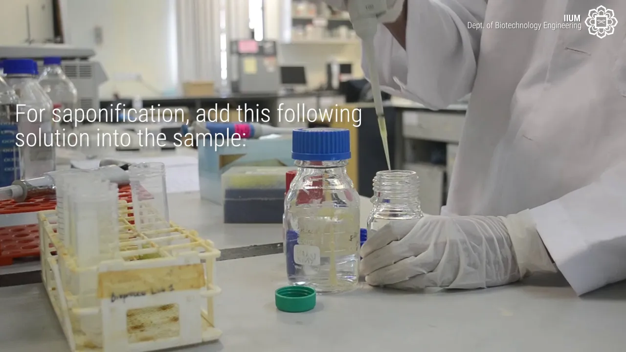
Extraction of microalgae and HPLC analysis
23.6K
This video demonstrates a complete method for extracting lutein from microalgae through sequential solvent extraction (7:3 acetone-methanol with calcium carbonate, followed by hexane partitioning), saponification, and purification, followed by quantification using HPLC with a reverse-phase C18 column and binary gradient elution (methanol-water 9:1 to ethyl acetate), where lutein is detected at 447 nm and quantified via standard curve analysis.
Click to watch →

Videodərs Müəllim:Səma Ələsgərova
104
Purity of retinoid esters is determined using chromatographic methods: (1) thin-layer chromatography is used to separate the sample, (2) the eluate is obtained, (3) spectrophotometry at 326 nm is performed, (4) purity is confirmed by maximum absorption at 326 nm, (5) impurities should not exceed 3% of the total. This combined chromatographic-spectrophotometric method ensures accurate purity assessment.
Click to watch →

Análise de Carotenoides em Alimentos
1.9K
Carotenoid quantification employs spectrophotometry and HPLC. Spectrophotometry uses light absorption at 400-450 nm based on the Beer-Lambert law, with results expressed as equivalent beta-carotene using specific absorption coefficients. Saponification removes interfering compounds like chlorophyll by breaking ester bonds with potassium hydroxide in methanol. HPLC provides detailed separation using C30 columns for superior isomer resolution compared to C18 columns. Detection at 450 nm with diode array detectors enables spectral identification. Method development optimizes mobile phase composition and gradient conditions. Replicates should be sufficient for statistical confidence, with triplicates common for routine analysis.
Click to watch →

2020 04 22 3교시 HPLC 2탄!
324
HPLC quantification uses several methods. The Peak Area Comparison Method compares peak areas between standards and unknowns. The Calibration Curve Method creates a linear relationship between concentration and peak area using standard samples. The Internal Standard Method, most widely used, adds a known internal standard to both standards and samples, using the ratio of analyte to internal standard peak areas for quantification. This method compensates for sample preparation variations and is essential for accurate results in complex matrices.
Click to watch →

SOBAT KIMIA: Pengenalan KCKT (Kromatografi Cair Kinerja Tinggi)
190
HPLC can determine compound purity by comparing the peak area of the compound of interest to total peak area: purity percentage = (Area of compound / Total Area) × 100. This provides quick assessment without additional analytical techniques. Sample extraction for pigment analysis involves homogenizing the sample, adding extraction solvent (e.g., acetone:water or methanol:water), filtering, and analyzing the extract. Different pigments have different solubilities in different solvents, allowing identification by extracting with various solvents and comparing with reference spectra.
Click to watch →
Streaking
Plate Sealing
Inoculation
Incubation Setup
Streaking
0:15- 1
Demonstrates aseptic quadrant streaking on agar to isolate colonies.
- 2
Flaming loop, picking colony, and diluting via successive touches.
Co-Cultivation and Non-Axenic Production Systems
While aseptic protocol is fundamental for maintaining pure (axenic) master cultures in a laboratory, strict sterility is often impractical, costly, and energy-intensive at an industrial scale. A major counterpoint to the pure-culture paradigm is the use of non-axenic cultivation and engineered co-cultures. In industrial biotechnology, microalgae are increasingly grown alongside beneficial bacteria or fungi (microbiome consortia). These symbiotic relationships can enhance microalgal growth, improve harvesting efficiency, and boost the yield of valuable metabolites like lutein through natural growth promotion and nutrient recycling. Furthermore, large-scale commercial production often utilizes open ponds or non-sterile photobioreactors, relying on selective environmental conditions (such as specific pH, salinity, or rapid growth rates) rather than sterile techniques to prevent contamination. Teaching students only aseptic monoculture overlooks these ecologically-driven, economically viable industrial alternatives that challenge the necessity of absolute sterility in microalgae biotechnology.
Fundamental principles of aseptic technique, including the correct usage of laminar flow hoods, Bunsen burners, and sterile handling tools.
Basic biology and physiology of microalgae, including their photosynthetic mechanisms, growth phases, and standard culture media requirements.
The theoretical concept of dilution streaking and how it isolates individual cells on solid agar to produce clonal colonies.
An introductory understanding of high-value bioproducts, specifically carotenoids like lutein, and why they are synthesized by microalgae.
Scale-up protocols transitioning microalgae from agar plates to liquid flasks, and eventually to large-scale photobioreactors (PBRs).
Environmental stress optimization techniques (such as nitrogen starvation or light intensity variation) to trigger and maximize lutein accumulation.
Downstream processing methods for microalgal biomass harvesting, cell disruption, and solvent extraction of lipophilic pigments.
Analytical methods, such as High-Performance Liquid Chromatography (HPLC) and spectrophotometry, to quantify lutein yield and purity.
67.8K views1.2Klikes7:42@bioproject2705Original Release: 2018-10-29
[Music] for streaking process we need a Bunsen burner inoculation loop tap agar plate sample and paraffin on all the process is done in a laminar air flow cabinet aseptically clean hands with alcohol this is crucial to avoid any contamination that can further affect the process flame the inoculation loop on Bunsen burner wait until the loop turns red then allow it to cool try to touch the loop on the agar to ensure that the loop is cool enough for streaking pick an isolated colony from sample agar immediately transfer the isolated colony to first quadrant of the agar [Music] flame the loop again and allow it to cool [Music] then continued the streaking from the previous area and extend the streaks repeat the step of flaming the loop and streaking until getting this streaking pattern [Music] make sure these lines touch during streaking process this technique is to make sure to dilute the concentration in order to get single colony lastly seal the plate by using the para film place the plate upside down while stored at room temperature under the light the plates must be stored in air-conditioned room to minimize the condensation that can contaminate the microalgae [Music] you [Music] for inoculation we need inoculation loop Bunsen burner sample parafilm and suspension media firstly remove the para film from petri dish then loosen the aluminium foil from conical flask [Music] flame the mouth of flask while rotating it in circular motion after that flame the inoculation loop until it turn red and let it cool take the isolated colony from the sample petri dish immediately transfer the colony into the suspension media after that flame the inoculation loop and mouse a flask again [Music] wrap the mouth of flask by using same aluminium foil use the parafilm to seal the flask don't forget to seal the sample plate again for future use label the flask based on name of the species and date press the stop button before open the lid [Music] remove the lamps and open lid carefully put the conical flask at the right size of holder close the lid and put the lamp on it set the Machine by adjusting the speed to 139 revolutions per minute and temperature to 23 points 9 degrees Celsius make sure the cooling system is switched on lastly press the start button you [Music]
Up Next

6:36
Astaxanthin Production: Algalif's Sustainable Microalgae Cultivation in Iceland
@TBDMediaGroup
14.6K views•2022-09-06

12:54
Triumph of Orthodoxy Icon: Byzantine Art & History Explained
@BenCallan
2.1K views•2024-08-06

9:49
FastAPI vs Flask vs Django: Choosing the Right Python Web Framework
@TechWithTim
302.5K views•2024-05-26

1:42
Game of Thrones Opening Credits: A Cinematic Analysis
@gameofthrones
46.3M views•2011-04-18
Related Study Plans & Knowledge Roadmaps
Structured learning paths in General & Interdisciplinary Studies
general-studies
Elevator Engineering: History, Physics & Cities
01From Hoists to Safety Brakes: The Dawn of Vertical Lift
02Hydraulic vs. Traction: The Engineering Physics of Elevators
03Modern Speed, Safety, and Algorithms
+ 2 more learning modules
12h5 modules
Explore Roadmap
general-studies
Rubber: Material Science, History & Geopolitics
01Origins of Natural Rubber & Mesoamerican History
02The Chemistry of Natural Rubber & Vulcanization
03The Geopolitics of the Rubber Boom & Colonization
+ 2 more learning modules
12h5 modules
Explore Roadmap
general-studies
Containerization: Evolution, Logistics & Trade
01Ancient Trade & Bulk Cargo: Amphorae to Barrels
02The Industrial Revolution & Cardboard Packaging
03The Containerization Revolution: Malcom McLean
+ 2 more learning modules
12h5 modules
Explore Roadmap
general-studies
Textile Tech: Evolution, Labor & Global Impact
01Pre-Industrial Textiles: Handloom & Cottage Industry
02Mechanization: The Spinning Jenny to the Power Loom
03The Factory System & Global Labor Transformation
+ 2 more learning modules
10h5 modules
Explore Roadmap