












![Brain Structures & Functions [AP Psychology Unit 2 Topic 6]](https://i.ytimg.com/vi_webp/7zwFI0k-JvE/maxresdefault.webp)










This lecture introduces the fundamental structure and organization of the brain, covering key anatomical terminology (dorsal, ventral, anterior, posterior, medial, lateral, ipsilateral, contralateral), planes of brain section (sagittal, horizontal, transverse), and the four cerebral lobes (frontal, parietal, temporal, occipital) with their primary functions. The brain is divided into the central nervous system (brain and spinal cord) and peripheral nervous system (cranial and spinal nerves), with the brain further organized into the cortex (outer layer with gyri and sulci), subcortical structures (limbic system, basal ganglia, thalamus, hypothalamus), and brainstem (pons, medulla, cerebellum). The brain is protected by the skull, meninges (dura mater, arachnoid, pia mater), and cerebrospinal fluid, while the blood-brain barrier regulates substance transfer between blood and brain tissue.
Basic Neuroanatomy: Brain Structures and Terms
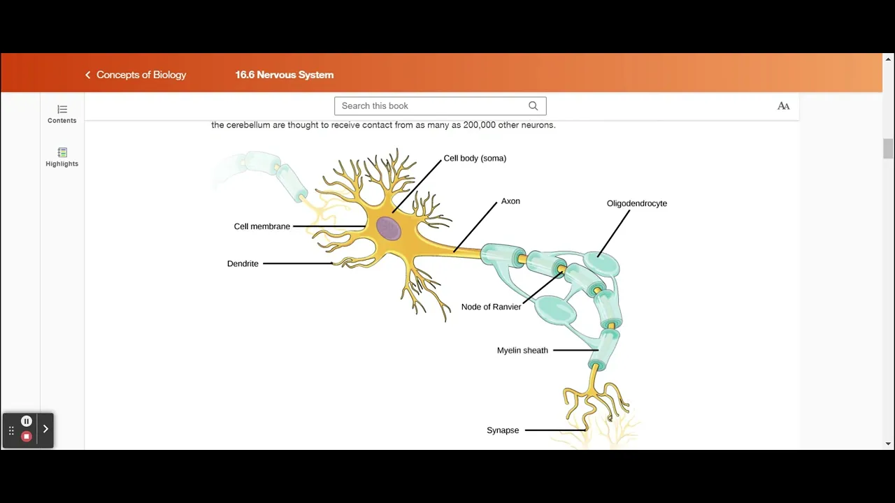
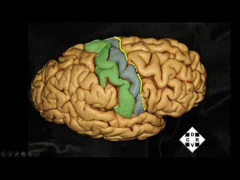
Added:introduction to neuroanatomy we're going to take a quick tour of the brain go over the big landmarks the structure and organization of the brain but first we're going to start out with some terminology we're going to start out with some terminology so that you know what i'm talking about when i say that something's anterior or if it's medial also we'll go through the structure of the nervous system the nervous system describes all of the nervous system in the body you can break it down into the central and the peripheral and then you can go further than that i'll also spend most of the time talking about the central nervous system and then a smaller amount of time talking about the peripheral nervous system they're both very important the one that we probably focus most attention on is the central so that will be the primary focus of this lecture as well okay so let's get started first we need to talk about where things are and how and the terminology that we use to describe structures in the brain not only and in the nervous system not only where they are located themselves but where they are located relative to other structures and the first thing we need to do is we need to think about um in your mind or you can watch this on the screen there is this line this imaginary line that goes from the center of the forehead now we're going to work with a quadruped here because this is where it was originally designed it goes through the center of the forehead into the middle the exact middle of the body all the way through and then exits through the tail this is called the neuraxis the narxis enters through the center of the forehead and then exits through the tail when we when we think about the neuraxis as far as where things are it simplifies things because anything that's located above the nerve axis we can say is dorsal anything that's located below the nerve axis is ventral anything located near the beginning or the front end of the neurocyst is rostral or anterior and anything located near the back end of the neraxis is posterior or caudal okay so that's the first thing but then it becomes more complicated because we don't walk on four legs we walk on two so you have to take this creature here and the neuraxis that goes along with it and then make it upright and when you do that what you see is that again you have the neuraxis it starts in the middle of the forehead and it it goes exactly through the middle of the body but then since we walk upright right about the middle of the brain it takes a 90 degree turn and starts going straight down so it's pretty much very similar to for the two-legged versus the four-legged except that when we're talking about different structures in the brain we may be talking about the occipital lobe which is located here in the back of the brain but we're still going to refer to it as dorsally located because see it's still on the top side of the neuraxis okay whereas it's a little so it's a little bit different in terms of a four-legged creature versus a two-legged creature and and it also confuses some of the terminology here because dorsal we know is anything towards the back and ventral is anything towards the belly but when we talk about humans now ventral is here in the front because it's still it's still on the belly okay it's below the nerve axis whereas dorsal it's still towards the back but now it's not exactly on the top okay hopefully that will make sense if you just think about that neuraxis and how it differs for quadrupeds versus bipeds like we humans okay the two um first descriptors or terms are anterior that's located near the head and posterior that's located near the tail now when we talk about humans the posterior then actually extends towards the feet anterior is the same posterior includes anything towards the feet the tail and the feet okay dorsal i mentioned that anything towards the back okay and so it usually means up above two as well but for humans towards the back is not necessarily up above all the time sometimes it's just towards the back ventral is the same thing you can see how it differs ventral here looks like it's below right because it's bel if we were describing um maybe the thalamus right here it's ventrally located because it's below the neuraxis but um but also the belly is located ventrally as well so just keep those those terms in mind and how they they differ a little bit for um animals or creatures with four legs and creatures with two okay the next one is superior and that is really a synonym with dorsal so superior and then inferior which is a cinnamon synonym with ventral you can use those interchangeably okay if we're trying to decide or describe a structure that is located more towards the center of the body versus one more located towards the side of the body or away from the neuroxis then we would use these terms lateral describes anything that is located more distally or way from the neuraxis here's the neuraxis here remember this little red line anything that's located away from the neuraxis is more lateral anything that's located more close to the neuraxis is more medial and we use these terms when we're describing the relative location of two structures so i'll just use structures in the face as an example when we want to describe where our eyes are what we could say is that our eyes are more lateral than our nose because our nose is closer to the midline of the body whereas if we were to talk about our eyes in relation to our ears our eyes are more medial to our ears right so they are turn they're terms that are used relatively um when you're trying to describe where a structure is in relation to another structure okay so here's a little animation that shows you medial structures are located more towards the midline or the nerve axis whereas lateral structures are located more towards the or more away from the neurosis or towards the side of the body okay also we need to be able to figure out or describe structures that are located on the same side of the body two structures that are on the same side versus structures that are on opposite sides if we're talking about two structures that are located on the same side of the body then we could use the word ipsilateral ipsilateral describes two structures that are located on the same side so my left eye and my left ear are ipsilaterally located or they're ipsilateral to each other now we're talking about structures that are located on opposite two structures that are on opposite sides of the body then we would use the term contralateral because they are located on opposite sides of the body so my right eye is contralateral to my left nostril for example because they are located on opposite sides of the body why is this important to know well because when we're talking about how our peripheral nervous system and our central nervous system are how they work together one of the really interesting things that you'll note that you'll find out if you don't already know is that you would think that the left side of the brain would control the left side of the body but it doesn't actually the left side of the brain controls the right side of the body and the right side of the brain controls the left side of the body so what we would say is that the brain the part of the brain that is involved in controlling movement for the left side of the body the right hemisphere is contralateral to the type of to the body or the side of the body that um that it controls now why does this happen it's believed that during early neural development we won't go into this in this class but during early neural development when the neural tube is forming the basis of our nervous system at some point the neural tube takes a 180 degree turn and so what ends up happening is that although the left part left side of the brain or the left hemisphere of the brain was originally going to control the left side of the body that turn in the development causes that contralateral wiring or setup organization of the brain and the part of the body or the side of the body that it controls okay so ipsilateral same side of the body contralateral or opposite sides of the body the next thing i want to talk about is um planes of view or or slices of brain tissue we will be looking at many of those as we go through the semester and so i want to you to have a good understanding of what we're looking at if somebody says this is a sagittal plane or a sagittal view of the brain then i want you to understand or recognize that what that means is that you're looking at a side view sagittal view is a side view this is an example of the inner of the part of the brain where we're looking at the inside and here's an example of a sagittal view when we're looking from the outside both of these are sagittal views because the thing that makes a sagittal plane or sagittal view sagittal is that you're looking at a half looking at um you know complete slice of the side of the brain so let me go through these the first one is the horizontal plane that's pretty easy right it's just a plane or a slice of brain that is parallel to the floor so the horizontal plane is this plane here if we took a little slice of brain out what it would look like is it would look something like this where we have the uh it looks like a slice of brain and we've got the front here and we've got the back of the brain here and you can see it's just it's a horizontal plane okay the next one is the one i was just talking about and that is the sagittal plane and the sagittal plane is where you're taking your it's a view of the side of the brain or a slice of a complete slice of the brain from the front to the back here where you're taking a slide it's a slice of the brain in the vertical plane so this is a horizontal plane this is a vertical plane and then the last one is the transverse and the transverse is sometimes called a coronal plane or a cornal cut and if you think about it um cutting so you know a roll of salami when you slice up that salami into the little round circles you're creating transverse cuts or transverse plane or slices that are in a transverse plane so here's the transverse plane if you were to cut it like a salami that's kind of gross but you would end up with cuts that look kind of like this where we have a slice of salami brain where we have the dorsal part the top of the brain and the ventral part and then we have the left and the right hemispheres here so a transverse plane actually is a cut that divides the front from the back and the the slice looks sort of like this okay so it's important to remember those because when we look at slices of brain tissue you'll need to be able to orient where that plane is in your brain or in the brain that you're looking at here's an example of a horizontal slice or a horizontal view so this is it's it's as if we were looking down someone just cut the top of the brain off and actually here's the skull so they've cut the top of the skull off with the brain and here's the front and here's the back and here is the the well actually it's reversed right and left this is often what happens when you're looking at imaging but you can see it's a horizontal plane and all we'll go through these different lobes in a slide coming up here's an example of a sagittal view or if this were a slice it would be a sagittal plane now which hemisphere of the brain are we looking at here the left or the right here is how you can figure out which hemisphere you're looking at first thing you have to do is find the cerebellum it's this little structure here it literally means little brain once you find the cerebellum then you know oh that's the back of the brain so then this must be the front of the brain and if that's the back and this is the front and this is the center then we know we're looking at the left hemisphere so when you're trying to figure out which hemisphere you're looking for or looking at find the cerebellum and that will give you an idea of where it is or what you're looking at now a sagittal section or sagittal view can either be you know it shows the the entire side of the brain it could be from the inside it could be a view like this where you're looking at the outside but this is a sagittal view okay now let's look at this last one this is a transverse section the transverse section is like if you took a slice out just like this square what you'd have these are the salami cuts remember and this is also called a coronal plane but you'd end up with a slice of brain where you have the top the dorsal part of the brain and the ventral part of the brain we would have the left hemisphere and the right hemisphere depending on which way it's oriented and then you'll see things like these little gaps or these little holes these are actually the lateral ventricles i'll talk about those in a few minutes but what's really cool about this transverse cut is that you can also see the gray and white matter in the brain so we see that you know the little bumps in the valleys the ridges in the valleys that we know as gyri and sulci i'll talk about those in a few minutes but we can also see inside the cortex here you can see what's called white matter oops white matter and gray matter gray matter as i'll mention in a few minutes is the neurons cell bodies and the white matter that's the way the neuron communicates information because and it's white matter because the part of the neuron the axon that communicates information is insulated with this sort of fatty tissue called myelin and so when you have a bunch of those axons together that have that myelin tissue covering them it makes it seem white in appearance and that's how you know these are the axons and these are the neural cell bodies here okay but this is an example again of a transverse cut or transverse plane also known as a coronal okay we're looking at a horizontal view now right because this is the top of the brain and with this you can see just like in the last slide you can see the differences between the different parts of the cortex so this entire part here is the cortex you don't typically see a brain that has all the the vascularity or the blood supply attached to it because usually when we're looking at brain they've removed that but here is a good view you can see just how much blood supply the brain gets it needs a lot if the if neurons in the brain are deprived of oxygen or sugar for more than four minutes then they start to shut down they start to die there's lots of ways you can kill neurons and that's one of them so blood supply is important okay so but what i wanted to refer to on this one is again you can see the gray matter now gray matter it's kind of pinkish gray in the fresh brain here the gray matter is all of the cortex that you can see all of the these little gyri and you know the ridges and the sulci the little valleys when you take a cut out you can see that they took a little slice out here then what you end up seeing is not only the gray matter which is the neuron cell bodies but you also see the white matter too and those are the axons that send the messages so you can see the brain is composed or it's the the structure of the brain is such that all of the neuron cell bodies form this outer layer of cortex and they send their wiring into the middle of the brain or towards the center of the brain they send it medially to send that to communicate whatever the neurons are trying to communicate um to the structures in the middle of the brain one of the structures of the court is the corpus callosum and i'll talk about that in a few minutes but gray matter that's neuron cell bodies and also because the neuron cell body and all of its little extensions um we call those dendrites because the early neuroanatomists thought that they looked a lot like branches tree branches and that's what the word dendrite means but the neuron cell bodies the gray matter contains or is it's made up of those cell bodies and the little extensions from the cell body which are dendrites whereas the white matter that's all of the the um the axons that are myelinated that you know with that fatty sort of lipid insulation to help make sure that message gets sent as fast as possible you can see the white matter on the inside here now every neuron only has one axon but there can be many many dendrites we'll talk a lot about that next time okay so we know that the brain is composed of neurons okay with their cell bodies and their axons but the other type of neural cell that the brain is composed of is glia or glial cells and it was once thought that glial cells only provided a structure like a scaffolding for neurons in the brain but actually we know more than that now we know that in just in the last 10 years glia glia play a much greater role in fact we know they're involved in communication between neurons they do more than just provide a structure and like scaffolding for the neurons sometimes they even re help to reabsorb neurotransmitters we know that they also play a role in feeding sugar and oxygen to neurons so they they play a supportive role but we know also now that they play also a role in communication between neurons if we think about the number of these cells these neurons in the brain and these glial cells anybody know how many neurons are currently believed to make up the human brain 86 billion good yeah right around 85 86 billion neurons and each of those neurons can be connected up to 10 000 times with other neurons it's which is mind-boggling um pun intended but um they but even with those 86 billion neurons we know that that is only 10 percent of the neural cells that you find in the brain because the other 90 whatever 909 yeah whatever the other 90 that's what i was looking for of cells are glial cells so they vastly outnumber the number of neurons so you can imagine how many there are quite a few and they play a very important role in supporting the neurons that we have okay um let me just show you a structure the structure of the nervous system really quickly the nervous system itself describes everything all of the neurons and all of the structures that make up the nervous system but you can divide it into two main parts and that is the central nervous system which is abbreviated as the cns and that is composed of the brain and the spinal cord and then the peripheral nervous system which is the pns and that is the cranial nerves spinal nerves and nerves that extend from the spinal cord and all other nerves in the body now i will spend most of my time today on the central nervous system hopefully we'll get to the peripheral nervous system as well but we spend most of our class topics on the central nervous system as well okay the central nervous system otherwise known as the brain and the spinal cord we're going to focus in on the brain right now when we look at a brain and obviously this is a drawing what we see is this surface that looks sort of like it's sort of bumpy it has those ridges and it has those valleys now those ridges are called gyri and the plural is or the singular is gyrus and the little valleys or indentations those are called sulci and the singular of that is sulcus so the cortex includes all of these gyri and all of these sulci that make up the brain and this is the outer covering of the brain or the outer surface of the brain does anybody know why it's got so many gyrian sulci why is it so bumpy it's to increase surface matter yeah actually it allows an increase in surface area so if you were to take the brain and you were to flatten it out completely it'd be about the size of you know if you take a newspaper if you remember those way back when and you spread it out that would be the surface area of the cortex when i teach in class i used to have this demonstration where i'd show a little box i don't know if you guys can see me i can't see you but i would show just a little box and i'd have a big piece of eight and a half by 11 paper and i'd say how are we going to get this paper into this little box because we have to do this and somebody would obviously say well you crumple it up into a little ball and then that will help it fit in the box that's kind of a metaphor for why we have all these little bumps in all these little ridges and sulci gyri and sulci in the brain because it allows all that surface area to fit into the box and this increase in volume and surface area is what makes us uniquely human the fact that we have so many neurons and we have it allows us to have so many other functions it's important that we have the space for all that so the cortex which earlier neuroanatomists thought this brain the outside of the brain looked kind of like tree bark and that's what cortex means that is the description of the outer covering of the brain you can also it's also called the cerebral cortex and it is that surface of the brain that has all those little root ridges and grooves each one of these ridges again is called the gyrus and gyri is the plural and each of the little grooves is called a sulcus and sulci is the plural now if they're really really deep they get they they get promoted to another word and that's a fissure it's like a deep sulcus and there are a couple in the brain that are really important to know and i'll point them out to you right now the first one is the central sulcus now it doesn't have it's not been promoted to fischer but it is still really important but the central sulcus is really the only sulcus that you'll notice goes that goes from the top very top of the brain all the way down continuously to where this part of the brain sort of juts out this is called the temporal lobe it goes all the way down you'll see the other ones may start out but they don't quite finish but this one it goes all the way from the center top part of the brain all the way down to this other sort of big sulcus and this one has been promoted to the word fisher this is the lateral fissure okay and here's the frustrating thing about neuroanatomy is that you'll find that the same structure or the same landmark in the brain can have up to four different names and here's one of them that you'll see used interchangeably sometimes in the literature this lateral fissure right here is also called the sylvian fissure now i will refer to it as the lateral fissure but others refer to it as a sylvian fissure these two sulci fissure in one case are really important because they help us um it establish where different lobes in the brain are okay so we need to know where the central sulcus is we also need to know where the lateral fissure is okay those will become important when we talk about the lobes of the brain okay let's go into a little bit more of the sulci and the gyri so if if we take a transverse cut a coronal cut slice of the brain that allows you to see just like in that other photo where there was just a little cut out of the brain you can see where the gray matter the pinkish gray matter is and where the white matter is and you can see that these gyri actually are pretty deep or they're pretty they go up pretty tall or they're pretty high and the sulci are actually pretty deep and that allows um as one very smart person hat was saying i wish i could see your face i don't know who it was but who said that that increases our ability to have surface area and the more surface area that you are allowed to have in the brain the more neurons you're allowed to pack in there so you can see how even though these are not on the surface of the brain these are also where the neuron cell bodies and dendrites are and all of this white matter is where all these neurons and cell bodies are sending their communications their axons because they and we know the axons are there because it has a whitish appearance from the myelin okay now the brain itself um is very soft it's kind of the cons the fresh brain it's the consistency of jello or soft butter and if you wanted to you could actually put your sorry you could actually put your hand right through it which seems kind of gross now why are the ones in jars so firm and you know you can shake the jar and it doesn't disintegrate it's because they've added a fixative and that fixative actually firms up the neural tissue but the fresh brain is actually not like that the brain that you have in your head is very delicate and very soft so there are some important ways that the brain has evolved to protect itself okay one of them is the blood-brain barrier the blood-brain barrier is not some structure something in the brain it actually describes a way that the blood supply in the brain is different from the blood supply in other parts of the body so the walls of your blood vessels are they they consist of these little cells called endothelial cells and they for they pack together and they form they join together to form a wall they kind of aggregate to form a wall the walls of your um the blood supply now in the rest of your body they're packed together but there's little gaps in between in the brain they are packed so tightly there's hardly any gap there at all and so what that does this tight packing of the cell endothelial cells in the brain's blood supply it prevents any substances from leaching out of the blood and into the neurons and anything from the neurons from going back into the blood now the rest of your body it's not like that you these cells are not packed so closely together and so it that is actually why drugs like that that use the patch work because if you put the patch on your skin your skin absorbs the drug and then eventually it gets absorbed into the bloodstream through those junctions because they're not as tightly packed as they are in the brain so the blood-brain barrier describes this tight organization or tight ju packing of those cells to to prevent any substances from going in and out of the blood supply now things like sugar and oxygen can still go through there and they do because your neurons need that to function but other things tox neurotoxins are prevented for the most part from crossing the blood-brain barrier okay so that when people talk about the blood-brain barrier that's what they're referring to they're referring to the way that the blood supply in the brain is very tightly packed the cells are tightly packed to prevent any substances from transferring across okay there is one part of the brain that does not have this tight junction and it is a part of the brain that is in the brain stem it's called the area postrema now the area pastrami is the one part of the brain that doesn't have this blood-brain barrier and it's for a very specific reason or it's thought to be for a specific reason and that's that you need to have a some part of the brain that's able to uh keep the keep the lookout for any sort of neurotoxins that might damage the rest of the body and the brain before um before the brain gets damaged before it actually shuts down the brain so here's an example of one that's legal and that's alcohol alcohol is a central nervous system depressant and what it will do in excess is it will shut down parts of the brain stem it will stop and the brain stems really important particularly the medulla it's in it's involved in remembering to make sure that you breathe and it's involved in keeping your heart beating so if you take a substance like alcohol in excess it'll shut down your ability to breathe and it shuts down your heart and that's the end of you so there is a this part of the brain the aeropastrami is there to kind of test the blood the you know test the waters test the blood supply to make sure that you're not taking in too many toxins that are going to kill you and what it does is it it sort of tastes or senses what's in the blood and if it senses a neurotoxin then it initiates the gag reflex so that you will evacuate whatever that is from your stomach that is causing um that is poisoning your brain and and threatening it to shut down so this is why if when you drink too much alcohol for example you get sick and you vomit because the aeropostrema is doing its job to keep you safe this is also why you need to remember that when you are drinking that you make sure that you don't drink too much alcohol too fast because if you drink too much alcohol too fast and it shuts down the central nervous system before the area of pastrama gets a chance to say hey it's too much we got to get rid of this then it can cause you to pass out and if there's too much in your blood supply and so much that it'll shut down your brain then that can cause you to die if the area pastrama is able to initiate the gag reflex after someone's passed out then you could choke on your vomit so not to be too gross but just to illustrate why it's really important to make sure that you keep track of your drinking and not drink too much alcohol too fast so that your brain will not be able to keep up okay public service announcement over but that is what the arie pastrama does it makes sure that if there is any toxins then it will help to get rid of that toxin to prevent the brain and the body from dying what's another way that the brain is protected uh well another way is that we have the bony skull and you can see this sort of porous tissue or porous looking thing here this is skull bone okay and the skull bone as you can imagine it's a very good protection for the brain it's not perfect i mean you can get a fractured skull but underneath the skull bone you have something called the meninges and the meninges are three layers of protective covering on the brain and i like to use the acronym pad you can use dap whatever you would like to use that helps you remember it but you need to remember that there's these three layers and they are a little they differ from each other in what they do but they encase the central nervous system in some parts of the central nervous system there's only two layers but we won't go into that much detail we need to know that there's three the first one and the one that's closest to the skull is the dura mater the dura mater is the toughest it's flexible it's the toughest of the other of all three anyway it's not completely impermeable i mean a gunshot wound a stab wound all a lot of those things can penetrate the dura mater but it is pretty strong it's the strongest of the three and it's the one that's closest to the skull the next one is the arachnoid and that's this um this little green layer here as you that you can see in the picture and it also includes all of this sort of spongy kind of tissue now the early neuroanatomists thought wow that looks kind of like a spider web and so hence the name arachnoid and they call that the arachnoid membrane now it's that spongy tissue because it's porous and it allows fluid to circulate this is where the cerebral spinal fluid circulates around the brain in the central nervous system and then the third one is the pia mater that's that this this little pink very delicate layer that lies just next to the cortex the surface of the cortex so between the pia mater and the arachnoid layer okay we've got this membrane this it's called the subarachnoid space this is where cerebral spinal spinal fluid is circulating around the brain but these are the three layers the dura the arachnoid and the pia that encase the brain and protect it okay so here's just another view where you've got the meninges and you've got the bony skull here we've got the dura mater it's the thickest the arachnoid and then you've got that subarachnoid space and then finally the pia mater which is that delicate layer that lies just on top of the cortex okay moving on to let's talk about this subarachnoid space a little bit more the subarachnoid space as i mentioned is the place where cerebral spinal fluid flows in the brain now cerebral spinal fluid if you ever wondered where that came from it actually is a product it's produced from the blood it's produced in the ventricles and i'll show you the ventricles in more detail in a few minutes there's a part of the ventricles that takes the blood supply and then pulls from it it's almost like plasma and it creates this clear fluid called cerebral spinal fluid and it creates a lot of it each day um upwards of 500 milliliters so you're constantly producing cerebral spinal fluid and then it circulates you can see that it's created it circulates it goes down into the spinal cord and back up and circulates around the brain and at some point it gets reabsorbed into the bloodstream so one of the things that cerebral spinal fluid does is it circulates to to remove toxins like plaques the kind of plaque that you would see in alzheimer's disease and once it circulates and removes those toxins or used up neurotransmitters the byproducts of neurotransmitters that have been broken down it gets reabsorbed in the bloodstream and then it gets excreted as waste so you've got this constant supply of blood that is being produced into cerebral spinal fluid which then flows around the in the central nervous system and then gets reabsorbed into the bloodstream we'll talk more about cerebral spinal fluid as we go through the semester because it does play a pretty important role okay the next thing i want to mention is the choroid plexus and that is the that is the factory inside the ventricles that is creating cerebral spinal fluid so choroid plexus it's in with it's within the ventricles of the brain it's highly vascular which means there's a lot of blood supply and that blood supply is important because that is what cerebral spinal fluid is is created from it's created from the blood it's it's that clear sort of fluid that um that circulates in the subarachnoid space in the meninges okay one thing i want to show you and this is an app that i think would be useful to have i want to show you because i'm going to talk about the ventricles next but it's really hard to visualize the ventricles with just this two-dimensional space let me go ahead and open up my ipad so that it's going to be a little bit easier to see but let me just tell you about the ventricles now there are a total of four and the first two are the lateral ventricles now they don't there's not a one and a two there's just you know here's the two this could be one and that could be two or vice versa but there's the lateral ventricles and these extend through each of the lobes so there's one in the left hemisphere and one in the right and then the next one is the third ventricle that's located in the center of the brain there's only one of those and then there is this like irrigation canal called the cerebral aqueduct that that carries fluid down into the fourth ventricle so there's a total of four ventricles the lateral ventricles there's two of those the third and then the fourth ventricle this is all the cerebral aqueduct here the cerebral aqueduct is what helps what carries that fluid throughout the the four different ventricles okay so this is the side view this is a sagittal view remember we're talking about the different planes and this is a transverse view so you can see how those ventricles sort of extend into the lobes of the brain again here's another sagittal view but it's really hard to appreciate what these look like i think as i was saying with the two dimensions so let me go ahead and show you a another thing this is an app that you can get it's called 3d brain and 3d brain allows you to take any of the structures that we're learning about in this class and it allows you to sort of visualize them and and move them around in a three-dimensional space so you get a better appreciation of just what they look like here is what the app looks like it's this little icon here 3d brain i'm gonna click on it and when you do you'll see that it's already opened up to the ventricles because i was just showing the last class but if we what you do is you can open up the ventricles and then you can rotate the brain so you can see here we've got the lateral ventricles right the left and the right and then you've got the third ventricle in the middle and then the cerebral aqueduct and then the fourth ventricle so if you've ever wondered if you want to know what these 3d structures look like that we're looking at in the class i highly recommend getting the 3d brain app i think there's a free version and then there's a 99 cent high definition version i think this is the high def version but it's well worth it if you want a better appreciation of what these structures look like and there's lots of structures that you can choose from to look at here's one of the hippocampus which we'll learn about when we have the week on learning in memory the hippocampus is that green structure and there's one in each hemisphere okay so i just want to give you a different view of the lateral ventricles and share that app with you in case you would like to be able to see these things in a different dimension as well now let's talk about the brain again so this is a horizontal view okay because we're looking down at the top of the brain and you're gonna see here's another really important brain landmark so we've got the lateral fissure which is on the lateral part of the brain that'll help you remember where it is we have the central sulcus right it's the only sulcus that goes from the top of the brain all the way down to the lateral fissure and then we have the longitudinal fissure and this goes the longitude of the brain and it's what really differentiates the left and the right hemispheres and the two cerebral hemispheres you'll see they're not really exactly symmetrical they are very very similar but they're not quite symmetrical because they do things slightly differently now they process the same information but they process it in a different way and you may be familiar with how most people in most people the left hemisphere process is language as the right hemisphere in most people processes more spatial information not spatial like you're so spatial but spatial like where things are in space and looking at the big picture of things that you're you're processing whereas the left hemisphere looks at specific details and processes things step by step so they process the same information they do it a little bit differently and then they share information through a structure called the corpus callosum i'll show you that in a minute okay but the cerebral hemispheres this is what you see when you're looking at a brain you see cortex and then you see how the cortex is divided up into the left and the right and the thing that divides it is this longitudinal fissure that goes from the front pole of the brain all the way to the back or the occipital pole it divides the brain in half and if you were to take some like your finger or some instrument and place it in the middle of the longitudinal fissure you would go down pretty deep until you hit that structure called the corpus callosum that i'll show you in just a few minutes so these are sort of symmetrical but they're not mirror images they're kind of sort of and they process the information in the world a little differently and that differs whether you're a righty or a lefty as well okay now i'm going to talk really generally about the four lobes of the brain that you need to know you need to know these four lobes you need to know the functions that are associated with them and also the primary processing cortices the the cortex the primary cortex that goes in each all right so the four lobes that you need to know are the frontal the temporal that's the green here frontal is the yellow in this picture the parietal that's this area back up here in the dorsal part and then the occipital lobe these are named because of the bones that are just covering that part of the brain oh one thing i want to mention is if you have a hard time remembering these then just remember the sentence freud took his pants off if you can remember that freud took his pants off then you'll know frontal for freud temporal for took pants is parietal and then occipital is off okay so that'll help you remember the four and then we have oops right down here we have the cerebellum we'll talk about that later on in the semester okay what does the frontal lobe do the frontal lobe in this picture is this whole area in green but it also includes everything anterior to the central sulcus now the central sulcus is this sulcus right here that goes from the top of the brain all the way down to the lateral fissure you can see the lateral fissure is um is denoted in this structure or in this drawing and it also the frontal lobe also includes this little purple gyrus that is part of the frontal lobe and that little purple gyrus plays a really important role because it is the primary motor cortex okay and it since it's in this gyrus that's just in front of the central sulcus it's called the pre-central gyrus the primary motor cortex is located in the pre-central gyrus anytime you are starting to initiate any sort of a movement this is the part of the brain that comes online because it's helping you to initiate what parts of your body are going to move all of this is done really outside of our awareness for the most part but this is the primary motor cortex and this is the part of the brain that's processing that movement now there are neurons in this precentral gyrus in the primary motor cortex that do specific jobs and they are dedicated to specific parts of the body and what you'll find or what they found is that parts of the body that are most important in movement and particularly in particular the mouth because communication is so important in humans there are a lot of neurons devoted towards the mouth for being able to produce speech and the movements of the face facial expressions a lot of neurons are devoted towards that also eating if we don't eat then we don't survive so a lot of these neurons are devoted towards the face and the mouth similarly for the feet and the hands you'll notice that there are when i show you the diagram in just a few minutes you'll notice that there is a very definite organization of the parts of the body that get mapped right onto the motor cortex and the parts of the body that are most important in movement will have more neurons devoted a higher volume of neurons devoted towards moving that part of the body you can map your entire body onto the neurons here so if you you know you could stimulate neurons in a certain part and you would be able to identify that they control movement in a certain part of the body again larger areas more neurons are devoted towards parts of the body that you it's really important for you to move and to move in the right way so like the mouth for example okay another part of the frontal lobe and i just mentioned this function another important part is broca's area and maybe you remember this from intro psych or from you know another source broca's area is this part of the brain it's located just adjacent to the primary motor cortex because it does a similar thing but it's involved in communication and as far as creating the sounds and the movements of the mouth to be able to create speech it's also involved in grammatical structure making sure that what we are saying makes sense grammatically and structure wise the little details of what we're saying now it is located anterior to the pre-central gyrus anterior means in front of and it goes along the lateral fishery here so the broca's area extends to the lateral fissure okay this is probably the most important i think the most important thing that the frontal lobe does and that is the prefrontal cortex which is involved in those behaviors and the functions that make us really uniquely human and we call those the executive functions these are our ability to pay attention these are our ability to not be impulsive our ability to organize to plan to have impulse control to not do the things that were that we know are bad for us to do and to do the things that we know we should be doing it also is involved in knowing the consequences of our actions so knowing if you you know say something mean to somebody out of frustration this part of the brain is the one that remembers that oh when you did that last time you got in trouble or you lost your job or whatever the negative consequence is also it remembers positive or pleasant consequences too it is what allows you to remember what happened last time so that you can adjust your behavior this time so the prefrontal cortex is hugely important and when you have dysfunction in the prefrontal cortex then you see a breakdown in these executive functions people who have concussions that cause damage in the pre in the frontal cortex in the prefrontal cortex they have a hard time sometimes until the concussion heals paying attention being able to focus being able to be organized and to do planning so prefrontal cortex dysfunction is is not a good thing to have okay so that's the frontal lobe the next lobe is the parietal lobe how do we identify that well it's all about the central sulcus again when we find the central sulcus we know that that divides the frontal lobe from the parietal lobe so the parietal lobe includes everything behind the central sulcus or posterior to the central sulcus and this if this is the pre-central gyrus then this is the post-central gyrus the post-central gyrus is the where the primary somatosensory cortex is located so this is the part of the brain that is involved in identifying physical sensations from your body it's also the part of the brain that is involved in knowing where your body is in space and where your body is moving in space so that's the primary somatosensory cortex and just like you can map on your entire body to the neurons in the primary motor cortex you can also map on your body into the primary somatosensory cortex and the parts of your body that are most important in being able to feel sensations those are the ones that they're going to have the most neurons the most volume dedicated to so you can imagine that when we're speaking it's not only important to have a lot of the primary motor cortex involved in moving the mouth right but also we need to know what it feels like when our mouth moves certain ways so that we know when to stop movement right and that is the somatosensory cortex it's that feedback into our brain that allows us to know what our body is doing what our mouth is doing so there are a lot of neurons in the primary somatosensory cortex that are devoted towards things like speech so you'll see that the mouth the area of the primary somatosensory cortex that controls the mouth or get sensations from the mouth is pretty big too okay so it's located on the post-central gyrus this is all again part of the parietal lobe the parietal lobe in general i mentioned so i'm this mapping let me go back this mapping of parts of the body onto the different places in these gyri that's called the homunculus because what people have done is they've taken the amount of neurons devoted to different body parts and they've created a little like human so if you look at the definition of homunculus it means like a little miniature small human when you map when you create a little small human or homunculus that has proportions that are the same as the number of neurons in each of these gyra you get this really misshapen weird looking creature that has a huge head and a huge mouth right because those are really important and there's a lot of neurons devoted towards those some people have said it looks sort of demonic it's it's kind of strange but if you go on the website i think there's a picture of the two homunculi there's one for the motor cortex and one for the somatosensory cortex but again the somatic primary somatosensory cortex it receives information about your body senses where you are in space position and movement okay the rest of the parietal lobe does a lot of other stuff um mostly what it's doing is processing it's processing the spatial location of your body of sounds of where you are in space where things are in space it does a lot of spatial processing and when you have damage in that area you see people that have deficits in being able to process things in there in space um there's something called hemi neglect which is associated with damage in the right posterior parietal lobe and that is when the person is just completely unaware of anything in the left side of space if you were to if you were to say hey what's going on over here on this right you know i'm sorry on the left side of space because we have that ipsilateral hookup they would not be aware of it until you tell them to look and then they'd be able to identify it it's just not something that's on their radar because that part of the brain the right posterior hemisphere is involved in making sure that you're aware of everything in your environment if you were to give somebody with hemi neglect a clock face like a round clock just a circle and say draw a clock a person without hemi neglect would draw all the numbers 1 through 12 and then they'd put the clock hands the person with hemi neglect would only draw the right side of the clock so you'd have numbers one well 12 through 6 but then everything on the left side of the clock would be bl would be blank but if you said hey what about the other side of the clock they would think oh okay and then they draw it in so it's not that they're not able to produce or know what should be on the left side of space they just don't do it because that's what the parietal lobe does it helps you to identify and pay attention to things in space okay here is a picture of the homunculi this is the mapping of different body parts onto these gyri here's this primary somatosensory cortex here's the primary motor cortex and you can see for the somatosensory we there are a lot of neurons that are devoted towards being able to sense where your mouth is in space what it's feeling what it's sensing as there are in the primary motor cortex there's a lot of neurons that are devoted towards movement of your mouth and and your lips and your face for example but you can map your entire body onto these two gyri okay moving on to the next one and that is the temporal lobe the temporal lobe if you can find the lateral fissure then you have found the temporal lobe because it is this entire structure here that sort of juts out now the problem with the temporal lobe and it's very similar to the parietal is that there's really no clear line of demarcation as far as where the parietal lobe ends and where the temporal lobe ends it's kind of a hazy thing here but for the rest of the temporal lobe it's really bounded by the lateral fissure now the temporal lobe does a couple really important things but one of them is that it processes auditory information so it contains the primary auditory cortex that's this little area here in pink that's where information that is auditory is processed first in the brain and then it goes to other places this is the primary cortex after processing auditory information here then it gets sent out to other parts of the brain we call those association cortex or association cortices so here's the primary motor here's the primary somatosensory here is the primary auditory cortex so receives information from the ears and it processes not only things like language but music and any other sort of sounds now it says receive sensory information from the ears that's not exactly true because all sensory information except for olfactory which is smell goes through another structure called the thalamus that's a subcortical structure that i will talk about hopefully if we have time in a few minutes okay the other main thing that the temporal lobe does is it's involved in memory and it's really the inferior part here right the lower part that's involved in that anytime you are identifying an object or a thing or even a person that's more more inferior but um the those are memory those involve things like the hippocampus and the hippocampus is located on the internal or the medial part of the temporal lobe i don't know if you can hear that my cat is just dying to get in here so but i've got to get through this so she'll be fine okay so the temporal lobe does not only primary auditory cortex but it also is involved in the identification of objects and people so memory the last one is the occipital lobe and the occipital lobe is located in the posterior part of the brain the most posterior part it contains the primary visual cortex because it processes visual information when visual information from our environment comes through goes to the thalamus first and then first place it goes is the primary visual cortex where it begins processing now there's lots of processing that happens after that but the first place the primary place is the primary visual cortex in the occipital lobe okay i mentioned the corpus callosum before remember it is that structure here is a sagittal view where you can see clearly the corpus callosum you can see where it's been actually cut it connects the left and the right hemisphere and it's got a whitish sort of appearance if you were to see it it's whitish because it contains the axons right remember the myelination the myelin sheath that lipid fatty structure because the the corpus callosum is a huge information superhighway that connects the left hemisphere with the right hemisphere so they can share information that sharing of the information helps us to get a better idea of whatever it is that we're processing in our environment so really the corpus callosum is this structure right here this whitish lightest structure and it connects the two hemispheres of the brain homologous parts so the temporal lobe in the right hemisphere is connected with the temporal lobe on the left hemisphere and etc okay and this is a bunch of axons interesting there was a lot of research several probably a couple decades ago where they found that the corpus callosi of women is much bigger than for men and they thought well maybe that's why women are so intuitive but then when they did a further investigation they noticed that yes it's bigger for women but it's denser for men and what i mean by that is not that men are dense but that the amount of axons are crammed in a much smaller space so really women and men have the same connections it's just they differed a little bit in what they looked like so it doesn't explain why women are more intuitive okay so we've gone over the cortex the cortical structures now we're going to talk about the subcortical region so the cortex is that outer covering of the brain the subcortical regions are what we're talking about now and i'm just going to talk about them very generally we will go into detail on them later in the semester but the structures that you need to know are those that are located in the brain but they're beneath the cortex they're beneath the cerebral cortex and they include the limbic system which really isn't a system but a a grouping of structures that do a similar thing in this case they are involved in emotion the basal ganglia again a grouping of structures that are involved in movement particularly voluntary movement and then lastly the thalamus and the hypothalamus now i mentioned the thalamus before because it is where all incoming sensory information goes first and then it's sent to the primary processing areas but i'll also briefly tell you about the hypothalamus let's see if i can do this in four minutes okay the limbic system these are again it's not a system but these are structures in the brain that are involved in the the processing of emotion and the expression and the experience of emotion now one thing that should be included here is the frontal lobes because we know the frontal lobes are involved in emotion as well and many people who study emotion agree that the frontal lobes are part of the limbic system structures the basal ganglia a group of structures that are involved in voluntary movement in movement disorders like parkinson's for example this is what starts to break down and that causes difficulties with movement so the basal ganglia are involved in voluntary movement the last is the thalamus it's these two little kind of bluish egg-shaped structures there's two thalami one in the left hemisphere one in the right and again this is the place where all incoming sensory information other than olfactory comes in and then is sent to the other parts of the brain for processing it's called thalamus because thalamus really means anti-room so it's like the front room you know the hallway when you go into a house that that area that um you have to go in that front room before you're sent to the room that you need to go to that's kind of like the thalamus it is the place where all incoming sensory information goes before it gets sent to other parts the hypothalamus plays a really really important role even though it's tiny and hypo means sub or below it's this little structure right below here there there are two actually but one of the things that you can use to help you remember it is that the hypothalamus is involved in the four f's okay feeding fighting fleeing and reproduction or fornication or whatever you want to substitute in there it actually does more than that but that helps you kind of remember what it does it's it's really involved in something called homeostasis which is a huge theme in this class and it's your tendency for your body to try and find keep your body in optimal processing mode okay so it identifies when you're hungry so that you know to eat it identifies when you're too cold so it starts you to shiver or get you to go feel cold so that you'll put on more clothing but the hypothalamus is really important it's also involved in the fight-or-flight reflex but that is the thalamus and the hypothalamus okay still have a little bit more time great let's talk about the brainstem also known as the hindbrain so we have the cortex here we have subcortical structures and then we have the brainstem and the brainstem is composed of three main structures the pons and that is this little bump here it actually means bridge because it was thought to bridge the this part of the brain the medulla with the rest of the brain and it's involved in lots of things including your sleeping being awake that not sexual arousal necessarily but with being awake and alert and paying attention it's also part of the reticular formation which is involved in allowing you to pay attention and process incoming stimuli and not be overwhelmed or be overwhelmed the second part of the brain stem is the medulla and i mentioned the medulla before because remember it does those really important things like helping us remember to breathe and keeping our heart beating we could call those vegetative functions that's what the medulla does it's the lowest part of the hindbrain here and it's involved in like i said essential things like remembering to breathe and then the last part is the cerebellum which is the little brain and that's the one that i said you should look for when you're trying to figure out which hemisphere of the brain that you're looking at it is probably the second most recognizable feature after the cortex and it does a lot of stuff that we're just finding out its role in brain processing has really elevated in the last few years because it can be involved in lots of things including not only movement but even memory it's also involved in coordination and being able to keep maintain direction and intensity it's also involved in learning motor skills okay let's see i'm on my last minute just to show you the pawns again is this little kind of eggy shaped structure it means bridge the medulla is the top or the lowest part of the brain in the brain stem and then last we have the cerebellum okay so the peripheral nervous system is the other part of the nervous system other than the central and it consists of lots of things including all of those nerves that allow us to move when we want to move and to experience different sensations in the body and also the cranial nerves you can divide the peripheral nervous system by function as well so we have the somatic or the sensory nerves and the motor nerves and then we also have the autonomic and that is the part of the peripheral nervous system that's involved in emotional response like the fight-or-flight response it prepares your body to respond to things that are stressful or highly emotional and then the other part is the so that's the sympathetic nervous system and the other part the parasympathetic is what allows us to relax and restore to prepare for the next time that we have to have an autonomic nervous system response here is a diagram of the structure of the peripheral nervous system you can divide it into the somatic again that's the cranial nerves which i'll show you in a moment and the spinal nerves which include the the motor neurons and also the sensory neurons that are allow us to get somatosensory information from the environment the other part of the peripheral nervous system as i mentioned in the last slide is the autonomic nervous system and that's involved in our fight or flight response and it includes both the sympathetic which is the fight and flight part and the parasympathetic which is the part of the autonomic nervous system that allows our bodies to restore and rest and recuperate for the next time we have to activate that fight-or-flight system so just in a little bit more detail the somatic nervous system again includes the spinal nerves which are motor neurons those operate the skeletal muscles to allow us to move to walk to sit and we we could refer to those as efferent because they are sending information from the brain out to the body and they allow us to cause an effect to our environment the other part is the sensory nerve neurons and they bring information about the senses in the sensory environment around us into the brain and they arrive from the you know the external environment they arrive into the brain so their afferent neurons a stands for arrhy or a begins the word arrive and a begins the word afrin so there that's the difference between efferent which causes an effect motor neurons cause us to move to cause an effect in our environment whereas sensory neurons bring the information from the environment into the brain where it arrives so we can process it and that's afferent neurons starts with an a and so does arrive if that helps you remember that the other part of the somatic nervous system is the cranial nerves and the cranial nerves are separate from the somatic nerves and these refer to nerves that are that innervate different parts of our face they all have different functions these are not something that you have to memorize for this class but i just want you to be aware of the cranial nerves and in fact there's another cranial nerve that is debated right now so far there are i believe 12 but there are it's thought that there might be an additional one a 13th we'll talk about that later in the semester let's talk a little bit more about the peripheral nervous system part of the or a specific part of the peripheral nervous system called the autonomic nervous system again this is your fight-or-flight system it's divided into two parts the sympathetic and the parasympathetic it regulates not only does it in is it involved in the fight-or-flight but it also regulates general activity levels in the body because most of your organs smooth muscle stomach blood vessels all of those things are innervated by the autonomic nervous system including the heart and the digestive system now these are totally separate systems the sympathetic and the autonomic often have opposite effects on those the parts of the body that they innervate so the sympathetic nervous system again that's the part that activates the fight-or-flight system whereas the parasympathetic is the part that activate when it's active it activates the relaxation and the restoring of um of the nervous system so that you you're prepared if you have to respond in a fight or flight situation next time both of these aspects of the autonomic nervous system innervate different parts of the body and often they have opposite effects so for example if you have the sympathetic nervous system that activates an increase in heart rate when the sympathetic nervous system is activated it causes your heart rate to increase now if the parasympathetic nervous system is activated it causes your heart rate to decrease so they often have opposite how can you remember the difference between the sympathetic and the parasympathetic well hopefully this will help sympathetic starts with an s and so does this little creature here if you remember it's his name is scrat so scrat and sympathetic scrat was a very nervous anxious little character and he was constantly hyper aroused very energetic moved around very quickly when your sympathetic nervous system is activated you have a higher increase or you have an increase in your general arousal you expend more energy because when that when the sympathetic nervous system is activated then it's preparing your body to respond to some sort of a threat and so it activates the body in a way that helps it to cope with whatever that threat is whether it's an emotional stressful threat or a physical threat maybe you are you walk out in the middle of the street and you're texting you don't realize that you walked out in front of a car all of a sudden you look up well you hear the horn and it activates your sympathetic nervous system to activate you to move out of the way of that car that's an example of activation of the sympathetic nervous system click on the link and you can see a little video that shows how scrat is and he is very clearly an example of an over-activated sympathetic nervous system so scrat starts with an s and sympathetic nervous system also starts with an s okay what about the parasympathetic nervous system the parasympathetic nervous system when it becomes active it actually slows the activity to help your body restore and recuperate and rest for the next time you might have to respond to some sort of an emotional or physical threat so when it is active it slows the activity of your organs and it it is it may seem counterintuitive but it is activated when you're relaxing it stimulates digestion so that you're able to take the nutrients from your food to get more energy and it helps you to prepare for the next time when you have to face any sort of a threat now i think about the typical panda bear that moves very slowly and very calmly and panda starts with a p and parasympathetic also starts with a p now don't think of kung fu panda because then that'll screw up the metaphor but if this helps you panda starts with a p and pandas are very slow and parasympathetic also starts with a p so that is the end of the basic neuroanatomy lecture we'll go into the details of these different structures and systems as we continue on through the semester but i just wanted to give you a quick overview of those parts of the nervous system so that you are prepared when we start talking about them in the future and that's it
Up Next

Xylem Transport in Plants: Endodermis to Xylem | CBSE Class 11 Biology
@LearnoHubClass1112
47.6K views•2015-01-16

Taste Matters: Why We Like the Food We Do | Professor John Prescott
@smagforlivet-video
409 views•2015-12-07

Enteric Nervous System Explained: The Gut's Brain | Neurobiology Lecture
@alumniu6029
438 views•2018-09-12

Bacteriophages: Earth's Deadliest Killers and Future Antibiotics
@kurzgesagt
34.6M views•2018-05-13
Related Study Plans & Knowledge Roadmaps
Structured learning paths in Biology