










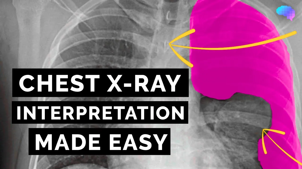





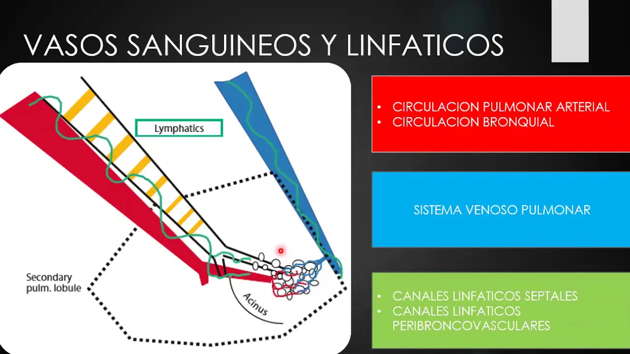


To properly interpret a chest X-ray, follow a systematic approach: first verify patient identity and technical quality (checking rotation, exposure, and breath-hold), then systematically examine the lungs from apices to bases in both upper, middle, and lower zones, assess the bronchus and carina for aspiration risks, evaluate the diaphragm and costophrenic angles for effusions, examine the heart and mediastinum including the cardiothoracic ratio, and finally review the bony thorax; always maintain clinical context when requesting or interpreting X-rays.
Chest X-Ray Interpretation for Beginners: A Systematic Guide
Added:Basic thoracic anatomy, including the structure and location of the lungs, heart, ribs, diaphragm, and mediastinum.
The fundamental physics of projectional radiography, specifically how different tissue densities (air, fluid, soft tissue, bone) appear on an X-ray film.
Understanding standard chest radiographic projections, particularly the difference between Posteroanterior (PA) and Anteroposterior (AP) views.
Basic medical terminology related to the respiratory and cardiovascular systems.
Identification of common chest pathologies, such as pneumonia, pneumothorax, pleural effusion, and congestive heart failure.
Application of structured interpretation algorithms (e.g., the ABCDE approach) to complex clinical scenarios.
Clinical correlation of radiographic findings with patient history, physical examination, and laboratory data.
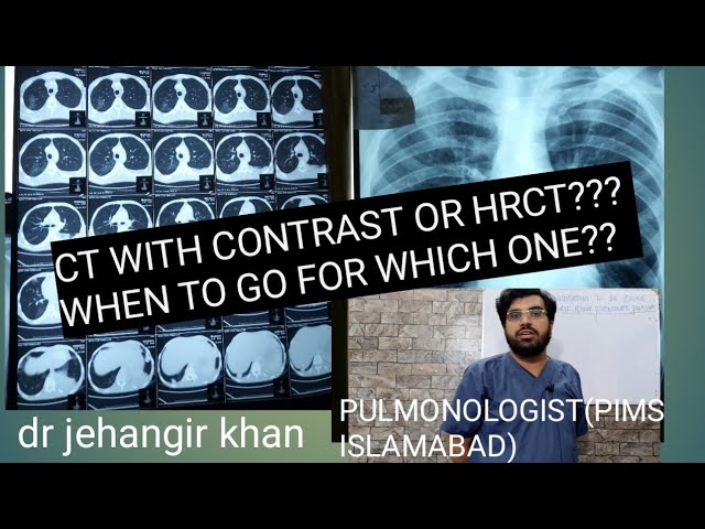
Understanding the indications for advanced thoracic imaging modalities, such as High-Resolution Computed Tomography (HRCT) of the chest.
66.1K views1.1Klikes23:20Original Release: 2022-04-10
[Music] hello and welcome back to my channel my name's dr james gill and you've joined me for something slightly to the side of clinical skills i've been joined by well a friend from ancient times hi so i knew adam before i went to medical school but we've ended up in a medical field so what does he do so i'm a radiographer more accurately a reporting radiographer now i frequently get radiologist and radiographer mixed up and i am in no way better about it which is the dark box dwellers are you so by trade i'm a radiographer so for about a decade i spent a long time taking x-rays and technically i'm a reporting radiographer so what that means is that i look at the x-rays and write down what's on them well i did that in a a yeah badly points to adam we need to have a score sheet for this one okay absolutely so we've got that bit cleared up and that kind of gives us an idea about why adam is here today so we've done plenty of work on clinical skills in terms of respiratory histories and things and respiratory examinations but today we're going to look at how to assess a chest x-ray and that's going to both is it any good in terms of having pressed the button and is there any problems on it in terms of looking at what you know the x-ray is showing us perhaps one freebie that you'll get for pressing the button okay just one um it's worth checking that i think the plan for today is an introduction to the chest x-ray and very very basic assessment of it um but hopefully if you guys like it we'll come back and look at it in a bit more detail absolutely so it's vitally important as a medical student or radiography student and junior doctors that we can assess a chest x-ray on the fly if you will now i'm relatively rusty at looking at chest x-rays whereas i was looking at them literally yesterday i will um do a radiology request and send it up say you know please assess this patient and i'll get back you know a whole epistle which basically says nothing found well exactly it's the question is what do you want to know because there's so much there that we can just sum up with no abnormality detected and actually i suppose that's a really crucial thing to highlight there it's about the asking the right question if i just send an extra request out to adam with please assess this patient what am i asking him to assess for i mean in terms of writing the forms what do you want to hit on it um what we want to know is what are the symptoms how long have they had them for and what do you think the problem is likely to be there will be always be incidental findings that will try and pick up but particularly with chest radiography there are so many appearances that can be equivocal without the clinical context that if we don't know the other side of the story we can't give you a decent answer from our end so just breathless patient query cause probably doesn't cut it it really doesn't i mean we'll do our best but in terms of providing the best answer to you and the best care that you can give to your patient the more you give us the more we can give back absolutely so i'm going to get that report but if we're on the wards and we need to be able to assess that x-ray ourselves we're going to go through that today so adam we've got a chest x-ray up here so what walk me through how we're going to assess this and how you know we're going to be able to talk about what we can see to a senior all right so first thing perhaps the most important thing is just the correct patience x-ray let's start at the very beginning you know and it's check everything first is it the right name is the right date of birth is it there right hospital number because you get patients with the same name and the same date of birth but they really shouldn't have the same identifying numbers in your own admin once we're certain that we're looking at the right x-ray have we got the right body part we're going to assume that in this case we wanted a chest x-ray once we know that we've got the right patient and the right part of the body we're off to a winning start then we need to make sure that we've got everything on that we need to see and that is presented in such a way that we can assess it one for the radiologist in the audience particularly the older ones is that there's a marker on so you know which side is the left side of the patient and you always look at an x-ray as if the patient is facing towards you this is why you must never ask a radiographer for directions because we will get left and right wrong the entire time in real life because we're used to looking at things the other way around and i think with that it's really worthwhile to highlight at this point that we know the heart is on the left but on the x-rays it's going to be on the right side of the screen and you can see that on scrubs where they got it wrong where they put the x-ray up the wrong way around thinking hold on my heart is on the left the heart was on the left on the screen there's always been a bugbear my heart my brain was wincing a little bit just saying it's on the left see the thing your heart is actually central but the left ventricle is bigger and noisier so it sounds like it's on the left and it is more of it on the left but it's really in the middle with a little bit over to the left it's not like it's just like it's down here well my apex beats all the way down here well that's something for you on a different medical professional okay so we're looking at the x-ray we've got the left side um identified because someone's put a nice helpful l up there okay so what do you want to include that's probably the best thing because you know you won't always get it all there let's start have we got everything on that we need to see um at the top of the image we want to see the apcs of the lungs at the bottom we want to see the bases of the lungs which you can see with the costophrenic angles and at the sides you want to see the lateral margins of the ribs there is an argument to be made that you want to see the skin ages but depending on the size of your patient and what you're looking for you can end up giving far more x-rays than you mean to if you wanted to include that edges and not least for we're limited by the size of the equipment that we can use there's only so much you can fit on so for this x-ray i can see edge of the clavicles i can see the top of the lungs and i can see the costophrenic angles at the bottom so i can see all of the lungs i'm happy to start trying to work out what's going on am i ready to look at it yet i've got more tonight we're getting there certainly so we've got everything on that we want to see then we need to make sure that the x-ray itself was of sufficient quality have we fired enough x-rays to get through the patient and essentially you want to be able to make out the lung markings both over the lungs and behind the heart and see some of the bony detail as well so in terms of the lung markings they were talking about those sort of beautiful sort of spider web type impressions on the lungs yes yeah they're down towards the middle and they get slightly fainter as uh they get smaller technically we want to be able to see the spinous process of t4 projected behind the mediastinum that's how you know you've got enough x-rays at it but i was worried that that was too much for episode one okay no it's fine we'll we'll work with that okay so i can see what it looks like i can see the lungs are in there i can see that um you've pressed the button hard enough oh god it doesn't work that way so we've got to go through a mental list so we've got our interest on we've given enough x-rays um is the patient standing straight up is it rotated that's uh something that i'm told you get taught at medical school so we check this by looking at the medial ends of your clavicles which are here and here and they want to be equidistant from the spinous process if it's rotated that could be a mythology if it's underexposed that could hide pathology so it's worth having a comment on the quality of the thing that you're looking at before you even think about the pathology not to go down too far in a rabbit hole but you say uh an abnormally rotated film called mimic pathology can you give me an example uh absolutely so um there's such a thing called mediastinal shift where the heart can move to one side to the other there are lots of things that could call this things like pneumothorax and effusions and collapse of the lung watch for those in episode two yes uh maybe three um and if the patient is turned you'll see the mediastinum go to the side if you do not realize that it's because the patient has turned you will see what you think is a horrible pathology patient might be normal they're just wonky okay i'll look out for my wonky patients so we've got the rotation covered we've covered the exposure and we've confirmed that we we can see what we want to see okay can i get into the medicine yet or we've still got a little bit i think we're getting there i mean we will need to consider what things can we actually see on the chest decks right but before we get to that let's talk a little bit more about how we obtained it because this isn't very blurry and the reason it isn't blurry is because the patient is holding their breath the patient has to hold their breath because you want to fully inflate the lungs and it stops them moving if you don't fully inflate the lungs they're shorter they're more compact you can get sort of artifactual compression of the lungs and it can sometimes when it collapse it will sometimes make the heart look larger than it is and measuring the width of the heart is a rule of thumb for checking for things like heart failure so in terms of that you said that that patient's going to take in a deep breath so to my mind i'm going to straight away look at the diaphragm then how am i going to determine a patient's taking a really deep breath versus someone who you know copd they've got hyperinflated lungs anyway well with when you have copd the problem is sorts itself out for you because they've got hyperinflated lungs so you don't need to worry about inflating them in terms of image quality but to make sure that they have breathing enough you count the ribs so we can look at the anterior ribs and the posterior ribs they're the same bone but there's a bit at the front and a bit at the back i was going to count these down now we can count one two three four five six seven eight and a hint of the ninth posterior rib that's how we know we've got enough so this this is the level of feedback i get from colleagues as a gp they're helping me count well you know can never underestimate people enough can you epoxy you and your epoxy radiology cupboard radiography covered and there's also and there's six anterior ribs as well uh essentially that's the mineral i think we've got slightly more than that on this x-ray so this patient is young and healthy and taken in a very nice deep breath okay so we've got a rotation we've got our exposure we know that everything's there we can see that we've got nicely inflated lungs and we've counted down the ribs right next i think we've done it for image quality now so i think we can actually start talking about whether or not there's any pathology okay let's talk about medicine yeah i have value once again what do you want to know about this patient james so for the sake of argument this is a normal chest x-ray required as part of a an insurance medical so we're not expecting to find anything but how can you show us that there's nothing on there well many are varied the short answer is the heart and mediastinal contours are within the normal limits and the lungs appear clear which is pretty much my standard form for a chest x-ray where there's nothing very interesting on it and but what we know from that is we're not worried about your heart we're not worried about your breathing and those are the things that generally get people's attention first okay so i can go along with that you've told me that the lungs seem fine and the heart seems fine but i'm in a e and you've just told me yeah these things fine how do i know that what do i have to do to be able to look at these lungs and look at the heart and say that they're normal okay um now when it comes to assessing the patient you can do it in any order you like but i would say always do it in the same order so you've learned to make sure that you've done it right i always like to start at the very top work my way down back around and back up again i would look at this once i've assessed that we've got sufficient technical quality i look at the apices of one lung move all the way down that lung start at the abcs of the other side move down that lung then look at the heart and media standard then go back into the look at the review areas so underneath the liver behind the heart back at the apices again then consider the bony thorax the important thing is that you do everything systematically and make sure that you take in everything and that you don't stop searching once you've seen something because some patients are unlucky and have more than one pathology oh dear so as you say um that that's of uh i think that's described as walking around the chest that way i've also seen it with an abcde approach so looking for airways breathing cardiac the diaphragms and then everything else so with that in mind let's walk around the chest exercise said so we'll start off looking at the apices and the lung fields walk me through this we don't say lung fields we only say fields if there's a tractor in it okay so the lungs themselves are divided into lobes there's three lobes on the right and there's two lobes on the left but when i am assessing a chest x-ray i think of them in terms of zones which are the upper middle and lower zones or you can be inferior middle and superior zones if you'd like to write longer words in your reports now with that are you separating the apices or are they considered considered in your upper zone both i would say it depends on the size of the thing that you've seen because if you've got a tiny pneumothorax that will just be in the apex but as something gets larger it takes up the middle the upper zone i would say it's roughly thirds and again the zones is not a thing that's based in anatomy it's based on the appearance of the x-ray itself the lobes themselves are an awkward mishmash of shapes that do not fit neatly into segments and they overlap each other we have a 2d representation of a 3d image a 3d structure i should say so it is easy to make a mistake of thinking something is in the wrong lobe but if you are describing it down the phone to someone who doesn't necessarily have the x-ray in front of them if you say it's in the upper zone they're going to know where to look if you say it's in the middle zone they're going to know where they look but if you say it's in the middle lobe it's a little more ambiguous okay that makes sense so we're looking over the lung zones and talk to me what i'm looking at in those zones in terms of the lung parenchyma the squidgy bit that we breathe exactly so the lungs themselves are a mishmash of vascular tissues and air and what we're looking at is the lung markings that are surrounded by uh and what we look for essentially is is there fluid where there shouldn't be fluid is there air where there shouldn't be a i think that's it like like ultimately dark gray good very dark gray bad light gray sometimes good light gray also might be bad essentially that's trite but you're looking at things other than how you expect them to be okay so for example if we were to see a space occupying lesion or something like that with a cancer again we're looking at those difference in densities as much as anything exactly with something like a lesion um it will typically be denser and it will be well defined whereas if this thing is more infective it's more likely to be more diffuse so we've looked over the uh the zones of the lungs then we need to go across to the middle for the tubes the breathy bits the bronchus what are we looking at there here was me worried i'd be too low brow so in the mediastinum at the very very top we have the bronchus attached to your mouth it's the tube through which you breathe and that comes down the center of the chest and it bifurcates a point called the carina goes off into your left and right bronchus and then into your bronchials that's something that i'm sure you can talk to me about is that you look for an aspiration because the right inferior bronchus is steeper than the rest so it's the place where something if something's fallen down there that's where it's gonna fall absolutely and if i've got a child that's come to the a e department with an acute onset shortness of breath sudden onset or an acute onset cough particularly that's hap whilst they're playing i need to have a look in the bronchus particularly so on the right side to see if they've breathed you know in a grape or something like that that's then lodged in there causing that sudden onset pathology okay so we've covered the lungs we've covered the bronchus i think that pretty much covers most of the airway and breathing other than the very bases uh with the costophrenic angles so what do we look at down there the crosstalking angles are just the very very edges of your lungs they should be nice and well defined they can become blunted which is often the manifestation of either thickening of the pleura which again anatomy will get to one another time or something called a plural effusion which is a buildup of fluid so uh we're now needing to have a look at the heart so centrally as you say yes so your heart consists of four chambers uh left and right atrium and left and right ventricles as i said before the ventricle is the noisy one and that pumps the blood through the rest of your body and the uh sorry the left ventricle pumps it through the rest of your body whereas the right ventricle pumps it into the lungs um your heart again it's in the center it's in a region that we'll call the mediastinum uh which contains all of the vascular structures just above the heart we can see a little lumpy bumpy bit sticking out um which is the arch of the aorta that goes up from the heart arches around and then goes back down and then through the diaphragm i remember at medical school people talking about seeing the aortic root unfolding on x-rays and things that's always been something i struggled to see um well you won't see it on this one because it's not um but unfolding is something that's as i understand things normal progression it is just part of the aging process um it's nice and straight when you're younger and as you age it can become a little more wobbly essentially fair enough fair enough so you mentioned the hyla regions there what's going on there because they automatic they look when you know first year medical school looking at chest x-rays i always was worried that there was something going on in the hilah because it looked different from the rest of the lung field well it's worth being worried about because assessing these bits is hard um they are complicated structures made up of lots of vascular structures that it's essentially where the blood is throwing from your heart throughout the lungs um ultimately they are at about broadly the same level the right one typically sits higher than the left and they should be of a uniform density and a similar shape one of the difficult things about looking at chest x-rays compared to looking at other parts of the body is their appearances are many and varied and what's normal for that patient it is unfortunately something you have to get your eye in on so it's like looking at patient uh back on patients ecg's can we review has there been any change from not their normal exactly yes so the heartland's sitting just over the diaphragm what comments can we make on that so the width of the heart typically should be no more than half of the width of the chest itself there's been some recent research about how good a cardiothoracic ratio is as a measurement tool for pathology but ultimately the rule of thumb is it shouldn't be any bigger than that okay so to my mind that's covered most of the things that we need to look at in terms of the actual chest pathology from a shorter breath patient what else do we want to look at what's the you know the summarizing things in the every part once we've looked at our heart and lungs um there's a lot more on that than just there there's a whole lot of bones um i mean when i started radiography i was shocked that we did more chest x-rays than anything else i thought it was all bony stuff um but it's worth having a look have they got any fractures have they got any lesions you can often pick up some degenerative changes on there it may not be relevant to what your patient has arrived for but there's a wealth of knowledge that that you can assess for okay so i think that's a an excellent walk around in terms of the chest x-ray we've got so just to recap that overview of the chest x-ray we've confirmed the patient's date of birth we've gone over the technical aspects and we're happy with the film we've looked over the airways in terms of looking at the bronchus and the karina we've looked over the parenchymer of the lungs looking over the zones not fields we've looked at the diaphragm we've talked about the costophrenic angles at the base of the diaphragm as well we've looked at the heart and we've determined that everything else is normal on that chest x-ray so i'd be happy with a student presenting that to myself from a radiology perspective what would you want one of your students to add in with that um so for mine i would look at the technical quality first certainly if it's a student radiographer it's their job to make sure that they've got a good image and we go i think through a thing called a 10 point check which we don't need all 10 points of today but ultimately is it of sufficient quality have you got everything on that you need to see is there anything else that you need to do okay well i think that's a reasonable point for us to end this overview of the chest x-ray um this is going to be an evolving series and we've got ideas about what we want to include covering pathology and things such as that so there's the medical bent to it but what do you guys want us to cover as well whether you're medical students whether or not you're radiologists radiography students or both sides i'm covering both sides so you know please drop a comment down below and we'll see if we can service those needs yeah say or are you any formal student or are you just interested you know yeah i'm quite happy to teach it to anybody um i had a point then it's flattered away um oh yes um there's a lot more to radiography than chest x-rays um we would like or i would like to discuss about all of them if times are missed so if you want to hear about those uh let us know below hey i certainly did from my perspective i'm distinctly looking forward to discussions about hip x-rays um because we see so many patients with hip pathology and hip problems it's like whereabouts we're drawing the lines and things like that so that should be very useful we can do that right well thank you very much if you consider if this video has been useful to you please hit the like button because that's how it tells youtube we're here and if you want notifications for when we're doing the next one please hit the subscribe button and we'll see you in the next one take care thank you [Music] [Music]
Up Next

127:29
Radiology Image Interpretation: Magic View Graphics for NEET-PG 2025
@marrowmed
53.6K views•2025-06-09

48:47
Graphic Medicine: Comics for Collaborative Healthcare Communication
@nationalpatientadvocate
189 views•2023-12-04

10:11
Cranial Nerve Examination | Clinical Skills OSCE Guide | Dr Gill
@DrJamesGill
5.7M views•2020-01-26

3:05
Stages of Labor and Vaginal Birth | Childbirth Animation
@nucleusmedicalmedia
52.1M views•2017-08-18
Related Study Plans & Knowledge Roadmaps
Structured learning paths in Medicine
medicine
Abnormal LFTs: Diagnostic Workup & Pathology
01Liver Anatomy and Basic Physiology
02Introduction to Liver Function Tests (LFTs)
03Hepatocellular vs. Cholestatic Patterns
+ 3 more learning modules
12h6 modules
Explore Roadmap
medicine
Analyzing Blood Smears: RBC, WBC & Platelets
01Introduction to Blood Components & Smear Preparation
02Normal Morphology of Blood Cells
03Erythrocyte Pathologies & Morphological Variants
+ 2 more learning modules
12h5 modules
Explore Roadmap
medicine
Mechanical Ventilation: Waveforms & Alarms
01Anatomy and Physiology of Breathing
02Introduction to Mechanical Ventilation & Core Modes
03Ventilator Settings and Waveform Interpretation
+ 2 more learning modules
12h5 modules
Explore Roadmap
medicine
Neuro Exam: Localization & Stroke Syndromes
01Foundations of Neuroanatomy and Vascular Supply
02The Neurological Examination: Step-by-Step
03Principles of Neurological Localization
+ 3 more learning modules
18h6 modules
Explore Roadmap