



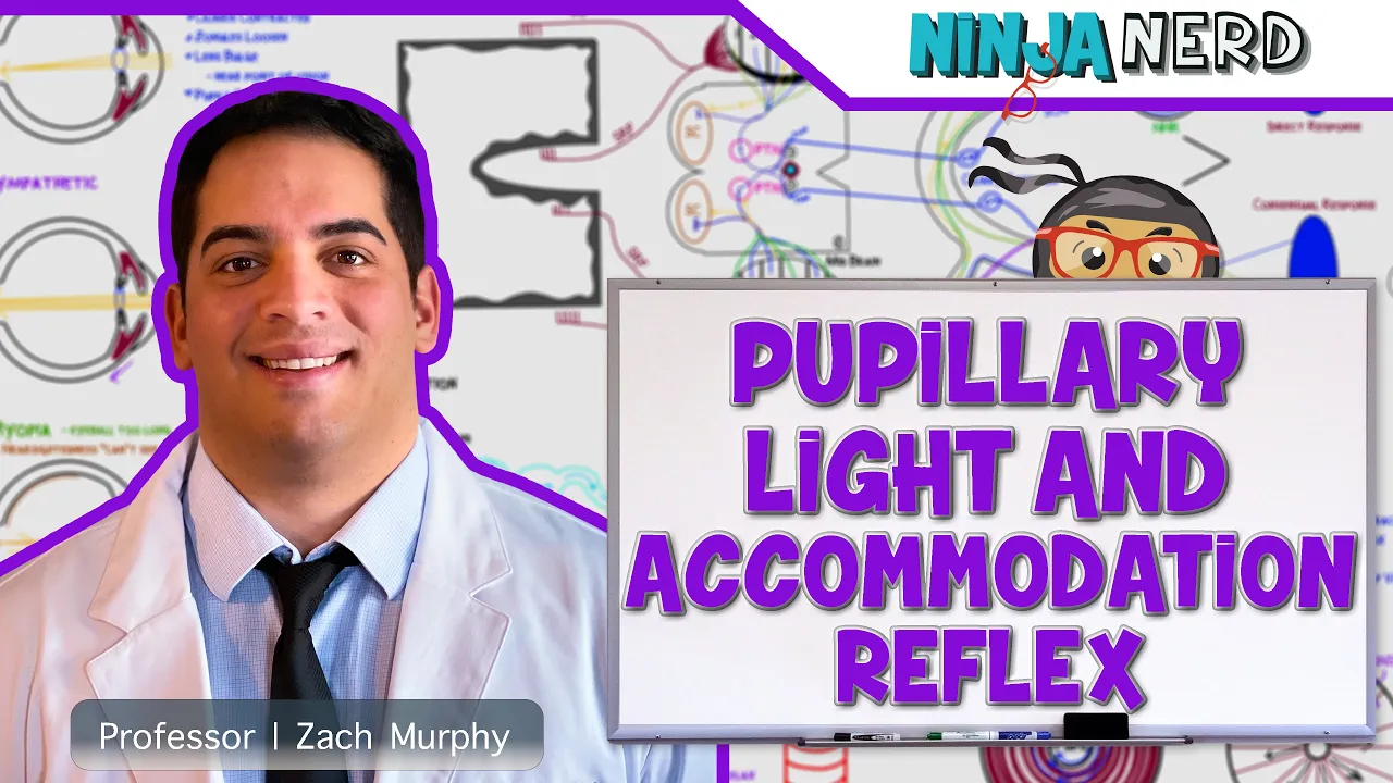



![[ASMR] Fastest Nurse Office Cranial Nerve Exam (Eyes, Ears, Hearing, Personal Attention)](https://i.ytimg.com/vi/mF25WlSDX5c/maxresdefault.jpg)


![Kranial sinirler (Nervus hypoglossus) - Bölüm 12 [Teorik]](https://i.ytimg.com/vi/B3jxmM-My-Q/maxresdefault.jpg)






![[ASMR] 30-Minute Realistic Head to Toe Medical Exam Roleplay 😴😴](https://i.ytimg.com/vi/9ELm0WFqAjA/maxresdefault.jpg)


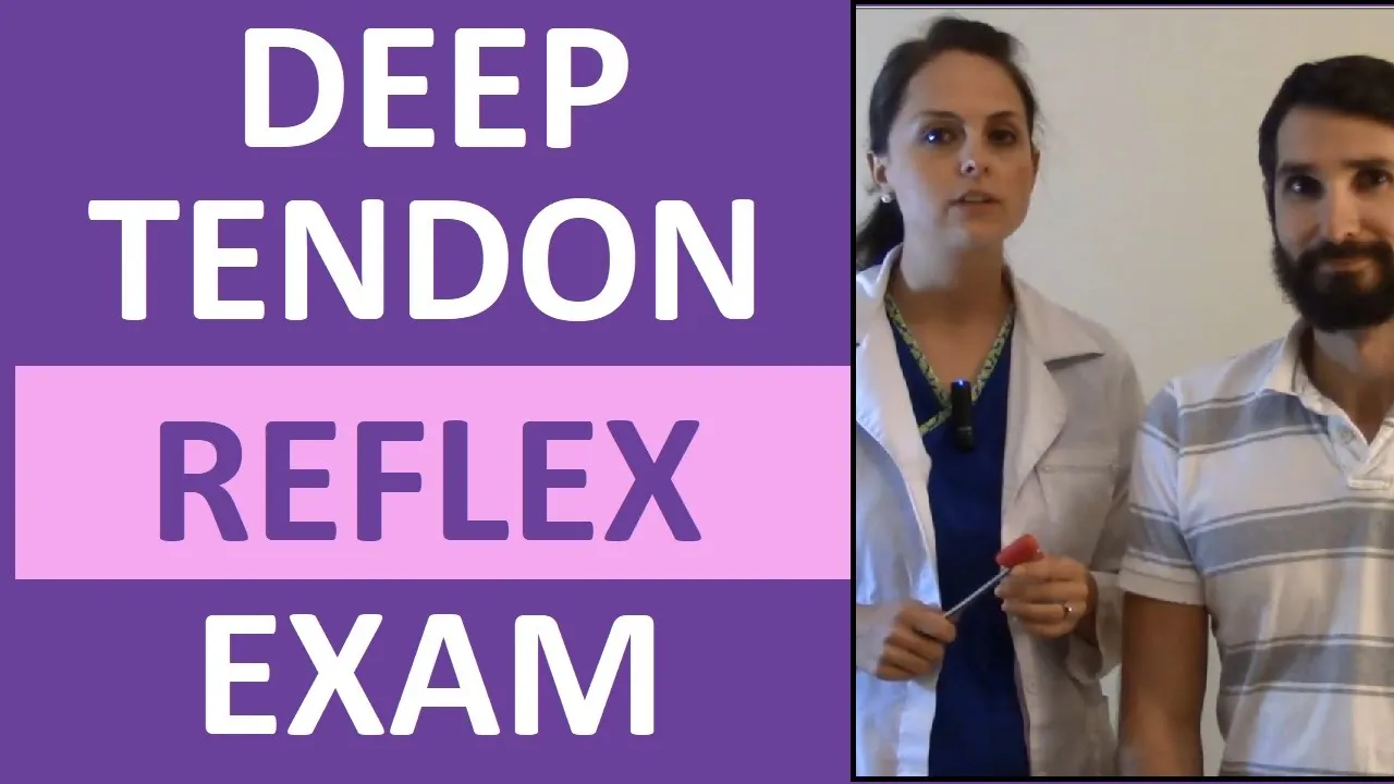
A comprehensive cranial nerve examination involves systematically testing twelve cranial nerves through various assessments including olfactory function (smell), visual acuity, peripheral vision, color vision, blind spot detection, facial sensation, facial muscle strength, hearing, and oral motor function, with each nerve evaluated using specific clinical tests such as the Snellen chart for vision, tuning forks for hearing, and cotton wool for sensation.
Cranial Nerve Examination | Clinical Skills OSCE Guide | Dr Gill
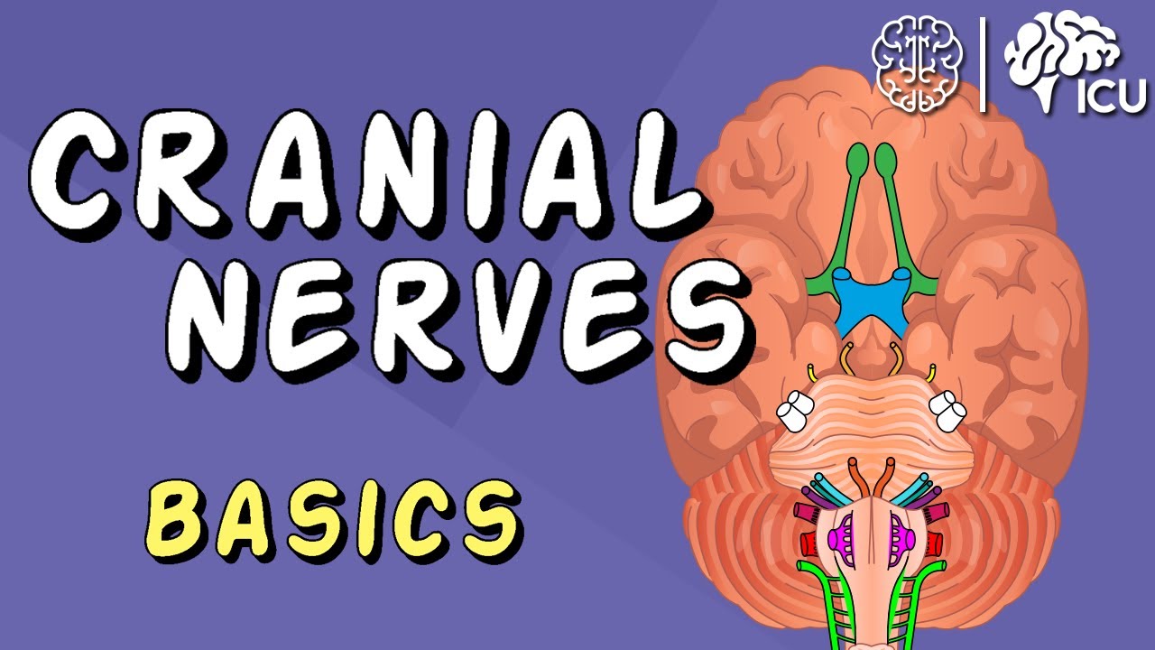
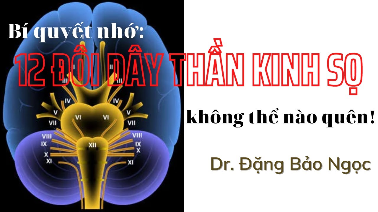
Added:Basic neuroanatomy of the 12 cranial nerves, including their names, numbers (I-XII), and whether they carry sensory, motor, or mixed fibers.
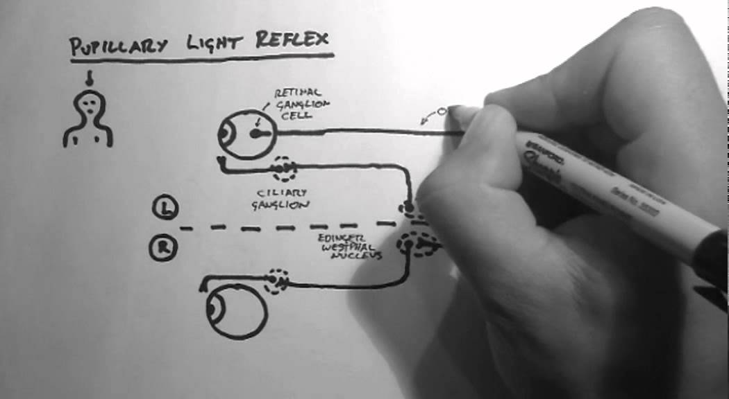
The physiological basis of key cranial nerve reflexes, such as the pupillary light reflex, corneal reflex, and gag reflex.
Familiarity with standard clinical examination tools, such as the ophthalmoscope, tuning fork (256Hz/512Hz), penlight, and Snellen chart.
Fundamental OSCE communication skills, including introducing oneself, confirming patient identity, and obtaining informed consent.
Interpretation of abnormal findings, such as recognizing pupillary defects, facial nerve palsy (upper vs. lower motor neuron lesions), and tongue deviation.
Clinical localization of neurological lesions within the brainstem (midbrain, pons, and medulla) based on patterns of cranial nerve deficits.
Integration of cranial nerve exam findings into clinical diagnostic frameworks for conditions like stroke, multiple sclerosis, Bell's palsy, and intracranial hypertension.
Mastering the complete head-to-toe neurological assessment, incorporating motor, sensory, cerebellar, and deep tendon reflex examinations.
5.7M views36.7Klikes10:11Original Release: 2020-01-26
[Music] my name's dr. Gill I've been asked to do an examination of your cranial nerves so the nerves that control your face and head I'll be okay so to start off could you confirm to me your name and date of birth please perfect so to start off we'll look at your sense of smell have you no send it changes with your nose at all okay so no blocked nose and eyes so if you could put a finger over one nostril and breathe in and relax and we'll do the same again on the opposite side super so I've got a substance here if you close your eyes and tell me what you can smell if anything coffee super okay that confirms everything's working there thank you so now we need to actually see how well you are your vision is so do you wear glasses normally yes okay do you have them with you okay and if you had any changes to prescription recently okay so if you look at my face is my guess I need black splotches my missing an eye perfect so what I'd like to do to start off with behind me is a snow on chart so could you cover your right eye for me okay and what's the lowest line you can read on there could you start from the left please perfect just swap eyes if you put your hand over your right eye what's the lowest line you could read get and stop from the right side of the paper perfect so you've got no problems there now I'm going to shine a light on your right okay so if you put a hand over your left eye forward please and then over the right eye okay and relax back to normal that's fine thank you so there no problems at all with that just walk over the same thing that's fine I'm just I'm the next thing if you'd look at my finger I know that was sealing behind me no matter the finger superb now in order to really get a good idea of how your eyes are working I actually need to have a look at the back would that be okay yeah so I'm going to shine the light directly in your eye but I'm gonna have to come quite close to do that take it off get elapsed ten I can focus it okay so if you just keep looking straight ahead for me okay if you could look up please and if you look down good left and look the right not directly into the light okay thank you I will just do a view up to society are you okay okay go into the same again okay look out for me and left and right I'm not great lead for the line please okay so now we're going to see how well your eyes work on the very side so we're going to look at your peripheral vision okay so I'm just gonna make sure on the right distance away so you can see my finger waggling yep tell one you can see my finger way yeah yep yep yep yep yeah so if it's all pies for me okay and you can see my finger way yep please stay looking in this eye I'm telling you see my finger yep yep yep yep yeah okay so right now I need to look over how your eyes will move so if you keep it head still for me and I want you to follow my finger tell me if there's any point any problems of that excellent so to finish off now I'm going to put Begich you prefer hand over one eye for me okay and I'm going to show you a pen top can you tell me what color that is right okay and if you could swap hands and this okay are they the same red on both sides okay so it doesn't seem to become paler a one eye compared to the other see that yeah now we're going to have a look for something called your blind spot so if you can put a hand over one eye for me okay and I want you to only look into this eye here and at some point as I move the pen across the pen will disappear or the pen top lon okay so if it's gone now I'm gonna carry on moving it across and tell when it reappears okay I'm here to get backwards says it okay now telling it reappears okay so I'm okay I'm gonna go up there okay and then down done super okay and that's one count back excellent so we swap over to the other riflemen again looking at why I hear it's a ton one it disappears done okay tell mine it reappears I just I want it displayed okay fine I'm not going up accent so your blind spot lines up well with mine there but thank you I appreciate that's a slightly difficult test today and now we're going to look at your sense of sensation on your face so I've got a little point here but it is blunt and if I could have a finger okay so you can see you can feel it but it isn't sharp okay if you close your eyes and tell mine you feel it yep yep yep okay that's terrific okay and now we're going to see what's the sensation on your eyes alike again so I've got a bit of cotton wool and if you could look all the way to the outside for me and I'm just going to bring this in from the side I'm just gonna touch the side of your eye that's fine we get to the same if you look over that way you seem to come in from the side okay super thank you very much now I'd like to tap on your forehead just relax on me okay so we've got a nice blink man that's good and finally if you could try and relax your jaw for me I'm just kind of tap over your jaw okay now we're gonna have a quick look but the muscles that control your face so if you could raise your eyebrows up as high as you can please zipper and screw your eyes tight don't let me open that's good future me teeth okay and blue cheeks out for me super now we're gonna have a look over your hearing have you noticed any problems with the area yeah good so we're going to start off if you could shrub the front of your ear please and I'm going to say word and I'd like you to repeat it better please 100 okay if we could stop over for me 99 excellent so we know that everything seems to be working well thanks thing we're going to do I'm going to get this tuning fork I'm going to strike it and I'll put it in your forehead okay now we're just going to do the same again so I'm just going to put this on your forehead and tell me if you can hit where you can hear the noise is it left right or middle okay super so we need to do an additional test on that which will involve putting this on the bone behind your ear when the sound stops please tell me and I got to move it round and I want to see if you can hear it then I'm gonna need to come around the back and let's do that and tell me when you can't hear the noise anymore okay and can you hear it now yeah excellent did the same on the side okay you can hear the noise okay tell one you can't you hear that yeah excellent now we need to have a look in your mouth like to take a sip of water and just so I please any problems that I home do you have any problems whether eating or drinking okay so no coughing afterwards actually so what we'd like to do now is I'm just going to have a look in the back of your mouths if you could open why please and nice long R please ah sip ever okay so I'm just going to test the powers now of the muscles in your head and neck so I had to put my hands on the side of your face and if you could push sip and we'll do the same any other side push push excellent now I'm gonna put my arms on your shoulders and push up for me okay that's excellent now the final bit you'll be able to stick your tongue up for me please okay and there's no problems there and put the tongue to the side of your mouth they have to push back I'm done the other side please I've never been having a reasonable conversation throughout but if you could just finish off by saying eh e io Anu please a excellent so if had no problems with your crane enough so it can give you a clean bill of health on that do you have any questions for me yeah super thank you for your time [Music] you
Up Next
12:40
Cranial Nerves Anatomy: 12 Pairs and Functions Explained
@Anatomyzone
1M views•2012-08-07

48:47
Graphic Medicine: Comics for Collaborative Healthcare Communication
@nationalpatientadvocate
189 views•2023-12-04

23:20
Chest X-Ray Interpretation for Beginners: A Systematic Guide
@DrJamesGill
66.1K views•2022-04-10

3:05
Stages of Labor and Vaginal Birth | Childbirth Animation
@nucleusmedicalmedia
52.1M views•2017-08-18
Related Study Plans & Knowledge Roadmaps
Structured learning paths in Medicine
medicine
Abnormal LFTs: Diagnostic Workup & Pathology
01Liver Anatomy and Basic Physiology
02Introduction to Liver Function Tests (LFTs)
03Hepatocellular vs. Cholestatic Patterns
+ 3 more learning modules
12h6 modules
Explore Roadmap
medicine
Analyzing Blood Smears: RBC, WBC & Platelets
01Introduction to Blood Components & Smear Preparation
02Normal Morphology of Blood Cells
03Erythrocyte Pathologies & Morphological Variants
+ 2 more learning modules
12h5 modules
Explore Roadmap
medicine
Mechanical Ventilation: Waveforms & Alarms
01Anatomy and Physiology of Breathing
02Introduction to Mechanical Ventilation & Core Modes
03Ventilator Settings and Waveform Interpretation
+ 2 more learning modules
12h5 modules
Explore Roadmap
medicine
Neuro Exam: Localization & Stroke Syndromes
01Foundations of Neuroanatomy and Vascular Supply
02The Neurological Examination: Step-by-Step
03Principles of Neurological Localization
+ 3 more learning modules
18h6 modules
Explore Roadmap