









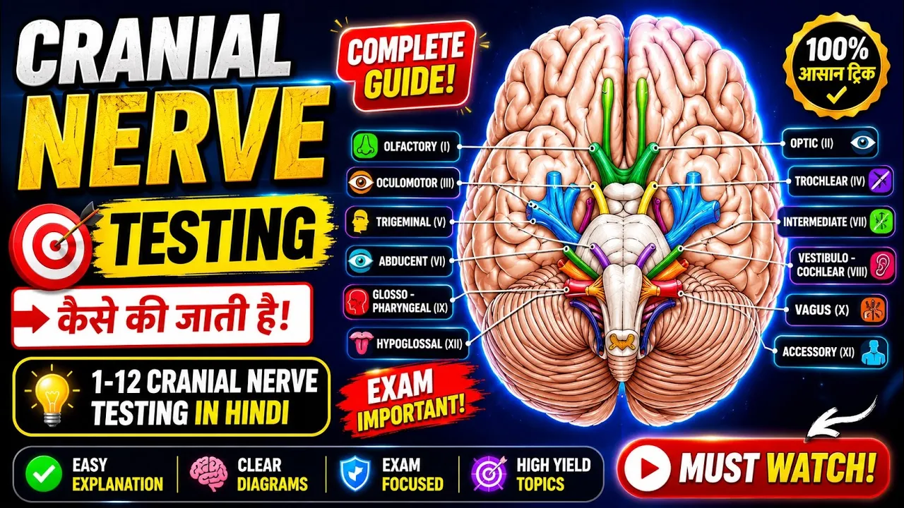








The 12 cranial nerves are nerves that emerge directly from the brain and brainstem, serving various sensory and motor functions including special senses (smell, vision, hearing, taste) and controlling muscles of the head, neck, and viscera; they originate from different brain regions (telencephalon/diencephalon for CN I-II, brainstem for CN III-XII) and can be remembered using mnemonics such as 'Oh Oh Oh to Touch and Feel Very Good Velvet Are Heaven' for their names and 'Sensory Motor Both' for their functional categories.
Cranial Nerves Anatomy: 12 Pairs and Functions Explained
Added:Basic division of the nervous system, specifically differentiating between the Central Nervous System (CNS) and Peripheral Nervous System (PNS).
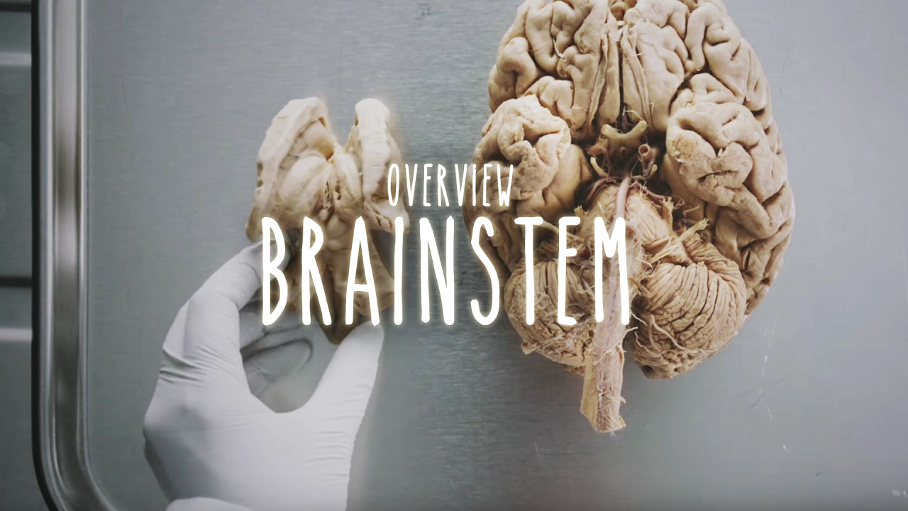
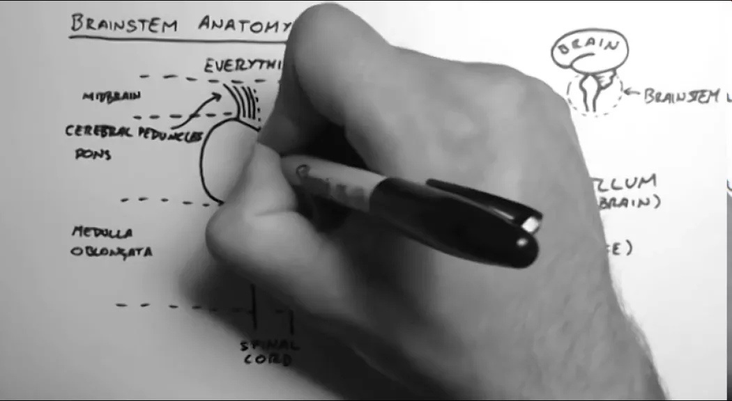
Gross anatomy of the brainstem, including the identification and major subdivisions: midbrain, pons, and medulla oblongata.
Fundamental neurological concepts of sensory (afferent), motor (efferent), and mixed nerve pathways.
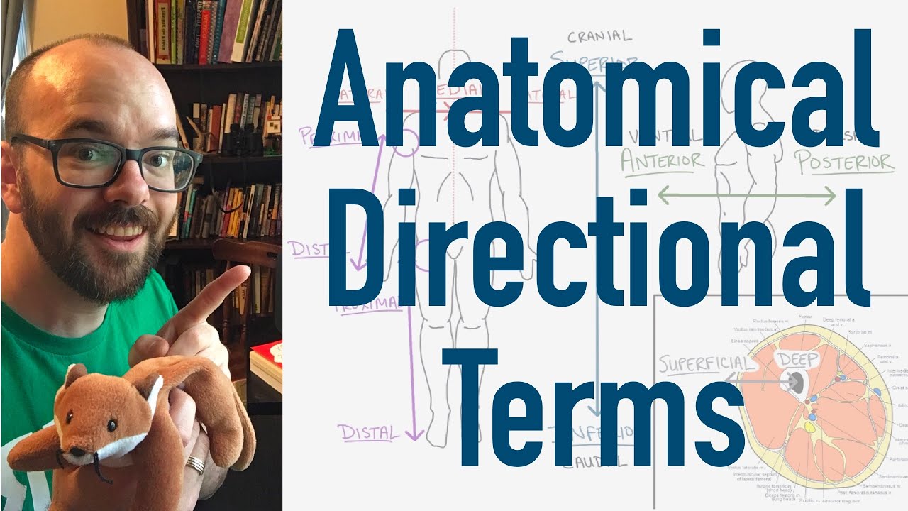
Standard anatomical directional terminology (such as anterior, posterior, lateral, medial, rostral, and caudal) to comprehend 3D spatial relationships.
Clinical methods and diagnostic tests used to assess cranial nerve function during a standard neurological examination.
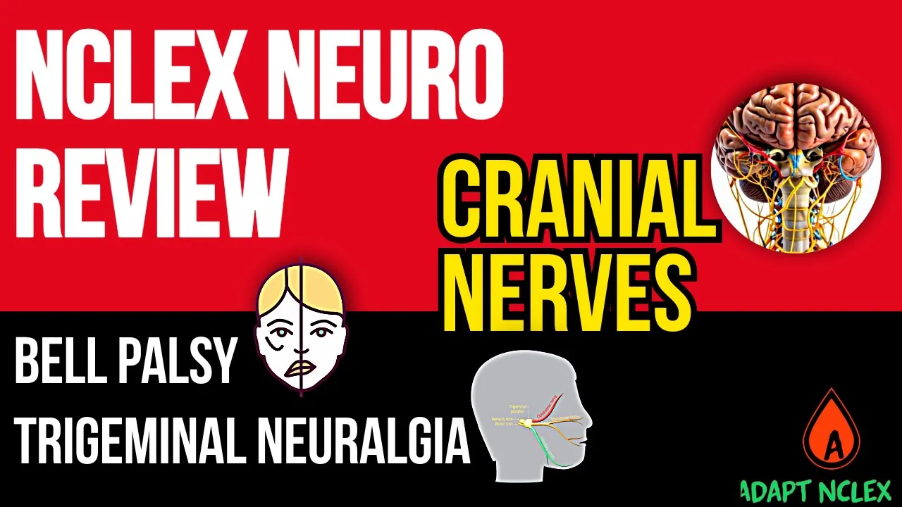
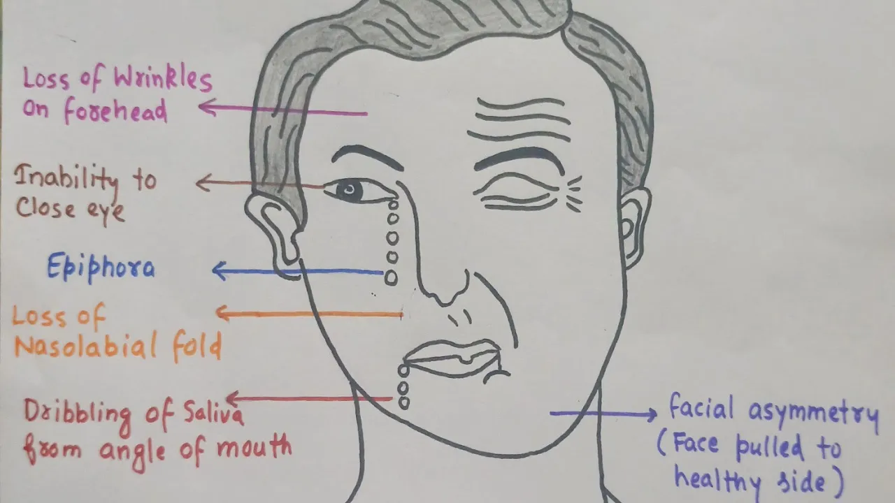
Pathological conditions and clinical correlates resulting from specific cranial nerve damage, such as Bell's palsy, trigeminal neuralgia, and ptosis.
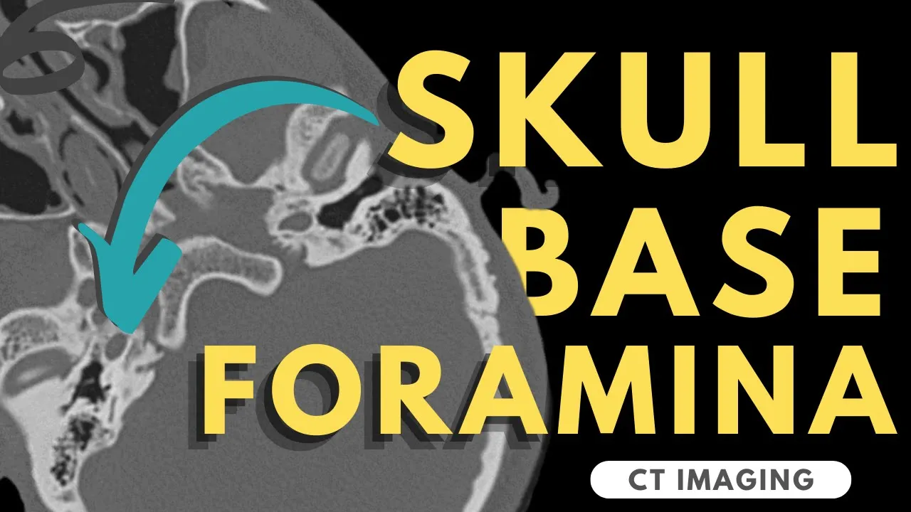
The specific foramina and fissures of the skull base through which each of the 12 cranial nerves exit the cranium.
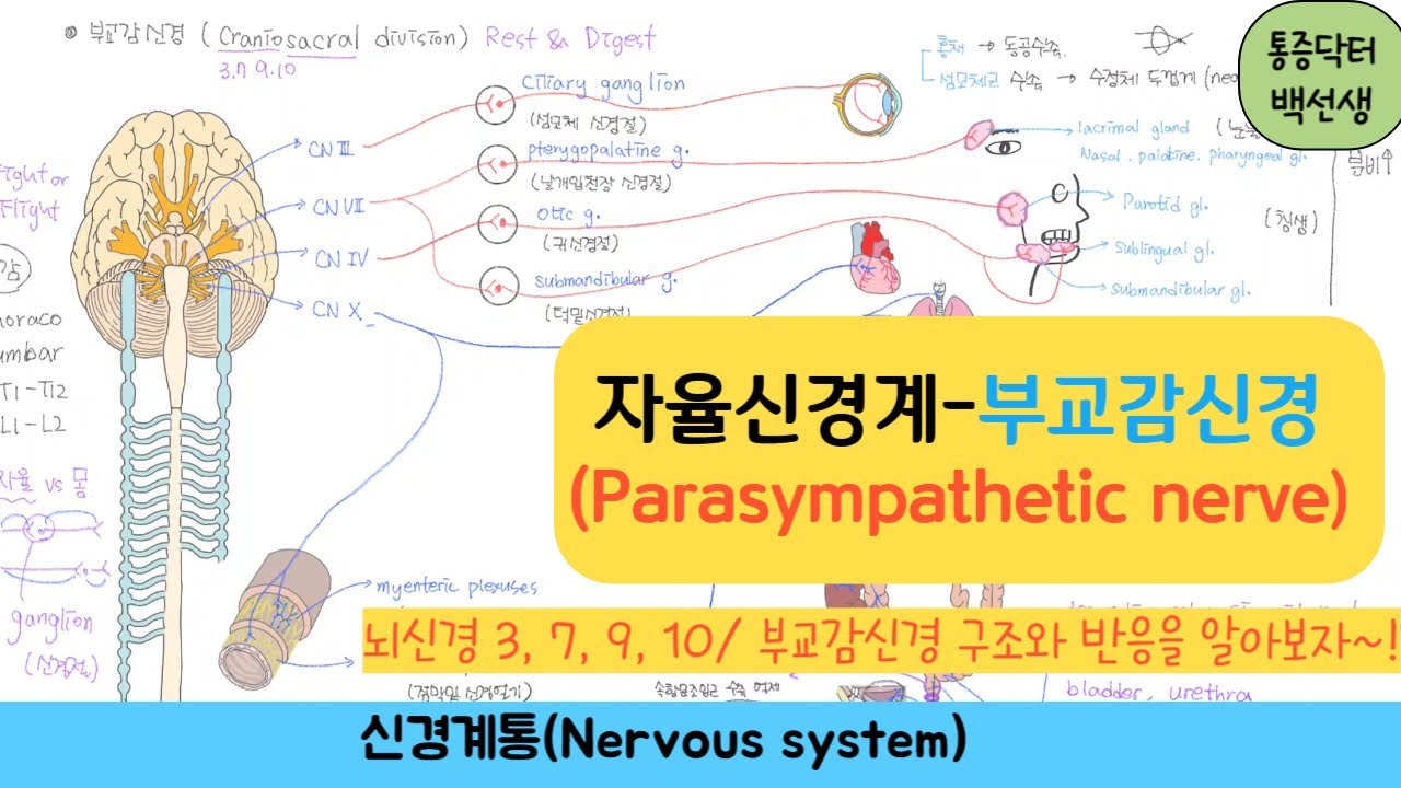
The autonomic nervous system pathways associated with cranial nerves III, VII, IX, and X, and their role in parasympathetic regulation.
1M views9.7Klikes12:40Original Release: 2012-08-07
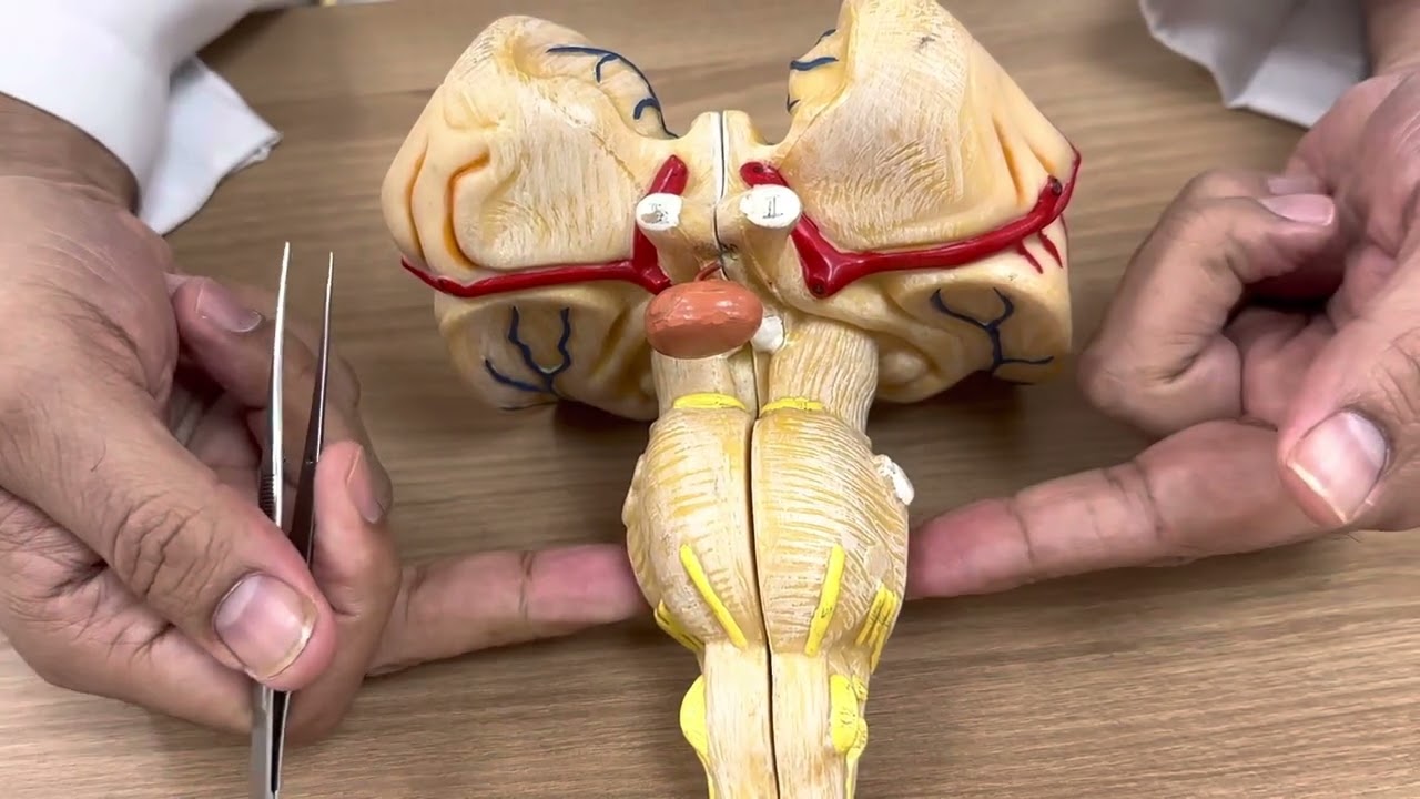
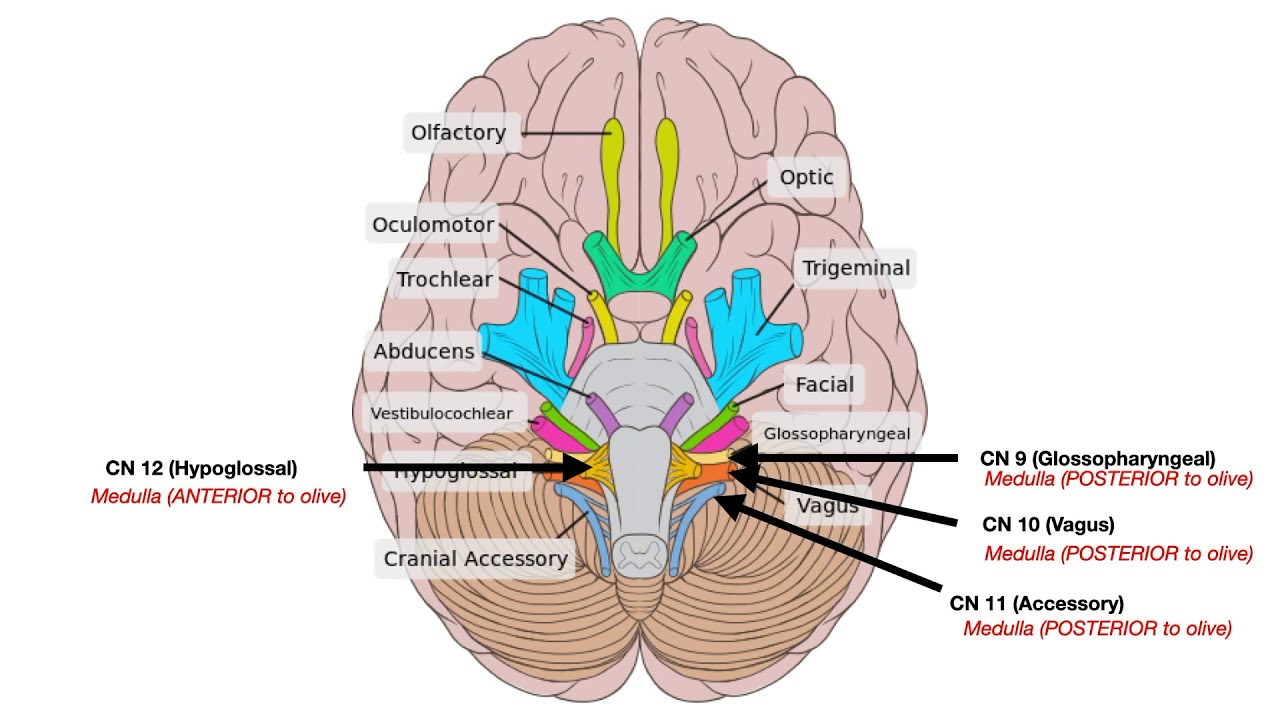
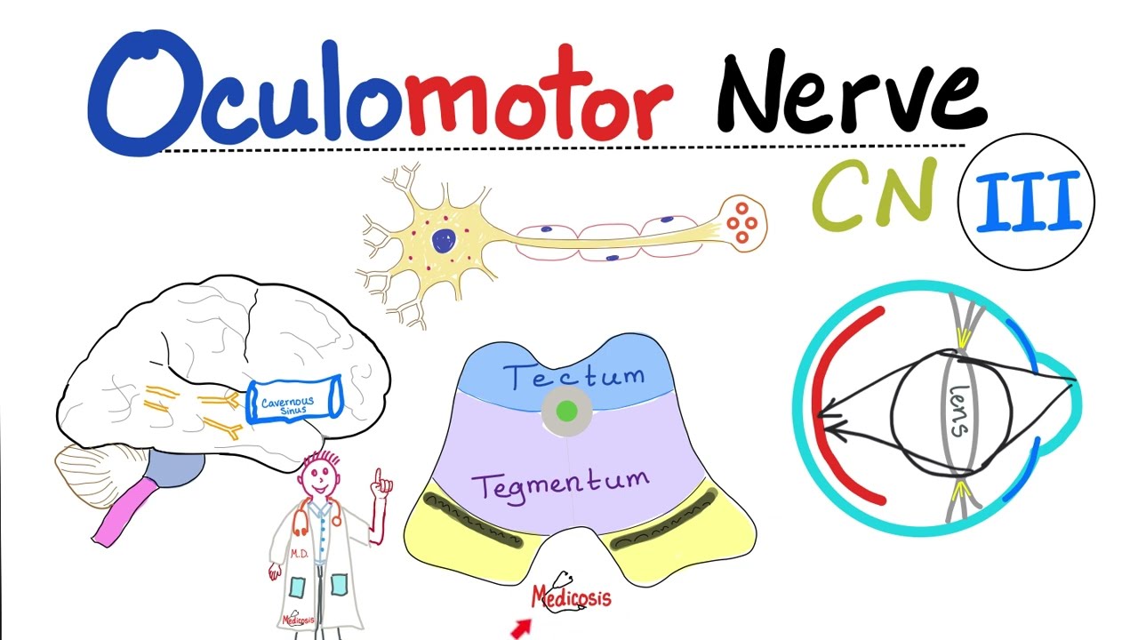
okay so this is a tutorial on the cranial nerves so cranial nerves the nerves that emerge directly from the brain and you've got 12 pairs of cranial nerves and they were part of the peripheral nervous system so all 12 pairs of part of the peripheral nervous system except cranial nerve number 2 which actually emerges which is kind of like an extension for of the brain so it's not technically a peripheral nerve so in this tutorial we're going to go through the 12 cranial nerves we're going to look at where they originate from in the brain and we'll talk a little bit about their function so the cranial nerves have lots of different functions you've got somatic and visceral components they've got motor components and there's some nerves which have special sensory component so components to do with the special senses like smell vision hearing bands and taste so if I just rotate the model of the brain round you can see so we're looking at the base of the brain and you can see all these nerves emerging from the brain so the first two nerves actually emerge from the telencephalon and dine kefflin but the rest of the nerves nerves 3 to 12 emerge from the brainstem so that's the midbrain the pons and the medulla so the nerves are sort of numbered from 1 to 12 from kind of top to bottom if you like so I'm just going to run through a list of the nerves so we've got the olfactory nerve number 1 optic oculomotor trochlear trigeminal Abdo --scent facial vestibular cochlea glosso pharyngeal Vegas accessory and hypoglossal nerves so they've got kind of strange names but there is a mnemonic for remembering these so I'll give you the clean version which is oh-oh-oh to touch and feel very good velvet are heaven so the dirty version is what probably most people are taught and it involves touching and feeling other things so you can use your imagination there so there's also a mnemonic for remembering the function of the cranial nerves so this is some say Mary money but my brother says big boobs matter more so s is sensory M motor B is both so central and motor so keep these mnemonics in mind when we go through the nerves so we'll start with cranial nerve number one which is the olfactory nerve and this this nerve is responsible for their m4 olfaction so the sense of smell so there are receptors in the nasal cavity and which pick up the sense of smell and the nerves pass through the nasal cavity up through the cribriform plate of the ethmoid bone and synapse on this the olfactory bulb so you can see this projection along the base of the brain so this is called the olfactory bulb and the olfactory nerves synapse onto this olfactory bulb so I've just switched over to this model and you can see these little nerves coming up to sign apps onto the olfactory bulb so if we just take a quick look at the cribriform plate we're looking at a and sides of the skull here so we're going to rotate the skull around and look at the inter of the skull so we're looking from a superior view at the inside of the skull so this is the anterior part and the cribriform plate lies here so it's not shown clearly here but the olfactory bulb runs along the cribriform plate and there are little holes in the cribriform plate called foramina where the olfactory nerves pass through so if I just rotate the model around you can see these see these olfactory nerves passing up through the Fermina to sign up something factory bulb so next we've got the optic nerve and you can see these two nerves on either side these big nerves coming up and you can see they cross at this area called the optic chiasm so these nerves the optic nerve nerve number two is responsible for vision so it carries sensory information from the retina to the brain so the third nerve is the oculomotor nerve so now we're coming this is the first nerve that originates from the brainstem so you can see its origin here in the interpeduncular fossa so you can see these two sort of pillars these are called cerebral peduncles and these are part of the midbrain so the oculomotor nerve originates anteriorly and on the midbrain in the floor of the interpeduncular fossa so you can see the oculomotor nerve originating here and this this nerve is responsible for innovating muscles that move the eye so extra ocular muscles and it also innovates the pupillary sphincter so now if number four is called the trochlear nerve so if we just zoom in a little bit to the brainstem I'll just remove the cerebellum and I'll remove the cerebral hemispheres so it just makes things a little bit more clear so we've just seen the oculomotor nerve here originating any interpeduncular fossa and if we follow the if we rotate the model around to look at the dorsal surface of the midbrain we can see the trochlear nerves originating so the trochlear nerve there's another nerve which is involved with movements of the eye so only innovates one eye muscle called the superior oblique muscle so it originates on the dorsal midbrain and it winds around the sides of the cerebral peduncles so the lower margin of the cerebral peduncle so next we've got the trigeminal nerve so this so you can see this big fat nerve coming off the side of the pons on the middle cerebellar peduncle so this is the trigeminal nerve so you can see it here overtaking laterally on the pons on the middle cerebellar peduncle so the cerebellar peduncle is a bit which connects the pons to the cerebellum so I'll just rotate around so you can see that a bit more clearly so you can see the big trigeminal nerve coming off the side of the pons and so this nerve is responsible for sensation from the face and and if you just follow the nerve along you can see this big bold so this is the trigeminal ganglion so this is a group of cell bodies of the sensory neurons and then it splits into three branches so if I just zoom in a bit you can see these three nerves coming off so you've got three branches of the trigeminal if you've got the ophthalmic maxillary and mandibular nerves so that's just worth remembering so as well as with providing sensation from the face the trigeminal nerve is responsible for the innovating the muscles of mastication if we just rotate the model around again so we're looking anteriorly at the brainstem just follow they're full of enzyme down we can see this nerve here originating at the border between the pons and the medulla so this nerve is called the abducens nerve and this is another muscle so nerve involved in innovating the extraocular muscles of the eye so innovates one muscle of the eye called the lateral rectus muscle so we've now talked about three muscles sorry three nerves responsible for innovating extraocular oculomotor nerve the trochlear nerve and now the abducens nerve so this originates at the border between the pons and medulla so if we just rotate the model again looking laterally if we follow follow the begging naturally we've got two nerves here and this part of the brainstem is called the cerebellopontine angle so just above the olive of the medulla you've got this nerve hair called the facial nerve so this originates and the cerebellopontine angle above the olive and it's responsible for muscles of facial expression and it receives taste from the anterior 2/3 of the tongue and it also has secreted motor function to the saliva glands and the lacrimal gland so just lateral to this now we've got the vestibular ocular nerve which also originates in this cerebellopontine angle and this nervous responsible for hearing and balance so it has two branches the vestibular part is responsible for balance and the cochlear part is responsible for hearing and then just below it we've got this nerve here which is called the glossopharyngeal nerve and this M originates just dorsal to the olive so you can see its position here just behind the olive and on the lateral margin of the medulla so this nerve is responsible for taste from the posterior third of the tongue and so remember the the facial nerve was responsible for taste from the anterior two-thirds the glossopharyngeal nerve is responsible for taste from the posterior third so it's responsible taste and it's got this secreto motor function to the parotid gland and it was also responsible for innovating muscles in the throat and carrying sensation and from various holes in the throat so that's nerve number 9 the glossopharyngeal nerve and now if number 10 originates just below it and this is the vagus nerve so this originates in the posterolateral sulcus of the medulla so just behind the olive and just below the just below where the glossy found jewel nerve originates so the vagus nerve innervates lots of laryngeal and pharyngeal muscle and it innovates a almost all of the abdominal and thoracic viscera so next we've got the accessory nerve so this is nerve number 11 and this nerve has cranial and spinal roots so it's got so this is this node here it isn't shown sort of it's not best illustrated by this model but you can see it's sort of it's got a lower origin and an upper origin so it's got a cranial and a spinal root so this muscle is responsible for innovating the sternocleidomastoid and the trapezius muscles and finally we've got the twelfth nerve so this is the hypoglossal nerve so you can see it here so this sits between the pyramids of the medulla so these are the medullary pyramids and it sits between these pyramids and the olives so you can see that here the hypoglossal nerve and this nerve is responsible for innovating get the muscles of the tongue so those are the the 12 cranial nerves you've got mnemonics remembering them and you've seen sort of where they originated on the brainstem in the and the thing
Up Next

14:01
Mini-Mental State Examination (MMSE): Clinical Guide
@DrMK7
7.9K views•2023-08-16

48:47
Graphic Medicine: Comics for Collaborative Healthcare Communication
@nationalpatientadvocate
189 views•2023-12-04

10:28
Neuroanatomy: Central and Peripheral Nervous System Divisions Explained
@AKLECTURES
136.2K views•2014-09-20

3:05
Stages of Labor and Vaginal Birth | Childbirth Animation
@nucleusmedicalmedia
52.1M views•2017-08-18
Related Study Plans & Knowledge Roadmaps
Structured learning paths in Medicine
medicine
Abnormal LFTs: Diagnostic Workup & Pathology
01Liver Anatomy and Basic Physiology
02Introduction to Liver Function Tests (LFTs)
03Hepatocellular vs. Cholestatic Patterns
+ 3 more learning modules
12h6 modules
Explore Roadmap
medicine
Analyzing Blood Smears: RBC, WBC & Platelets
01Introduction to Blood Components & Smear Preparation
02Normal Morphology of Blood Cells
03Erythrocyte Pathologies & Morphological Variants
+ 2 more learning modules
12h5 modules
Explore Roadmap
medicine
Mechanical Ventilation: Waveforms & Alarms
01Anatomy and Physiology of Breathing
02Introduction to Mechanical Ventilation & Core Modes
03Ventilator Settings and Waveform Interpretation
+ 2 more learning modules
12h5 modules
Explore Roadmap
medicine
Neuro Exam: Localization & Stroke Syndromes
01Foundations of Neuroanatomy and Vascular Supply
02The Neurological Examination: Step-by-Step
03Principles of Neurological Localization
+ 3 more learning modules
18h6 modules
Explore Roadmap